Small Incision Osteotomy: An Innovative Approach for Removal of Impacted Kuntscher Nail
Vishal Kumar1, Chirag Arora2, Sameer Aggarwal3
1 Associate Professor, Department of Orthopaedics, PGIMER, Chandigarh, Punjab, India.
2 Senior Resident, Department of Orthopaedics, PGIMER, Chandigarh, Punjab, India.
3 Professor, Department of Orthopaedics, PGIMER, Chandigarh, Punjab, India.
NAME, ADDRESS, E-MAIL ID OF THE CORRESPONDING AUTHOR: Dr. Chirag Arora, C 2/74, Ashok Vihar, Phase-2, Delhi-110052, India.
E-mail: aro.chirag@gmail.com
The use of Kuntscher nail has been one of the most important advancement in trauma surgery. Removal of impacted Kuntscher nail represents one of the difficult problems in orthopaedics today. A 53-year-old male presented with pain and implant back out following Kuntscher nail done 20-years back. The fracture had clinically and radiologically united. Implant extraction was done by an innovative technique described subsequently as small incision osteotomy. Gigli saw wire was passed through the slot of Kuntscher nail by a small bony window near the greater trochanter, another window was made at the tip of the nail and wire end extracted out. Subsequently, osteotomy of femur was done without exposing the whole length of the femur. Osteotomy was opened with a small osteotome and nail was extracted through the entry site.
Fracture, Gigli saw wire, Trauma surgery
Case Report
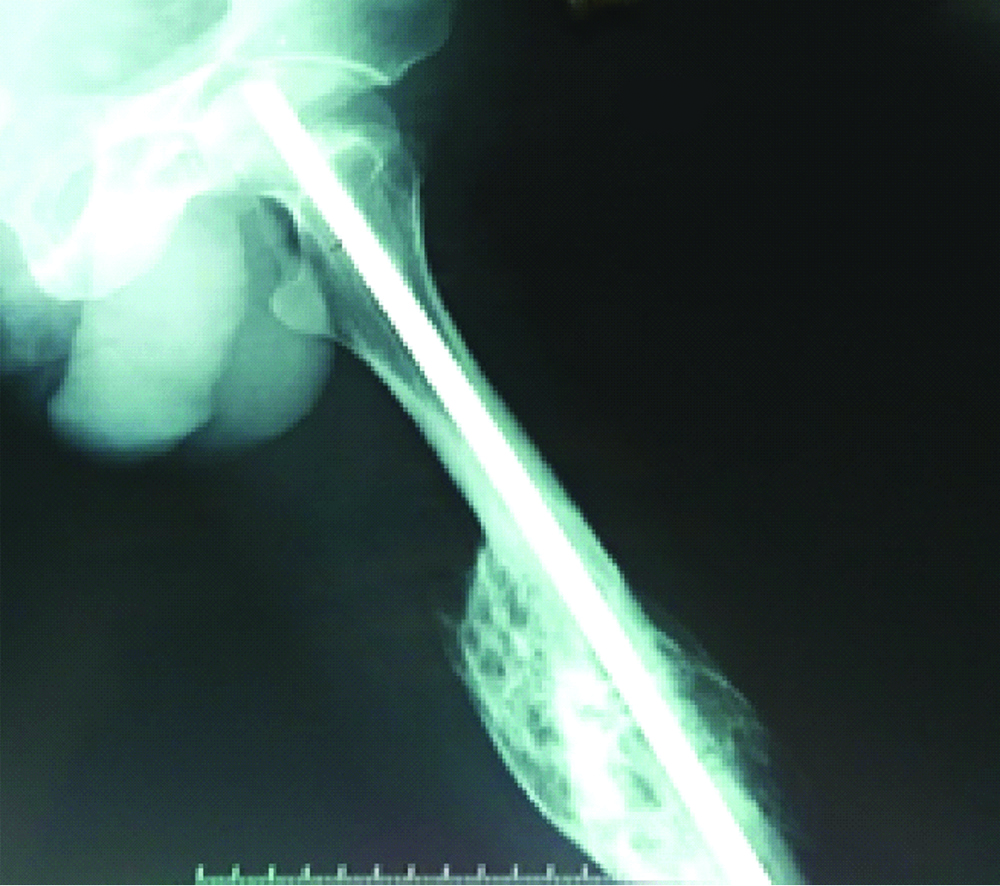
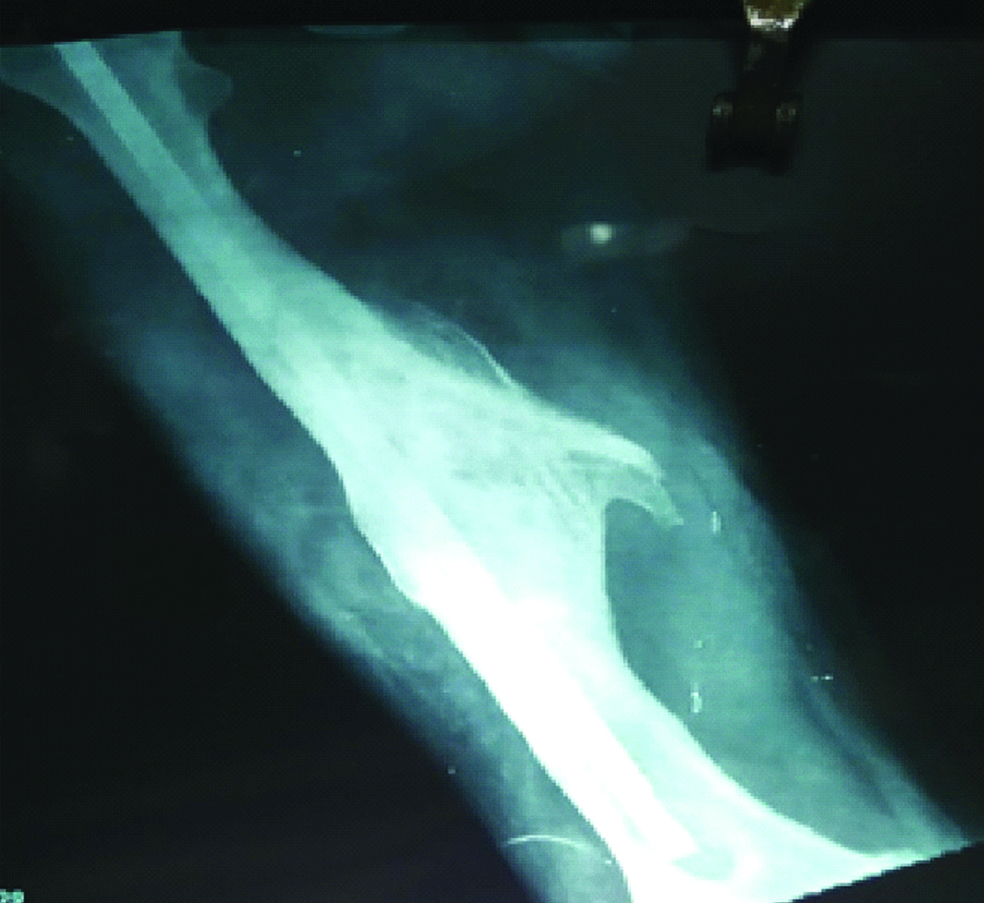
A 53-year-old male attended the outpatient department with complaints of persistent pain over the right gluteal region and swelling due to prominent implant. The patient met a car accident 20 years back and sustained a closed femoral shaft fracture treated with an open reduction internal fixation with Kuntscher nail. The patient started complaining of pain since the past one year and had developed swelling over the gluteal region with palpable proximal end of the nail. Radiographs revealed united fracture with an extensive callus formation and implant backing out [Table/Fig-1a,b].
Showing preoperative radiograph AP views.
Showing preoperative radiograph Lat views.
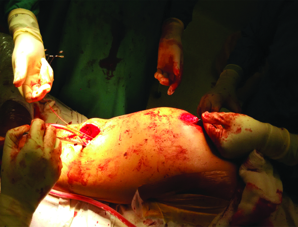
He was administered spinal anesthesia and placed in a lateral position with affected side upwards on the operating table. A 5 cm incision was made centred over the proximal end of nail and bone was exposed over the lateral aspect of femur. Another incision was made at lateral aspect of distal thigh at the level of nail tip under image guidance. Bone exposed, window created and a gigli saw wire was passed through the nail slot on anterolateral aspect and taken out from the distal end of the nail. Osteotomy was done with gigli saw wire and handles without exposing the whole length of femur, corresponding to the length of nail [Table/Fig-2]. Subsequently, small osteotome is introduced at osteotomy incision site and praying of cortex was done to complete the osteotomy.
intraoperative photograph showing the technique of small incision osteotomy.
The nail was hammered to remove it through the entry point. Nail was inspected to confirm that there was no breakage. The patient was discharged the next day with full mobilisation [Video-1].
Discussion
The use of Kuntscher nailing system for the fractured shaft of femur has been a major technological break through in nailing design. It is closed anterograde technique but subsequently, most Kuntscher nails were done retrograde, thereby opening the fracture site [1]. Removal of a Kuntscher nail is considered a routine procedure once its purpose has been served, but could become really challenging and may turn into a nightmare. Kuntscher nail is a cloverleaf-shaped hollow tubular nail with an anterolateral slot which allows for some plasticity in situ, there is an eye at the proximal end to engage the hook for extraction. Most commonly, pulling off the nail from the entry point is determined as a primary procedure for removal; however, slippage of the extraction device remains a major course of failure, in these cases impacting the nail from fracture site can be used as an alternative [2,3].
The availability of interlocking nails, improved surgical instruments and closed nailing techniques has favoured the exodus of Kuntscher nails [4]. Most of the nail removals done today are being done for nails inserted a decade ago and as the experience with nail removal and implantation decreases, the complications are bound to rise. This article presents a case of an incarcerated Kuntscher nail and describes a salvage procedure for the nail extraction after all previously described methods have failed.
The incidence of broken impacted Kuntscher nails has been reported from 1% to 3.3% [4,5]. The major reasons cited for incarceration have been bony ingrowth, excessive callus formation and/or bent or broken nails. Impacted Kuntscher nails are difficult to remove, therefore a number of techniques and instruments have been described for its removal [Table/Fig-3,4]. Universal nail extraction sets are available but they frequently fail and alternate techniques should be known. Georgiadis GM et al., described one technique using a carbide drill to create a hole in proximal fragment to attach the hook, but requires large soft tissue dissection to expose the proximal part of the nail [6]. Various other techniques have been described to extract the Kuntscher nails using hooks or stacked wires inserted through a window in the lateral femoral cortex, or Ender nails, which must be placed beyond the distal tip of a broken nail and then hooked through the locking holes of the nail [3,7,8].
Showing nail removal techniques used by various authors [2,3,6-12].
| Authors | Technique | No. of Patients | Complications | Recommendations |
|---|
| Mari R et al., [2] | Longitudinal femoral osteotomy | 1 | Non-union and redisplacement | Remove early |
| Liodakis E et al., [3] | Retrograde mobilisation through arthrotomy | 1 | Arthrotomy | Useful |
| Georgiadis GM et al., [6] | Creating new hole with carbide drill | 3 | Avoid embedding metal shavings | Recommended carbide drill bit/ conical extraction device. |
| Marwan M et al., [7] | Cerclage wire | 3 | Slippage | NA |
| Weinrauch PC et al., [8] | Proximal stacked wire | 1 | NA | Not useful for solid nails |
| Steinberg EL et al., (9) | Steinman pin and 10 mm k nail | 1 | NA | Adequate reaming |
| Park SY et al., [10] | Bent and hooked guide wires | 1 | Frequent slippage | NA |
| Maini L et al., [11] | Ender nail | 1 | Check the size of ender nail carefully | Not for larger nails |
| Rohilla et al | Bending the nail 90 | 1 | Breakage | NA |
The various techniques of intramedullary nail removal [6,7,13-21].
| Hollow Nails |
|---|
| High-speed drill to cut a hole in the nail [6] |
| Cerclage wire [7] |
| Custom-made hook [13] |
| Beaded guide [14] |
| Modified Kuntscher reaming guide [15] |
| Vise grip locking pliers [16] |
| Multiple guidewires [17] |
| Corkscrew extractor [18] |
| Solid Nails |
| Percutaneous osteotomies and grasping device [13] |
| K-wire mounted on a concave instrument [19] |
| Push-out technique [20] |
| Synthes extraction kit |
| Laparoscopic forceps [21] |
Retrieval of broken Kuntscher’s nail is difficult from other nails, such as interlocking nails, because of the presence of a slot in the nail. Thus, the methods listed above may become technically difficult or unsuccessful. Various techniques have been described such as use of Cerclage wire [7] multiple wire jamming [8] and Steinman pin technique [9], however these cannot be used with Kuntscher nail because of nail architecture.
The described technique of small incision osteotomy has a number of advantages like minimal soft tissue stripping, less morbidity and early recovery, in addition being a simple innovative technique which can be done using minimal equipment available in all orthopaedic operating rooms and no specialised or custom-made devices are required. To the best of authors’ knowledge and extensive literature search, the technique of small incision osteotomy has not been described prior to this.
Conclusion
Small incision osteotomy for Kuntscher nail removal is an innovative technique with many advantages such as shorter operative time, lesser blood loss, lesser morbidity, better cosmesis and early postoperative recovery. However, longer series is recommended to know the results and difficulties faced during removal. There are very few cases published in the literature using this technique for nail removal in a difficult stuck situation.
[1]. Knothe U, Knothe TML, Perren SM, 300 years of intramedullary fixation-from Aztec practice to standard treatment modalityEur J Trauma 2000 26:217-25.10.1007/PL00002445 [Google Scholar] [CrossRef]
[2]. Marí R, Valverde VD, A technical note for extracting an incarcerated femoral kuntscherNail J Orthopaedic Case Reports 2016 6(3):10-12.10.2174/187432500161001001227053972 [Google Scholar] [CrossRef] [PubMed]
[3]. Liodakis E, Krettek C, Hankemeier S, A new technique for removal of an incarcerated expandable femoral nailClin Orthop Relat Res 2010 468:1405-09.DOI 10.1007/s11999-009-1022-410.1007/s11999-009-1022-419655211 [Google Scholar] [CrossRef] [PubMed]
[4]. Bone LB, Johnson KD, Treatment of tibial fractures by reaming and intramedullary nailingJ Bone Joint Surg 1986 68A:87710.2106/00004623-198668060-00009 [Google Scholar] [CrossRef]
[5]. Olerud S, Karlstrom G, The spectrum of intramedullary nailing of tibiaClin Orthop 1986 212:101-12.10.1097/00003086-198611000-00012 [Google Scholar] [CrossRef]
[6]. Georgiadis GM, Heck BE, Ebraheim NA, Technique for removal of intramedullary nails when there is failure of the proximal extraction device: a report of three casesJ Orthop Trauma 1997 11:130-32.10.1097/00005131-199702000-00012 [Google Scholar] [CrossRef]
[7]. Marwan M, Ibrahim M, Simple method for retrieval of distal segment of the broken interlocking nailInjury 1999 30:333-35.10.1016/S0020-1383(99)00092-3 [Google Scholar] [CrossRef]
[8]. Weinrauch PC, Blakemore Extraction of intramedullary nails by proximal stacked wire techniqueJournal of Orthopaedic Trauma 2007 21(9):663-64.10.1097/BOT.0b013e3181583b3417921843 [Google Scholar] [CrossRef] [PubMed]
[9]. Steinberg EL, Luger E, Menahem A, Helfet DL, Removal of a broken distal closed section intramedullary nailJ Orthop Trauma 2004 18:233-35.10.1097/00005131-200404000-00007 [Google Scholar] [CrossRef]
[10]. Park SY, Yang KH, Yoo JH, Removal of a broken intramedullary nail with a narrow hollowJournal of Orthopaedic Trauma 2006 20(7):492-94.10.1097/00005131-200608000-00007 [Google Scholar] [CrossRef]
[11]. Maini L, Jain N, Singh J, Singh H, Bahl A, Gautam VK, Removal of a multisegmental broken nail by close technique using a TEN nailThe Journal of Trauma 2009 66(6):E78-E80.10.1097/01.ta.0000236051.08735.eb18277264 [Google Scholar] [CrossRef] [PubMed]
[12]. Singh R, Siwach R, Rohilla R, Kaur K, Innovative technique for extraction of a Kuntscher nail penetrating into the knee jointEuropean Orthopaedics and Traumatology 2011 2(1-2):51-53.10.1007/s12570-011-0048-8 [Google Scholar] [CrossRef]
[13]. Cohn BT, Bifield L, Fatigue fracture of a tibial interlocking nailOrthopedics 1986 9:1215-18. [Google Scholar]
[14]. Incavo SJ, Kristiansen TK, Retrieval of a broken intramedullary nailClin Orthop 1986 210:201-02.10.1097/00003086-198609000-00028 [Google Scholar] [CrossRef]
[15]. Yoslow W, LaMont J, Alternative method for removing an impacted AO intramedullary nailClin Orthop 1986 202:237-38.10.1097/00003086-198601000-00034 [Google Scholar] [CrossRef]
[16]. Sivananthan KS, Raveendran K, Kumar T, A simple method for removal of a broken intramedullary nailInjury 2000 31:433-34.10.1016/S0020-1383(00)00015-2 [Google Scholar] [CrossRef]
[17]. Franklin JL, Winquist RA, Benirsche SK, Hansen ST, Broken intramedullary nailsJ Bone Joint Surg Am 1988 70:1463-71.10.2106/00004623-198870100-00004 [Google Scholar] [CrossRef]
[18]. Wise DJ, Hutchins PM, Novel method for removal of a broken GK femoral nailInjury 1996 27:294-95.10.2106/00004623-198870100-00004 [Google Scholar] [CrossRef]
[19]. Krettek C, Schandelmaier P, Tscherne H, Removal of a broken solid femoral nail: a simple push-out technique. A case reportJ Bone Joint Surg Am 1997 79:247-51.10.2106/00004623-199702000-00013 [Google Scholar] [CrossRef]
[20]. Charnley GJ, Farrington WJ, Laparoscopic forceps removal of a broken tibial intra-medullary nailInjury 1998 29:489-90.10.1016/S0020-1383(98)00094-1 [Google Scholar] [CrossRef]
[21]. Schmidgen A, Naumann O, Wentzensen A, A simple and rapid method for removal of broken unreamedtibial nailsUnfallchirurg 1999 102:975-78.10.1007/s00113005051310643398 [Google Scholar] [CrossRef] [PubMed]