Materials and Methods
It was a prospective, cross-sectional study conducted in Department of Radiodiagnosis, Vinayaka Missions Medical College, Salem Tamil Nadu, India. Study was conducted for a period of one year from September 2012-September 2013. A total of 250 patients with routine antenatal check-up scan were included in the present study that fulfilled the inclusion and exclusion criteria. Institutional Ethical Committee Clearance was obtained for the study (IRB number: 2023). The details of each patient were entered in PCPNDT form and informed consent was obtained from every subject. Gestational age was assessed by menstrual history (last menstrual period) and in conjunction with BPD, HC, AC, and FL measured using pre-installed Hadlock obstetrics software in the machines.
Inclusion criteria: Cases with known last menstrual period, gestational age between 12 and 40 weeks and singleton pregnancy were included.
Exclusion criteria: Patients with multiple pregnancies, severe oligohydramnios, pregnancy-related complications such as pre-eclampsia, gestational diabetes, intrauterine growth retardation, those foetuses with suspected anomalies were excluded from the study.
Scanning Protocol
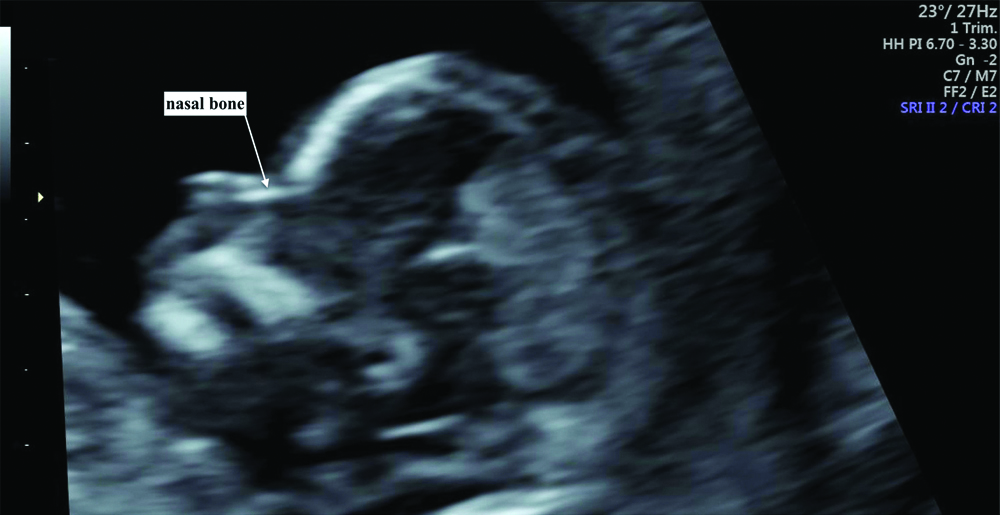
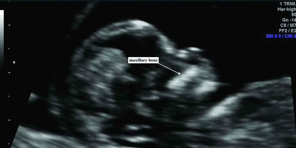
Curvilinear probe of 3.5 MHz of two GE ultrasound machines with similar pre-installed biometry software was used for the study. After completion of the measurement of the BPD, HC, AC and FL, identification and measurement of the nasal and maxillary bones were done. The plane of the nasal bone was strictly done in the median midsagittal plane of foetal face [Table/Fig-1]. Care was taken to keep the angle of insonation close to 45° to 135°. After the identification of the correct plane, three independent measurements were obtained and averaged to obtain the final measurement. From the midsagittal view, the probe was slightly moved posteriorly to visualise the maxillary bone which is rod-shaped [Table/Fig-2]. Similar to the nasal bone, three measurements of the maxillary bone were taken and averaged to obtain the final measurement. The scan was done by a single observer, to eliminate interobserver variation.
Plane of measurement of nasal bone.
Plane of measurement of maxillary bone length.
Statistical Analysis
Following data collection, statistical analysis was performed using Stats Direct Statistical software (version 2.7.8). The 5th, 50th and 95th percentiles of each parameter were described for each gestational age. Pearson’s correlation coefficients were calculated to examine the strength of the linear relationship between the nasal and maxillary bone with gestational age, BPD, HC, AC, and FL. For each parameter, regression analysis was used as a dependent variable to calculate the equation of the linear fitted function with each of the independent variables.
Results
In 250 cases, the maternal age ranged from 17 years to 37 years (24.20±4.15 years). The gestational age by LMP ranged from 12 weeks to 40 weeks. The mean nasal bone length increased linearly with gestational age from 2.9 mm at 12 weeks to 13.1 mm at 40 weeks. Nomograms for nasal and maxillary bone lengths were tabulated. The mean maxillary bone length increased linearly with gestational age from 8.7 mm at 12 weeks to 15.6 mm at 40 weeks [Table/Fig-3]. Both NBL and MBL increased linearly with increasing gestational age. According to Pearson’s correlation, they were found to have significant positive correlation with gestational age, BPD, HC, AC and FL [Table/Fig-4,5].
| Gestational age (Weeks) | Nasal bone length (Mm) | Maxillary bone length (Mm) |
|---|
| 5% | 50% | 95% | 5% | 50% | 95% |
|---|
| 12-13 | 2.5 | 2.9 | 3.3 | 7.7 | 8.7 | 9.7 |
| 14-15 | 4.0 | 4.2 | 4.4 | 9.9 | 10.1 | 10.3 |
| 16-17 | 4.7 | 5.5 | 6.3 | 10.4 | 10.6 | 10.8 |
| 18-19 | 6.1 | 6.3 | 6.5 | 11.0 | 11.2 | 11.4 |
| 20-21 | 6.6 | 6.8 | 7.0 | 11.4 | 11.6 | 11.8 |
| 22-23 | 7.2 | 7.4 | 7.6 | 11.2 | 11.4 | 11.6 |
| 24-25 | 8.2 | 8.6 | 9.0 | 11.9 | 12.1 | 12.3 |
| 26-27 | 8.9 | 9.1 | 9.3 | 12.7 | 13.1 | 13.5 |
| 28-29 | 9.5 | 9.7 | 9.9 | 13.4 | 13.8 | 14.2 |
| 30-31 | 10.2 | 10.4 | 10.6 | 12.8 | 13.2 | 13.6 |
| 32-33 | 9.5 | 10.3 | 11.1 | 12.5 | 13.7 | 14.9 |
| 34-35 | 10.4 | 10.8 | 11.2 | 14.2 | 14.6 | 15.0 |
| 36-37 | 10.9 | 11.1 | 11.3 | 13.4 | 14.8 | 16.2 |
| 38-39 | 11.6 | 12.0 | 12.4 | 15.1 | 15.3 | l5.5 |
| 40 | 12.9 | 13.1 | 13.3 | 15.4 | 15.6 | 15.8 |
Regression equation for NBL.
| Gestational age | 1.158-0.011×Gestational age (r=0.989; p<0.0001) |
|---|
| BPD | 0.053+0.124×BPD (r=0.989; p<.0001) |
| HC | 0.035+0.0365×HC ([r=0.989; p<.0001) |
| AC | 0.033+0.154×AC ([r=0.986; p<.0001) |
| FL | 0.209+0.137×FL (r=0.982; p<.0001) |
Regression equation for MBL.
| Gestational age | 1.4742-0.0075×Gestational age (r=0.989; p<.0001) |
|---|
| BPD | 0.699+0.088×BPD (r=0.989; p<.0001) |
| HC | 0.688+0.0252×HC (r=0.963; p<.0001) |
| AC | 0.769+0.0234×AC (r=0.966; p<.0001) |
| FL | y=0.807+0.097×FL (r=0.947; p<.0001) |
The ratios of nasal bone length and maxillary bone length with BPD, HC and AC, FL remained virtually constant throughout the gestation [Table/Fig-6,7].
Ratio for nasal bone length.
| GA | NBL |
|---|
| Total Number of Cases | 50th % (mm) | SD | NBL/BPD | NBL/HC | NBL/AC | NBL/FL |
|---|
| 12-13 | 10 | 2.9 | 0.2 | 0.13 | 0.03 | 0.04 | 0.19 |
| 14-15 | 14 | 4.2 | 0.1 | 0.14 | 0.03 | 0.04 | 0.22 |
| 16-17 | 17 | 5.5 | 0.4 | 0.14 | 0.04 | 0.04 | 0.21 |
| 18-19 | 15 | 6.3 | 0.1 | 0.14 | 0.03 | 0.04 | 0.21 |
| 20-21 | 17 | 6.8 | 0.1 | 0.13 | 0.03 | 0.04 | 0.19 |
| 22-23 | 33 | 7.4 | 0.1 | 0.12 | 0.03 | 0.04 | 0.19 |
| 24-25 | 28 | 8.6 | 0.2 | 0.13 | 0.03 | 0.04 | 0.18 |
| 26-27 | 19 | 9.1 | 0.1 | 0.13 | 0.03 | 0.04 | 0.18 |
| 28-29 | 15 | 9.7 | 0.1 | 0.13 | 0.03 | 0.03 | 0.17 |
| 30-31 | 10 | 10.4 | 0.1 | 0.13 | 0.03 | 0.04 | 0.17 |
| 32-33 | 18 | 10.3 | 0.4 | 0.12 | 0.03 | 0.03 | 0.16 |
| 34-35 | 22 | 10.8 | 0.2 | 0.12 | 0.03 | 0.03 | 0.16 |
| 36-37 | 20 | 11.1 | 0.1 | 0.12 | 0.03 | 0.03 | 0.16 |
| 38-39 | 7 | 12.0 | 0.2 | 0.13 | 0.03 | 0.03 | 0.16 |
| 40 | 5 | 13.1 | 0.1 | 0.14 | 0.04 | 0.03 | 0.14 |
Ratio for maxillary bone length.
| GA | MBL |
|---|
| Total Number of Cases | 50th % (mm) | SD | MBL/BPD | MBL/HC | MBL/AC | MBL/FL |
|---|
| 12-13 | 10 | 8.7 | 0.5 | 0.40 | 0.11 | 0.13 | 0.58 |
| 14-15 | 14 | 10.1 | 0.1 | 0.34 | 0.09 | 0.11 | 0.53 |
| 16-17 | 17 | 10.6 | 0.1 | 0.28 | 0.07 | 0.09 | 0.45 |
| 18-19 | 15 | 11.2 | 0.1 | 0.25 | 0.06 | 0.08 | 0.37 |
| 20-21 | 17 | 11.6 | 0.1 | 0.22 | 0.06 | 0.07 | 0.33 |
| 22-23 | 33 | 11.4 | 0.1 | 0.19 | 0.05 | 0.06 | 0.30 |
| 24-25 | 28 | 12.1 | 0.1 | 0.18 | 0.05 | 0.06 | 0.26 |
| 26-27 | 19 | 13.1 | 0.2 | 0.19 | 0.05 | 0.06 | 0.26 |
| 28-29 | 15 | 13.8 | 0.2 | 0.18 | 0.05 | 0.05 | 0.25 |
| 30-31 | 10 | 13.2 | 0.2 | 0.17 | 0.03 | 0.04 | 0.22 |
| 32-33 | 18 | 13.7 | 0.6 | 0.16 | 0.04 | 0.04 | 0.21 |
| 34-35 | 22 | 14.6 | 0.2 | 0.17 | 0.04 | 0.05 | 0.23 |
| 36-37 | 20 | 14.8 | 0.7 | 0.16 | 0.04 | 0.04 | 0.21 |
| 38-39 | 7 | 15.3 | 0.1 | 0.17 | 0.04 | 0.04 | 0.21 |
| 40 | 5 | 15.6 | 0.1 | 0.17 | 0.04 | 0.04 | 0.21 |
Discussion
The second-trimester sonographic screening for Down syndrome is based on morphologic and biometric parameters, like nuchal fold thickening, hyperechoic bowel, pyelectasis, short humerus and femur. The importance of absent nasal bone or shorter nasal bone length for gestation age was observed in foetuses with Down syndrome [7,8]. Nomogram is most essential to differentiate normal from abnormal parameter in any population. A few studies have assessed the foetal nasal bone lengths at different gestational age [Table/Fig-8] [10-14]. Sonek JD et al., had studied the normal range of nasal bone length in 11th to 40th week gestations in 3537 patients which ranged from 2.3 mm to 12.1 mm in length [10]. In the present study, the nasal bone was evaluated from 12 to 40 weeks gestation and was 2.9 mm at 12 to 13 weeks and 13.1 mm at 40 weeks. In a previous study by Cicero S et al., the absence or hypoplasia (defined as 2.5 mm) of the foetal nasal bone was the single most sensitive and specific second-trimester marker for Down syndrome, with a detection rate of 61.8% and a false positive rate of only 1.2% [7]. In comparison with the studies done in the Indian population [13,14], the nasal bone measurements in the present study were slightly higher.
Comparison with other studies on nasal bone [10-14].
| Gestational age included in the study (weeks) | Total number of patients studies | Range of nasal bone length (mm) |
|---|
| This study | 12-40 | 250 | 2.9-13.1 |
| Sonek JD et al., [10] | 11-40 | 3537 | 2.3-12.1 |
| Persico N et al., [11] | 16-24 | 135 | 4.1-7.1 |
| Gamez F et al., [12] | 19-22 | 2035 | 6.19-6.77 |
| Narayani BH et al., [13] | 16-26 | 2962 | 3.3-6.65 |
| Kashikar SV et al., [14] | 17-22 | 486 | 3.7-7.0 |
Goldstein I et al., evaluated maxillary bone lengths throughout gestation between 13 to 40 weeks [15]. The maxillary bone length at 50th percentile was 10 mm at 14 weeks. The mean range of maxillary bone length was 9.97 mm to 14.84 at 14-40 gestational weeks. In the present study, the maxillary bone was 10.1 mm at 14 weeks and 15.6 mm at 40 weeks. In comparison, the maxillary bone length was similar to 14-34 weeks gestational age and present study showed a higher MBL from 34-40 weeks.
The ratio of NBL/FL at 40 weeks GA was skewed and so was the ratios between the MBL and BPD, HC, AC and FL in the early second-trimester. This was probably attributed to the less of number of cases in the group.
Limitation
The present study included a small group of patients and was confined to one group population. A large population and a multicentric study in the Indian population to assess the sensitivity and specificity when using our normal reference range as cut-off values are required.
Conclusion
Normative data for ultrasonographic measurements of the foetal nasal and maxillary bone lengths throughout pregnancy are provided. Both nasal and maxillary bone length are dependent variables of gestational age as determined by the last menstrual period. There was significant correlation with biparietal diameter, head circumference, abdominal circumference and femoral length. These data potentially allow the prenatal diagnosis of abnormal nasal and maxillary bone length which is a marker for chromosomal defects.