Overjet Reduction with the Use of the ELIBA Device: A Case Report
Eleonora Ortu1, Davide Pietropaoli2, Ruggero Cattaneo3, Mario Giannoni4, Annalisa Monaco5
1 DDS, PhD, Department of MeSVA, University of L’Aquila, Italia.
2 DDS, PhD, Department of MeSVA, University of L’Aquila, Italia.
3 MD, Department of MeSVA, University of L’Aquila, Italia.
4 Professor, Department of MeSVA, University of L’Aquila, Italia.
5 Professor, Department of MeSVA, University of L’Aquila, Italia.
NAME, ADDRESS, E-MAIL ID OF THE CORRESPONDING AUTHOR: Eleonora Ortu, Department of MeSVA, University of L’Aquila, P.le Salvatore Tommasi-67100, L’Aquila, Italia.
E-mail: eleortu@gmail.com
The dental overjet, in subjects with skeletal class II, division I, malocclusion represents one of the difficult problems to resolve for the orthodontist. We describe a case of dental overjet reduction, treated with the Lingual Elevator by Balercia (ELIBA). This orthodontic approach, convenient for patients, can help the orthodontist to solve complex cases of dental overjet.
Malocclusion, Orthodontist, TENS, Teeth
Case Report
A healthy eight-year-old Caucasian girl, had a chief complaint of forwardly placed upper teeth. Medical history of the child was insignificant. The dental history was also insignificant.
Extraoral examination revealed that the patient had skeletal class II subdivision, mandibular deficit and was breathing through mouth. Intraoral examination revealed that the patient had molar class II relation on right side and molar class I relation on left side, incorrect swallowing with tongue interposition, low tongue posture, exhibiting an overjet of 6 mm and presence of caries in the deciduous teeth.
A dental panoramic radiograph was prescribed that [Table/Fig-1] revealed a lack of space in the upper arch for the eruption of the teeth (especially for the canines and for the rotated lateral incisors), extraoral and intraoral photos were taken (frontal, right and left) [Table/Fig-2a-e] and alginate impressions of both the dental arches were made to plan the treatment according to the IOTN index described by Brook PH et al., [1]. According to the IOTN, the patient displayed a grade 3 treatment requirement (Increased overjet >3.5 mm but ≤6 mm).
Dental panoramic radiograph of the patient revealing the lack of the space for the eruption of the teeth.

The extraoral and intraoral images of the patient before the treatment with the ELIBA. a) Patient’s right profile. b) Frontal image of the patient. c) Patient’s left profile. d) Intraoralimage showing the overjet of 6 mm prior to treatment. e) Frontal aspect of the teeth prior to the treatment.

The treatment modalities were discussed with the parents of the child and it contemplated the use of the ELIBA device for the rehabilitation of the tongue (ELIBA reproduced perfectly the oro-lingual space) and the consequent reduction of the overjet, and the use of the Schwarz devices for the complete eruption of the teeth. Bimler device was used to finalise the case. The dental and oral health of the patient was good, infact the patient was also under the care of the dental hygienist of the dental clinic. An informed consent was obtained from the parents before the treatment.
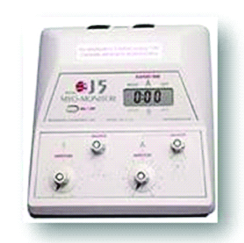
After analysing the dental stone models of the dental arches, physiologic free oro-lingual space impression, for the construction of the ELIBA, was recorded using Myoprint (Bosworth Sapphire, Boswort Company). This is an acrylic resin that on hardening does not change its consistency over the time under Ultra Low Frequency-Transcutaneous Electrical Nervous Stimulation (ULF-TENS) [2]. The low-frequency neurostimulator generates a repetitive synchronous and bilateral stimulus through pulsed currents with adjustable amplitude of approximately 0-24 mA, duration of 500 ms, and a frequency of 0.66 Hz. The two TENS electrodes were placed bilaterally over the cutaneous projection of the notch of the fifth pair of cranial nerves, located between the coronoid and condylar processes and it was retrieved by manual palpation in the zone anterior to the tragus; a third grounding electrode was placed in the centre of the back of the neck [3]. Based on the literature, central nervous system stimulation was obtained by sensory stimulation of cranial nerves V and VII with low-frequency TENS. Low-frequency TENS is typically applied at higher intensities that produce motor contractions on purpose to lead lower jaw in a neutral position in which, it is no more conditioned by occlusal adjustments [4,5]. J5 Myomonitor® TENS Unit device (Myotronics-Noromed, Inc., Tukwila, WA, USA) [Table/Fig-3] with disposable electrodes (Myotrode SG Electrodes®, Myotronics-Noromed, Inc., Tukwila, WA, USA) was used. Patient was asked to open her mouth and Bosworth sapphire resin (Sapphire is a custom acrylic material for taking peripheral impressions) was put. Sapphire extends to the appropriate height without slumping. This soft material records peripheral detail without distortion and sets to become a rigid part of the tray in physiologic free oro-lingual space. The patient bites in protrusion touching the incisive papilla with the tip of the tongue all the time for precise impression taking. So, ulf-TENS was raised to motor threshold until lower jaw begins to move (7/10 spikes), then it was lowered to sensorial threshold until Myoprint was totally stiffened [Table/Fig-4a-c]. ELIBA was formed in dental laboratory, completely reproducing the Myoprint impression of physiologic free oro-lingual space, Crozat clasps were used on first permanent lower molar to adequately stabilise the oral device [Table/Fig-4c-e]. ELIBA must be totally passive and should not touch oral mucosa [4-9].
J5 Myomonitor® TENS Unit device.

The ELIBA device and its production. a) Dental stone models of dental arches. b) Myoprint impression with Sapphire resin over the dental cast. c) Myoprint impression of the perfect physiologic free oro-lingual space. d) Eliba device with Crozat clasps on first permanent lower molars. e) Eliba device over the dental cast.

The pretreatment overjet (6 mm) reduced to 2 mm after 12 months as assessed between the maxillary and right mandibular central incisors by using an orthodontic ruler. After 12 months of using the ELIBA device, the IOTN index passed from grade 3 to grade 1, as shown in [Tables/Fig-5a-f]. Schwarz devices were used by the orthodontist to complete the case and allow the complete eruption of the teeth. To finish, a Bimler was used to finalise the case. The patient completed the orthodontic treatment with a satisfactory alignment of both dental arches, and the tongue function was rehabilitated (final swallowing with the tongue in its position).
The extraoral and intraoral images of the patient after the treatment with the ELIBA. a) Patient’s right profile. b) Frontal photograph of the patient. c) Patient’s left profile. d) Intraoral right image that shows the reduction of the overjet e) Frontal image of the teeth. f) Intraoral left image of the patient that shows the reduction of the overjet.

Discussion
Class II, division I malocclusion is more frequent in Caucasian population and the identification of the aetiological causes is more important for the success of the orthodontic treatment. Infact, there is certainly a genetic factor, but the tongue position, lip and cheek, oral respiration are majorly related to this malocclusion [10]. A low tongue posture is associated with mouth breathing and developing inflammatory diseases such as otitis, tonsillitis, sinusitis and adenoid hypertrophy [11-14]. Last but not least, mouth breathers with a low tongue posture often suffer from chronic neck pain and poor posture [2].
The first step for the resolution of this malocclusion, is the rehabilitation of the tongue posture and the consequently reduction of the dental overjet. Dental overjet is the horizontal overlapping of incisal ridges on sagittal plane. According to Grainger RM, a value of overjet more than 6 mm is detected in 23% of pedodontic patients from 6 to 12 years and in 15% of people from 12 to 17 years [15]. The objective of an orthodontist is to plan a correct treatment to enhance patient’s masticatory function and aesthetics. There are several orthodontic devices, both fixed and removable, which are used by the orthodontist based on patient’s clinical features [16].
The patient described in this case was treated with the ELIBA, a device that allow the complete rehabilitation of the tongue and the reduction of the dental overjet [1]. Lingual Elevator by Balercia was discovered according to meticulous laws of neuromyofascial occlusal dynamics. This oral device was chosen because it is not heavy and large and this enhances patients’ compliance. This device was designed by Professor Balercia on purpose to treat the “atypical swallowing”. ELIBA seizes the patient’s physiologic free oro-lingual space, defined by lower jaw anteriorly and laterally, by oral floor inferiorly and superiorly by ventral surface of the tongue. The ELIBA modifies the pattern of swallowing by mainly acting on the inter-component of the trigeminal system and partly on the autonomic nervous system, bringing improvements to the skull-sacral system. The Medium Longitudinal File (FLM) allows a direct connection between the various cranial nerves: it is a very important way of connecting the upper part of the spinal cord and the mesencephalus. This device, through a mechanism of proprioception, interception, stimulates the trigeminal system, which in turn, for the mechanisms described above, stimulates the occulomotor system [17]. Literature shows that tongue and neck muscles are related because of common proprioception by a common trunk from ansa cervicalis through the hypoglossal nerve [18]. It appears that several factors account for the persistence of incorrect swallowing patterns and that tongue thrust plays an important role in the aetiology of open bite as well as in the relapse of treated open bite patients. Today, the researches with regard to swallowing lead us to conclude that “it is not possible to modify it” because swallowing is subcortical, precisely at the ventral part of the rostral part of the trunk of the brain [19,20]. The lingual elevator by Balercia, through a mechanism of proprioception and interception, properly stimulates the trigeminal system which in turn, for the mechanisms described above, also stimulates the posture. ELIBA device helps to keep healthier posture and more free movements of tongue. Apex of the tongue touching its spot during swallowing together with centripetal force of perioral muscles act as an orthodontic device to enhance reduction of the overjet, with functional and aesthetic improvements. In literature, other authors used the lingual elevator to modify the position of the tongue in the orthodontic objectives [21,22].
Conclusion
Enhancing the dynamic function, and thereby the cause of the malocclusion, makes orthodontic relapse much less likely and thus maintains stable occlusion. The present approach with Eliba can be easily used by the orthodontist and it is also comfortable for the patients.
[1]. Brook PH, Shaw WC, The development of an index of orthodontic treatment priorityEuropean Journal of Orthodontics 1989 11(3):309-20.Epub 1989/08/0110.1093/oxfordjournals.ejo.a0359992792220 [Google Scholar] [CrossRef] [PubMed]
[2]. Monaco A, Petrucci A, Marzo G, Necozione S, Gatto R, Sgolastra F, Effects of correction of Class II malocclusion on the kinesiographic pattern of young adolescents: a case-control studyEuropean Journal of Paediatric Dentistry: Official Journal of European Academy of Paediatric Dentistry 2013 14(2):131-34.Epub 2013/06/14 [Google Scholar]
[3]. Annalisa Monaco and Ruggero Cattaneo, “La TENS per usoodontoiatrico” 2016 Futura Publishing Society [Google Scholar]
[4]. Ortu E, Pietropaoli D, Mazzei G, Cattaneo R, Giannoni M, Monaco A, TENS effects on salivary stress markers: A pilot studyInternational Journal of Immunopathology and Pharmacology 2015 28(1):114-18.Epub 2015/03/3110.1177/039463201557207225816413 [Google Scholar] [CrossRef] [PubMed]
[5]. Monaco A, Cattaneo R, Spadaro A, Marzo G, Neuromuscular diagnosis in orthodontics: effects of TENS on the sagittal maxillo-mandibular relationshipEuropean journal of paediatric dentistry: official journal of European Academy of Paediatric Dentistry 2008 9(4):163-69.Epub 2008/12/17 [Google Scholar]
[6]. Castroflorio T, Farina D, Bottin A, Debernardi C, Bracco P, Merletti R, Non-invasive assessment of motor unit anatomy in jaw-elevator musclesJournal of oral rehabilitation 2005 32(10):708-13.Epub 2005/09/1510.1111/j.1365-2842.2005.01490.x16159347 [Google Scholar] [CrossRef] [PubMed]
[7]. Falla D, Dall’Alba P, Rainoldi A, Merletti R, Jull G, Location of innervation zones of sternocleidomastoid and scalene muscles-a basis for clinical and research electromyography applicationsClinical Neurophysiology: Official Journal of the International Federation of Clinical Neurophysiology 2002 113(1):57-63.Epub 2002/01/2210.1016/S1388-2457(01)00708-8 [Google Scholar] [CrossRef]
[8]. Ortu E, Lacarbonara M, Cattaneo R, Marzo G, Gatto R, Monaco A, Electromyographic evaluation of a patient treated with extraoral traction: a case reportEuropean Journal of Paediatric Dentistry 2016 17(2):123-28. [Google Scholar]
[9]. Ortu E, Pietropaoli D, Adib F, Masci C, Giannoni M, Monaco A, Electromyographic evaluation in children orthodontically treated for skeletal Class II malocclusion: Comparison of two treatment techniquesCranio: The Journal of Craniomandibular Practice 2017 :1-7.Epub 2017/11/17 [Google Scholar]
[10]. Partal I, Aksu M, Changes in lips, cheeks and tongue pressures after upper incisor protrusion in Class II division 2 malocclusion: a prospective studyProgress in orthodontics 2017 18(1):29Epub 2017/09/2610.1186/s40510-017-0182-028944417 [Google Scholar] [CrossRef] [PubMed]
[11]. Ortu E, Sgolastra F, Barone A, Gatto R, Marzo G, Monaco A, Salivary Streptococcus Mutans and Lactobacillus spp. levels in patients during rapid palatal expansionEuropean Journal of Paediatric Dentistry: Official Journal of European Academy of Paediatric Dentistry 2014 15(3):271-74.Epub 2014/10/13 [Google Scholar]
[12]. Ortu E, Pietropaoli D, Ortu M, Giannoni M, Monaco A, Evaluation of cervical posture following rapid maxillary expansion: a review of literatureThe Open Dentistry Journal 2014 8:20-27.Epub 2014/05/0710.2174/187421060140801002024799964 [Google Scholar] [CrossRef] [PubMed]
[13]. Ortu E, Giannoni M, Ortu M, Gatto R, Monaco A, Oropharyngeal airway changes after rapid maxillary expansion: the state of the artInternational Journal of Clinical and Experimental Medicine 2014 7(7):1632-38.Epub 2014/08/16 [Google Scholar]
[14]. Pietropaoli D, Tatone C, D’Alessandro AM, Monaco A, Possible involvement of advanced glycation end products in periodontal diseasesInternational Journal of Immunopathology and Pharmacology 2010 23(3):683-91.Epub 2010/10/1510.1177/03946320100230030120943037 [Google Scholar] [CrossRef] [PubMed]
[15]. Grainger RM, Orthodontic treatment priority indexVital Health Stat 2 1967 (25):1-49.Epub 1967/12/01 [Google Scholar]
[16]. Aprile G, Ortu E, Cattaneo R, Pietropaoli D, Giannoni M, Monaco A, Orthodontic management by functional activator treatment: a case reportJournal of Medical Case Reports 2017 11(1):336Epub 2017/12/0310.1186/s13256-017-1505-y29195511 [Google Scholar] [CrossRef] [PubMed]
[17]. Marchili N, Ortu E, Pietropaoli D, Cattaneo R, Monaco A, Dental occlusion and ophthalmology: a literature reviewThe Open Dentistry Journal 2016 10:460-68.Epub 2016/10/1410.2174/187421060161001046027733873 [Google Scholar] [CrossRef] [PubMed]
[18]. Edwards IJ LV, Paton JF, Yanagawa Y, Szabo G, Deuchars SA, Neck muscle afferents influence oromotor and cardiorespiratory brainstem neural circuitsBrain Struct Funct 2015 220(3):1421-36.10.1007/s00429-014-0734-824595534 [Google Scholar] [CrossRef] [PubMed]
[19]. Monaco A, Cattaneo R, Masci C, Spadaro A, Marzo G, Effect of ill-fitting dentures on the swallowing duration in patients using polygraphyGerodontology 2012 29(2):e637-44.Epub 2011/09/2010.1111/j.1741-2358.2011.00536.x21923894 [Google Scholar] [CrossRef] [PubMed]
[20]. Monaco A, Cattaneo R, Mesin L, Ortu E, Giannoni M, Pietropaoli D, Dysregulation of the descending pain system in temporomandibular disorders revealed by low-frequency sensory transcutaneous electrical nerve stimulation: a pupillometric studyPloS one 2015 10(4):e0122826Epub 2015/04/2410.1371/journal.pone.012282625905862 [Google Scholar] [CrossRef] [PubMed]
[21]. Seo YJ, Kim SJ, Munkhshur J, Chung KR, Ngan P, Kim SH, Treatment and retention of relapsed anterior open-bite with low tongue posture and tongue-tie: A 10-year follow-upKorean Journal of Orthodontics 2014 44(4):203-16.Epub 2014/08/1910.4041/kjod.2014.44.4.20325133135 [Google Scholar] [CrossRef] [PubMed]
[22]. Kim YS, Kown SY, Park YG, Chung KR, Clinical application of the tongue elevatorJournal of Clinical Orthodontics: JCO 2002 36(2):104-06.Epub 2002/03/21 [Google Scholar]