Twain Impaction-A Novel Orthodontic Approach
Karnati Kumar Reddy Praveen1, Priyank Seth2, Karnati Chaitanya3, Gopuvaram Gayathri4, Myint Wei5
1 Senior Lecturer, Department of Orthodontics, Faculty of Dentistry, SEGi University, Petaling Jaya, Selangor, Malaysia.
2 Senior Lecturer, Department of Orthodontics, Faculty of Dentistry, SEGi University, Petaling Jaya, Selangor, Malaysia.
3 Senior Lecturer, Department of Orthodontics, Faculty of Dentistry, SEGi University, Petaling Jaya, Selangor, Malaysia.
4 General Dental Practitioner, Private Practice, Frisco, Texas, USA.
5 Senior Lecturer, Department of Oral Surgery, Faculty of Dentistry, SEGi University, Petaling Jaya, Selangor, Malaysia.
NAME, ADDRESS, E-MAIL ID OF THE CORRESPONDING AUTHOR: Karnati Kumar Reddy Praveen, Jalan Teknologi Kota Damansara, Petaling Jaya, Selangor, Malaysia.
E-mail: kpkr823@gmail.com
Primary failure of eruption (PFE) contend one of the varied rationale for etiology of malocclusions with eruption sequence disturbances. In a contemporary world of modernisation and advanced technological innovations, orthodontics is being revising its literature with new dimensions fortifying the biomechanics with biological considerations. The Poul Gjessing universal spring which is a distinguished and highly efficient spring assured simultaneous three dimensional control of teeth, which being a sculler achievement of many specialists. This seldom case report details a unique in its incidence with ectopically erupted and impacted first premolar interfering the late eruption pathway of canine, leading to twain impaction.
Canine impaction, Premolar impaction, PG Spring, Primary eruption failure, 3-D tooth movement
Case Report
A 20-year-old, Chinese origin male patient reported to the clinic with a chief complaint of gaps between his teeth and inability to chew on his right side, which made him disturbed during peer gathering. His medical history was non-contributory to the malocclusion and no familial expressions were noticed to patient consciousness apart from mild irregularities among siblings with proper recorded consent.
Diagnosis
On clinical examination, extra oral features were balanced with only increase in the lower facial height, shallow mentolabial sulcus and potentially incompetent lips [Table/Fig-1a]. Intraorally the hygiene was fair with full complement of permanent teeth except right permanent maxillary canine (13) and first premolar (14) with their predecessors, retained deciduous canine (53) with first molar (54) and the third molars [Table/Fig-1b]. There was no evident reason inferred either from the case history or clinical examination for the retention of primary teeth. A shallow crater like feature was evident both clinically and radiographically in relation to 54, with inward sloping of adjacent alveolar bone depicting the localised focal area of “fold-in” effect of anterior and posterior right maxillary alveolar segments suspecting a non-responding or negative inward pull force from within the alveolar bone, leading to lateral open bite [Table/Fig-1b,c].
However, generalised anterior spacing was seen in both the arches with normal overjet, overbite and end on posterior inter-occlusal relationship on right side. Occlusal wear facets were evident as a feature of pathologic canine guidance during left side excursion. A comprehensive diagnosis revealed Angle’s Class II division 1 subdivision dento-alveolar malocclusion on Class-I skeletal bases with class I soft tissue profile and retained primary teeth, impacted permanent teeth, increased lower facial height, dental midline incongruence, proclined-protruded incisors and strained mentalis muscle on closure of lips [Table/Fig-2].

Pretreatment cephalometric tracing values.
| Parameters | Normal | Obtained | Inference |
|---|
| SNA | 82° | 79° | Retrognathic Maxilla |
| SNB | 80° | 77° | Retrognathic Mandible |
| ANB | 2° | 2° | Class I Skeletal bases |
| MPA | 32° | 41° | High |
| UI-NA (Angle) | 22° | 30° | Proclined Incisor |
| UI-NA (Linear) | 4 mm | 11.5 mm | Protruded Incisor |
| LI-NB (Angle) | 25° | 33° | Proclined Incisor |
| LI-NB (Linear) | 4 mm | 12 mm | Protruded Incisor |
| UI-LI (Angle) | 131° | 110° | Proclined Incisors |
| “S” Line-UL | 0 mm | 3 mm | Protrusive Lip |
| “S” Line-LL | 0 mm | 6 mm | Protrusive Lip |
| Wit’s Appraisal | -1 (Males) | -1 | Class I Skeletal bases |
Treatment Objectives
The objectives were to correct the dental component to establish function and aesthetics with good alignment and levelling of the arches. Extraction of 53 and 54 was planned to facilitate passive eruption of impacted teeth thereby, uprighting of 14 can provide space for canine eruption.
Treatment Plan and Procedure
Surplus space deemed non-extraction approach with 0.022 MBT prescriptions (adequate control on force and arch stability). Aligning and levelling was achieved sequentially till 0.019X0.025 NiTi. The PG Universal Spring (PGUS) [1] made of 0.016 X 0.022 SS was preferred to upright the first premolar. Once the objective of uprighting, retraction and proper bucco-palatal root position achieved, the spring was discontinued and 0.017X0.025 NiTi was loaded as continuous arch wire with open coil spring to prevent relapse and maintain the space [Table/Fig-3]. The impacted canine was stationary in its position requiring surgical exposure.
Midtreatment opg showing the uprighting of 14 (a) with PGUS and achieving three dimentional stability compared to pre (b) and mid treatment (c) periapical radiographs.

Discussion
Primary failure of eruption (PFE) contend one of the varied rationale for aetiology of malocclusions with eruption sequence disturbances resulting in partial or complete unerupted teeth in the absence of a mechanical obstruction. The manifestations were first described by Proffit WR and Vig KW, [2]. Its prevalence being 0.06% with the posterior teeth most commonly affected, was grouped into three different types depending on the features presented either mesial to or distal aspect of involved tooth and severity as described by Frazier-Bowers SA et al., [3]. Later, It was also stemmed into primary failure of eruption (PFE), mechanical failure of eruption and indeterminate failure of eruption depending upon the cause by Sylvia A et al. However, genetic mutation in the PTH1R gene is also one systemic factor resulting in PFE [4]. In this case, the primary failure of eruption of 14 leads to mechanical interference and failure of eruption of 13, contemplating the chronologic and eruption sequence of 13.
In a contemporary world of modernisation and advanced technological innovations, orthodontics, is being revising its literature with new dimensions fortifying biomechanics with biological considerations [5-8]. The bio-friction between the root and bundle bone generated, spread the transparency over experimenting materials, bracket prescriptions, mechanics, designs, accessibility and their combinations to achieve optimal forces for three dimensional control and conceptual placement of tooth along with anchorage configurations [9-11]. The Poul Gjessing (1985) universal spring, commonly used for retraction, intrusion and uprighting of anterior teeth has other clinical applications also such as alignment of the buccal teeth and extrusion movements. All these applications are achieved by the sectional wires to avoid the untoward effects of a continuous arch wire; either includes en-masse incisor retraction or individual canine retraction [11-13]. A novel approach was presented by uprighting first premolar by this distinguished and versatile spring which assured simultaneous three dimensional controls of teeth in the antero-posterior plane and retraction, so that adequate space will be provided for passive eruption of canine [Table/Fig-4]. Most of the closed loop and open loop springs which are designed for retraction have some degree of uprighting mechanics or can also be converted into uprighting springs by incorporating a gable bend or by angulating the alpha or beta bends thereby, generating a rotational moment in the vertical plane [14,15]. An accentuated downward “v” bend to generate couple is placed mesial and distal to the loop which also prevented the force decay due to fatigue of the bends while loading and activation of the spring in the occluso-distal direction, in this condition to upright the premolar [Table/Fig-4b]. However, PG spring retained the uniqueness for its efficiency. The spring also achieved a good bucco-palatal root position concurrently, as the tooth was also bucco-palatally inclined and a sculler achievement for many specialists. The literature was seldom furnished with the bucco-lingual control of tooth which was clearly explained by the inventor [12]. Twisting of the posterior extension for about 45° bucco-palatally initiated the 3-dimentional control of tooth directing the force palatally. The spring design, extra length of wire incorporated and the rectangular wire in twin bracket made it possible to achieve bucco-palatal torque, there by reducing the total treatment time many folds.
As no signs of ankyloses observed along with individual tooth abnormalities like dilaceration for both the teeth, alternative treatment plans like apicotomy were ruled out [16]. A regular closed flap technique with bonded attachment and traction was preferred and immediate traction was also suggested in adult patient inspite of anticipation [17]. Once the crown had clinical appearance [Table/Fig-5] box elastics were used to level the occlusal plane for achieving a good intercuspation [Table/Fig-6].
Diagramatic representation of simultaneous uprighting and retraction of first premolar (a-c). Followed by vertical traction eruption of canine after adequate space generation (d-f).

Mid treatment intraoral photograph.

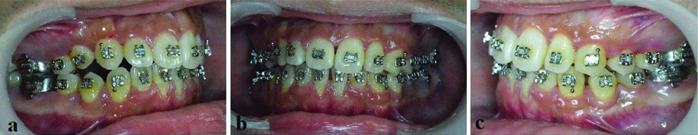
Mid treatment intraoral photographs showing the clinical progress to attain functional occlusal plane.
Conclusion
Ectopic eruption, primary eruption failure and impacted teeth have diversified factors which can lead to tooth size-arch length discrepancies (TSALD) beyond functional occlusal plan disclusion contributing as one of the primary local etiologies for malocclusion.
The clinician should possess sound knowledge with experience in critically understanding and analysing such etiologies to minimise the risk for failures. They should also be highly skilled with force and moment generating orthodontic materials to get the desired outcome in positioning the tooth in its most neutral zones thereby minimising the drawbacks.
[1]. Gjessing P, A universal retraction springJ Clin Orthod 1994 28(4):222-42. [Google Scholar]
[2]. Proffit WR, Vig KW, Primary failure of eruption: a possible cause of posterior open-biteAm J Orthod 1981 80:173-90.10.1016/0002-9416(81)90217-7 [Google Scholar] [CrossRef]
[3]. Frazier-Bowers SA, Koehler KE, Ackerman JL, Proffit WR, Primary failure of eruption: further characterization of a rare eruption disorderAm J Orthod Dentofac Orthop 2007 131:57810.1016/j.ajodo.2006.09.03817482073 [Google Scholar] [CrossRef] [PubMed]
[4]. Sylvia A, Bowers F, Long S, Tucker M, Primary failure of eruption and other eruption disorders-Considerations for management by the orthodontist and oral surgeonSemin Orthod 2016 22:34-44.10.1053/j.sodo.2015.10.006 [Google Scholar] [CrossRef]
[5]. Krishnan V, Nair SV, Ranjith A, Davidovitch Z, Research in tooth movement biology: the current statusSemin Orthod 2012 18:308-16.10.1053/j.sodo.2012.06.009 [Google Scholar] [CrossRef]
[6]. Alqerban A, Jacobs R, Fieuws S, Nackaerts O, Willems G, Comparison of 6 cone-beam computed tomography systems for image quality and detection of simulated canine impaction-induced external root resorption in maxillary lateral incisorsAm J Orthod Dentofacial Orthop 2011 140(3):e129-39.10.1016/j.ajodo.2011.03.02121889061 [Google Scholar] [CrossRef] [PubMed]
[7]. Bariani RC, Milani R, Guimaraes JCH, Moura W, Ortolani CF, Orthodontic traction of impacted upper canines using the VISTA techniqueJ Clin Orthod 2017 L1(2):76-85. [Google Scholar]
[8]. Faber J, Berto PM, Quaresma M, Rapid prototyping as a tool for diagnosis and treatment planning for maxillary canine impactionAm J Orthod Dentofacial Orthop 2006 129(4):583-89.10.1016/j.ajodo.2005.12.01516627189 [Google Scholar] [CrossRef] [PubMed]
[9]. Perinetti G, The friction concept must acknowledge the biology of tooth movementAm J Orthod Dentofacial Orthop 2008 134(4):468-69.10.1016/j.ajodo.2008.08.00718929260 [Google Scholar] [CrossRef] [PubMed]
[10]. Lane DH, Nikolai RJ, Effects of stress relief on the mechanical properties of orthodontic wire loopsAngle Orthod 1980 50(2):140-45. [Google Scholar]
[11]. Chang A, Chang C, Roberts WE, Simplified open-window technique for a horizontally impacted maxillary canine with a dilacerated rootInt J OrthodImplantol 2015 39:76-84. [Google Scholar]
[12]. Dinçer M, Gülşen A, Türk T, The retraction of upper incisors with the PG retraction systemEu Orthod Soc 2000 22:33-41.10.1093/ejo/22.1.3310721243 [Google Scholar] [CrossRef] [PubMed]
[13]. Rohit SK, Ragni T, Pratik C, Canine retraction: A systemic review of different methods usedJ Orthod Sci 2015 4(1):1-8.10.4103/2278-0203.14960825657985 [Google Scholar] [CrossRef] [PubMed]
[14]. Ribeiro GLU, Jacob HB, Understanding the basis of space closure in orthodontics for a more efficient orthodontic treatmentDental Press J Orthod 2016 21(2):115-25.10.1590/2177-6709.21.2.115-125.sar27275623 [Google Scholar] [CrossRef] [PubMed]
[15]. Kumar V, Sundareswaran S, The begg’s uprighting spring-revisitedJ Orthod Sci 2015 4(1):30-32.10.4103/2278-0203.14961525657990 [Google Scholar] [CrossRef] [PubMed]
[16]. Osório LB, Osório LB, Ferrazzo VA, Serpa G, Ferrazzo KL, Apicotomy as treatment for failure of orthodontic tractionC Rep Dent 2013 2013:16823210.1155/2013/16823224383010 [Google Scholar] [CrossRef] [PubMed]
[17]. Gracco A, Maltoni I, Maltoni M, Zoli L, Eruption of a labially impacted canine using a closed-flap technique and orthodontic wire tractionJ Clin Orthod 2012 46(10):625-30. [Google Scholar]