Identification of sex of the skeletal remains is often required for the creation of the biological profile of an individual for the legal as well as anthropological and archaeological studies [1-3]. The forensic anthropological experts know that there are wide variations in the skeletal characteristics among people of different race and geographic location and the upper limb bones have been found to help in sex determination in different populations [4,5]. The humerus is among the long bones which have been found to remain in better condition after death of the individual and can be used for analysis of sex of the individual. Recently, there has been much interest in the study of humerus features especially its metric characteristics for sex differentiation of skeletal remains [2,3,6,7]. The present study was done to assess the role of multivariate analysis of humerus metric parameters for sex differentiation of adult male and female humerus.
Materials and Methods
This cross-sectional observational study was done on 176 adult human humeri of known sex available in the bone bank of the Department of Anatomy, Government Medical College, Aurangabad, Maharashtra, India. The study includes humeri available from bone bank in a teaching hospital in Marathwada region of Maharashtra state. Although the convenience sample may not be representative of reference population from the region, it provides an important data related to humerus features and its utility in sex differentiation. All the humeri were dry, free of damage or deformity and were fully ossified. The personal records of all the humeri for age, sex were available with the bone bank. The instruments used for the measurements of various parameters of the humerus were: scale, osteometer, sliding vernier calliper, standardised and flexible steel tape, scientific balance and weight, non-elastic threads, marker pencils and pens. The following measurements were taken for each humerus [Table/Fig-1]:
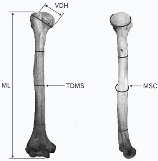
Posterior and mediolateral views of the left humerus showing some parameters used [2].
VDH: Vertical head diameter; TDMS: Transverse diameter at the middle of the shaft; ML: Maximum length or Total Length (L); MSC: Mid-shaft circumference.
Weight (W): Weight of each dried humerus was recorded with the help of scientific balance and weight. It was recorded in grams.
Total Length (L): The functional length of the humerus, i.e., the distance between the upper and lower end in anatomical position was recorded with the help of osteometer. It was measured by applying lower end to osteometer wall, and the sliding pointer was used to mark the head, and the length was recorded in mm. The midpoint of the shaft was marked simultaneously. It may also be called as maximal length.
Vertical Diameter of Head (VDH): This is the maximum diameter of the head in the vertical plane (coronal), it was measured with the help of vernier calliper in mm.
Transverse Diameter of the Head (TDH): This is the maximum diameter of the head, in the transverse plane of the head of the humerus. It was also measured by vernier calliper, in mm.
The Circumference of Midshaft (MSC): It was measured with the help of non-elastic thread at the midpoint of the shaft by the procedure as for the other circumferences. Length of the thread was measured on the scale, in mm.
Length of the Shaft of Humerus (l): It is measured between two lines; upper and lower. Upper line was drawn at a distance of 1 cm below the lowest point on articular margin of head; this point was in line with medial epicondyle. Lower line was drawn at upper concavity of olecranon fossa.
Circumference of Head at Anatomical Neck (CA): The circumference of anatomical neck of humerus was measured by marking a fixed point at groove opposite the Greater tubercle on anatomical neck with a marker pencil and running the non-elastic thread along the groove starting from the fixed point and back to it. The length of thread then recorded on scale in mm.
Circumference of Surgical Neck (CS): It was measured at a point 1 cm below the lowest point on margin of articular surface of head of humerus; the point was in line with medial epicondyle. It was measured with the help of non-elastic thread by same method as that of CA.
Maximum Width of Upper End of Humerus (WDU): It was recorded by placing the upper end of humerus transversely with lesser tubercle facing upwards in osteometer and recording the distance shown on osteometer scale in mm.
Width of Bicipital Groove (WDG): It was the distance between two lips of biciptal groove measured at the level of surgical neck with the help of vernier calliper.
Anteroposterior Diameter of Midshaft (APMS): the anteroposterior distance of midshaft of humerus is measured at the level of midpoint of shaft with the help of vernier caliper in mms.
Transverse Diameter of Midshaft of Humerus (TDMS): The maximum transverse diameter of midshaft is measured at midshaft point by holding humerus in anatomical position by vernier calliper in mm.
Bi-Epicondylar Distance (BED): Distance between two epicondyles of lower end of humerus is measured with the help of vernier calliper in mm.
Trochlear Width (TWD): It was measured by vernier calliper as anteroposterior width of trochlea at medial margin of medial flange of trochlea, recorded in mm.
Capitulum Width (CWD): It was measured as maximum anteroposterior distance of capitulum. It was recorded in mm on vernier calliper scale with limbs of vernier calliper parallel to humerus.
Width of Articular Surface of Lower End (WDASL): It was measured with vernier calliper as a maximum width of articular surface at lower end of humerus. While measuring, the limbs of vernier calliper remain parallel to humerus.
Height of Medial Flange of Trochlea (HT): It was measured with the help of vernier calliper as maximum length of medial flange of trochlea on inferior aspect.
Circumference of Shaft Distal to Deltoid Tuberosity (CDT): It was measured at a point 1 cm distal to midshaft point with same method as above. It was also defined as second one-third portion of the humeral diaphysis, distal to deltoid tuberosity (minimum circumference).
Trochlear Distance (DTC): The measurement from the location of the known minimum circumference to the trochlea. It was measured with the help of non-elastic thread, the length of which was measured on scale.
Distance of Articular Margin from the Apex of Greater Tubercle (L1N): It was measured with the help of vernier calliper as a distance between highest point on greater tubercle and nearest point on articular margin.
Distance between the Nearest Point of Margin of Lesser Tubercle and Articular Margin of Head (L2N): It was measured with the help of vernier calliper as distance between nearest point on lesser tubercle and articular margin.
Length Index (LI): This was obtained by dividing the functional length with the length of shaft.
Length Index (LI) = Functional length (L)/Length of shaft (l)
Circumference Index (CI): This was obtained by dividing the circumference of anatomical neck with the circumference of mid shaft.
Circumference Index (CI)=Circumference of Anatomical Neck (CA)/Circumference of Mid Shaft (CMS)
Statistical Analysis
All the values were tabulated and analysed statistically by usual statistical methods. The values of range, mean and standard deviation were obtained, demarking points were calculated and subsequently, Welch’s unpaired t-test applied to each of these parameters for evaluating the statistical significance as there was an unequal number of male and female humeri available. The demarking point in the present study represents the values of the measurement values unique for male and female humerus bones. A discriminant score higher than the upper demarking point indicates a male humerus bone whereas a lesser than lower demarking point score indicates a female humerus bone. The values between these two demarking points may represent either a male or female humerus bone i.e., it forms the overlap range [2]. After univariate analysis, “multivariate linear discriminant analysis” was applied using SPSS to assess the accuracy of a combination of variables for discriminating male from a female humerus. Based on the review of literature and ease of measurement, authors identified five parameters for multivariate analysis i.e., weight, total length, vertical diameter of head, transverse diameter of the head, and the circumference of midshaft [2,8-10]. These parameters were used for multivariate discriminant analysis and the rate of correctly identified humeri using the combination of these parameters was calculated.
Results
A total of 176 humeri were studied, 46 female and 130 male humeri. [Table/Fig-2,2,3,4,5, and 6] describe the statistics of humerus measurements among the males and females used for discriminant analysis. [Table/Fig-7] describes the values for the other 18 parameters studied. It can be inferred from the data that except for height of medial flange of trochlea and circumference index, all other studied parameters showed a statistically significant difference between male and female humerus bones.
Statistical analysis of weight of male and female humerus.
| Details of measurements | Male (g) | Female (g) |
|---|
| Mean±SD | 107.82±18.10 | 71.96±13.31 |
| Range | 67-155 | 49-97 |
| Demarking points | >112 | <53 |
| Overlap range | 53-112 |
| Welch’s unpaired t-test | p<0.001 |
Statistical analysis of length of humeri.
| Details of measurements | Male (mm) | Female (mm) |
|---|
| Mean±SD | 312±14.78 | 283±17.91 |
| Range | 283-358 | 247-325 |
| Demarking points | >337 | <267 |
| Overlap range | 267-337 |
| Welch’s unpaired t-test | p<0.001 |
Statistical analysis of vertical diameter of head of humerus.
| Details of measurements | Male (mm) | Female (mm) |
|---|
| Mean±SD | 43.28±2.26 | 37.41±2.78 |
| Range | 38-50 | 33-43 |
| Demarking points | >46 | <36.51 |
| Overlap range | 36.51-46 |
| Welch’s unpaired t-test | p<0.001 |
Statistical analysis of transverse diameter of head of humerus.
| Details of measurements | Male (mm) | Female (mm) |
|---|
| Mean±SD | 40.12±2.03 | 34.33±2.63 |
| Range | 35-46 | 30-41 |
| Demarking points | >42 | <34 |
| Overlap range | 34-42 |
| Welch’s unpaired t-test | p<0.001 |
Statistical analysis of circumference of midshaft of humerus.
| Details of measurements | Male (mm) | Female (mm) |
|---|
| Mean±SD | 60.84±3.72 | 53.28±4.71 |
| Range | 52-70 | 45-66 |
| Demarking points | >67 | <49 |
| Overlap range | 49-67 |
| Welch’s unpaired t-test | p<0.001 |
Statistical analysis of male and female humerus parameters.
| Parameter | Males (mm) (Mean±SD*) | Females (mm) (Mean±SD) | p-value** |
|---|
| Length of Shaft (l) | 238.62±12.77 | 220.41±14.74 | <0.001 |
| Circumference of Head at Anatomical Neck (CA) | 131.83±6.15 | 115.2±7.88 | <0.001 |
| Circumference of Surgical Neck (CS) | 90.84±6.3 | 79.98±7 | <0.001 |
| Maximum Width of Upper End (WDU) | 47.86±3.47 | 41.57±3.8 | <0.001 |
| Width of Bicipital Groove (WDG) | 10.54±1.47 | 9.37±1.37 | <0.001 |
| Anteroposterior Diameter of Mid Shaft (APMS) | 19.29±1.36 | 17.11±1.66 | <0.001 |
| Transverse Diameter of Midshaft (TDMS) | 18.84±1.46 | 16.04±1.5 | <0.001 |
| Bi-Epicondylar Distance (BED) | 59.95±3.45 | 52.57±4.36 | <0.001 |
| Trochlear Width (TWD) | 24.56±1.39 | 21.22±1.71 | <0.001 |
| Capitulum Width (CWD) | 23.23±1.23 | 20.98±1.96 | <0.001 |
| Width of Articular Surface of Lower End (WDASL) | 40.82±2.08 | 35.3±2.85 | <0.001 |
| Height of Medial Flange of Trochlea (HT) | 7.98±1.41 | 8.28±1.28 | >0.05 |
| Circumference of Shaft Distal to Deltoid Tuberosity (CDT) | 60.03±3.46 | 52.22±4.35 | <0.001 |
| Trochlear Distance (DTC) | 126±13.7 | 113.74±7.73 | <0.001 |
| Distance of Articular Margin from the Apex of Greater Tubercle (L1N) | 10.12±0.96 | 9.13±0.98 | <0.001 |
| Distance between the Nearest Point of Margin of Lesser Tubercle and Articular Margin of Head (L2N) | 18.79±1.32 | 17±2.47 | <0.001 |
| Length Index (LI) | 1.31±0.03 | 1.29±0.03 | <0.001 |
| Circumference Index (CI) | 2.17±0.13 | 2.17±0.15 | >0.05 |
* SD: Standard Deviation ** p-values for Welch’s unpaired t-test (unequal sample size)
The [Table/Fig-8] describe the demarking points for male and female humerus for the described parameters.
Demarking points of male and female humerus parameters.
| Parameter | Males (mm) | Females (mm) | Overlap Range |
|---|
| Length of Shaft (l) | >264.64 | <200.30 | 200.3-264.64 |
| Circumference of Head at Anatomical Neck (CA) | >138 | <113 | 113-138 |
| Circumference of Surgical Neck (CS) | >101 | <72 | 72-101 |
| Maximum Width of Upper End (WDU) | >53 | <37 | 37-53 |
| Width of Biciptal Groove (WDG) | >13.68 | <6 | 6-13.68 |
| Anteroposterior Diameter of Mid Shaft (APMS) | >22 | <15 | 15-22 |
| Transverse Diameter of Midshaft (TDMS) | >21 | <14 | 14-21 |
| Biepicondylar Distance (BED) | >66 | <49 | 49-66 |
| Trochlear Width (TWD) | >26 | <20 | 20-26 |
| Capitulum Width (CWD) | -- (Not obtained) | <19 | --- |
| Width of articular Surface of Lower End (WDASL) | >44 | <34 | 34-44 |
| Height of Medial Flange of Trochlea (HT) | >12 | <3.76 | 3.76-12 |
| Circumference of Shaft Distal to Deltoid Tuberosity (CDT) | >65 | <49 | 49-65 |
| Trochlear Distance (DTC) | >136 | <85 | 85-136 |
| Distance of Articular Margin from the Apex of Greater Tubercle (L1N) | >12 | <7 | 7-12 |
| Distance between the Nearest Point of Margin of Lesser Tubercle and Articular Margin of Head (L2N) | >24 | <15 | 15-24 |
| Length Index (LI) | >1.37 | <1.23 | 1.23-1.37 |
| Circumference Index (CI) | >2.62 | <1.77 | 1.77-2.62 |
It was observed that 119 of male and 40 of the female humerus were accurately identified by multivariate discriminant analysis and the total number of humeri identified correctly by multivariate discriminant analysis was 159 out of 176.
The accuracy rate for sex determination by discriminant analysis using the five parameters i.e., weight, total length, vertical diameter of head, transverse diameter of the head and the circumference of midshaft of the humerus bone was 91.53% for males and 86.95% for females with an overall accuracy of 90.34%.
Discussion
The objective of the study was to assess the role of multivariate analysis of a combination of humerus metric parameters for sex differentiation of adult male and female humerus and study the differences in various humerus parameters in male and female humerus bones. The metric values of all parameters were higher in males as compared to females in the present studied humeri. Research from India and other parts of the world also reflects that the dimensions of the humerus are larger in males as compared to females [2,5,8-10]. The bone remodelling differs in males and females with more cortical bone development in the adolescent boys. Males attain maturity at a later age and have an additional time frame of a year or two for bone growth. Also, the different pattern of work-related physical force requirements has been speculated to be the reason behind this difference [11-13].
Multivariate analysis of the humerus parameters, i.e., weight, maximum length, vertical head diameter, transverse head diameter and circumference of midshaft done in the study sample bones correctly identified 91.53% male bones and 86.95% female bones with overall accuracy being 90.34%. Similar to present study, Ogedengbe OO et al., studied the sex differences in humerus among the KwaZulu-Natal South African population [2]. They found that multivariate discriminant analysis of maximum length, vertical head diameter, the lower half of shaft transverse diameter and circumference of midshaft were able to identify 87.7% of humerus bones correctly as male or female [2]. Bašić Ž et al., studied the sex differences in humerus among ancient and contemporary Croatian population and found that multivariate discriminant analysis of maximum length, vertical head diameter, epicondylar width and maximum and minimum diameter at midshaft was able to identify 84.8% of humerus bones correctly as male or female [9]. In a recent Indian study from the same region, i.e., Maharashtra, stepwise discriminant analysis of humerus dimensions showed higher accuracy with the weight of humerus, maximal length, transverse diameter of humerus head, midshaft circumference, the width of trochlea and capitulum being the most discriminating parameters. The accuracy found was 100% for male humerus bones and 95% for female humerus bones and an overall 98.1% accuracy. The humerus dimensions observed in their study on 265 bones were similar to present study results [14]. Maximal length of humerus found in their research was 310.79±14.10 mm in males and 278.15±15.43 mm in females. The weight of humerus was 104.50±14.50 g in men and 68.48±12.54 g in women. Vertical diameter of the head in their study was 42.97±1.93 mm in males and 37.06±3.03 mm in females. Transverse diameter of the head was 39.84±1.62 mm in men and 33.75±2.61 mm in women. The circumference of mid-shaft in their study was 60.87±3.69 mm in males and 51.71±3.74 mm in females [14]. However, the humerus dimensions were on the higher side for both men and women in population from a European country, i.e., Croatia [9] and also in a population from KwaZulu-Natal province in South Africa. However, the humerus length was found to be on the lower side in a Korean population [5]. Soni G et al., study conducted on 40 male and 40 female right humeri measured six parameters. The mean values of five out of these six measurements were significantly lower in females as per results of univariate analysis. The combination of parameters of vertical head diameter of the shaft and epicondylar width provided 85% accuracy in male and 90% accuracy in female humerus bones [15]. The trochlear width in present study was 24.56±1.39 mm in males and 21.22±1.71 mm in females; anatomical neck circumference in males was 131.83±6.15 mm and in females was 115.2±7.88 mm and the BED in males was 59.95±3.45 and 52.57±4.36 mm in females. The study by Reddy B and Doshi MA reported similar trochlear width of 24.70±1.12 mm in males and 20.96±1.58 mm in females; the anatomical neck circumference reported in males was 131.27±5.51 mm and in females was 112.78±8.07 mm; the BED in males was 60.5±3.05 mm and 52.17±3.78 mm in females [14]. In an Egyptian study, the BED in males was 60.7±1.6 mm and 57.4±3.3 mm in females [16]. In a study from the Croatian population, the epicondylar width in females was 56.15±4.98mm. The study further reported overlap ranges for humerus measurements. For epicondylar width, value of <53 mm was found to be only in females, value of >65 mm was found to be only in males whereas 53-65 mm was found to be the overlap range for males and females [9]. In the present study, BED overlap range was 49-66 mm with value of <49 mm found only in females and value of >66 mm found only in males. Similarly, in Croatian population, overlap range of maximum humerus length was 247-385 mm, maximum vertical head diameter was 38-46 mm, and maximum diameter at midshaft was 20-23 mm [9].
Thus, the present data regarding humerus measurements described are in line with literature from Indian population with differences in humerus measurements data from other continent populations. The difference in measurements across different population groups has been attributed to difference in diet patterns, genetic differences and environmental factors affecting the growth patterns [17]. Multivariate analysis with similar parameters has been found to be able to differentiate male and female humerus bones with reasonable accuracy in different population groups.
Limitation
The significant limitations of the present study include a small sample and from a limited geographic area. There was an unequal number of male and female humerus bones available with majority of bones belonging to males and the results should be interpreted with this limitation in the perspective. Further studies with a large sample from diverse regions across India will help in the better analysis of sex differentiation and population-specific data related to humerus bone.
Conclusion
The present study results reflect that multivariate analysis of a group of metric parameters of male and female humerus bones can be helpful for identification of sex from the humerus bones with reasonable accuracy. Multicentre studies with large sample across the geographical regions of India need to be done to assess the role of multivariate analysis of various osteometric parameters of the humerus for the differentiation of male and female humeri.
* SD: Standard Deviation ** p-values for Welch’s unpaired t-test (unequal sample size)