Bilateral Radicular Dens Invaginatus in Mandibular First Premolars-Report of Two Rare Cases
YM Karuna1, Shailaja Datta2, BS Suprabha3, Arathi Rao4, Ravikiran Ongole5
1 Assistant Professor, Department of Pedodontics and Preventive Dentistry, Manipal College of Dental Sciences, Mangalore, MAHE, Manipal, Karnataka, India.
2 Former Postgraduate Student, Department of Pedodontics and Preventive Dentistry, Manipal College of Dental Sciences, Mangalore, MAHE, Manipal, Karnataka, India.
3 Professor, Department of Pedodontics and Preventive Dentistry, Manipal College of Dental Sciences, Mangalore, MAHE, Manipal, Karnataka, India.
4 Professor, Department of Pedodontics and Preventive Dentistry, Manipal College of Dental Sciences, Mangalore, MAHE, Manipal, Karnataka, India.
5 Professor and Head, Department of Oral Medicine and Radiology, Manipal College of Dental Sciences Mangalore, MAHE, Manipal, Karnataka, India.
NAME, ADDRESS, E-MAIL ID OF THE CORRESPONDING AUTHOR: Dr. Arathi Rao, Professor, Department of Pedodontics and Preventive Dentistry, Manipal College of Dental Sciences, Manipal Academy of Higher Education, Light House Hill Road, Mangalore-575001, Manipal Academy of Higher Education, Manipal, Karnataka, India. Karnataka, India.
E-mail: arathi.rao@manipal.edu
Dens invaginatus is a developmental dental anomaly which is clinically seen as a furrow on the palatal aspect of the tooth either limiting to the coronal pulp or extending to the radicular apex. It occurs most commonly in females affecting crowns of the maxillary lateral incisors. This case report presents two bilateral cases of Type II radicular dens invaginatus in mandibular first premolars. It was an incidental diagnosis on an intraoral periapical radiograph when a 12-year-old male patient and a 12-year-old female patient reported with their respective chief complaints. Conservative management in the form of strict follow up of the cases was planned.
Anomaly, Developmental, Odontogenesis, Root
Case 1
A 12-year-old female patient reported to the Department of Pedodontics and Preventive Dentistry with the chief complaint of gum pain on the lower right back tooth region since three days. Past medical history and past dental history of the patient was not significant. On intraoral examination, patient was having eruption gingivitis of #47. Upon examination of the area of complaint (premolar region) the tooth #45 was erupting. No abnormality was detected in relation to tooth #44 [Table/Fig-1]. Upon horizontal and vertical percussion there was no tenderness, mobility in relation to both #44 and #45. The gingiva covering was healthy and uninflamed. However, an IOPAR was taken out to rule out any other underlying pathology. No relevant findings related to the chief complaint were present on IOPAR, but incidentally it was found that the tooth #44 has an open apex with a well-defined invagination in the apical third of the root canal, suggestive of radicular dens invaginatus [Table/Fig-2a]. An IOPAR of the contralateral premolar (#34) was taken to rule out the similar finding that showed the presence of radicular dens invaginatus involving the tooth #34 [Table/Fig-2b]. As both the involved teeth had an open apex, a differential diagnosis of root bifurcation was also made and it was decided to carry out further evaluation using focal Cone Beam Computed Tomography (CBCT). Treatment for eruption gingivitis was provided in the form of local irrigation with saline and symptomatic pain relief by prescribing analgesics. Appointment was given for obtaining CBCT images. However, patient reported after eight months of initial IOPA due to unforeseen personal circumstances. At this visit, the obtained CBCT images (Ultra low dose CBCT on a planmeca CBCT machine) [Table/Fig-3] gave a definitive diagnosis of Type II radicular dens invaginatus in relation to #34 and #44 [1]. Tooth anomaly being part of any known a syndrom was ruled out by the paediatrician. The prescribed symptomatic treatment for the eruption gingivitis had already relieved the chief complaint of the patient (pain near the premolar region), suggesting the source of pain to be erupting #45. Since both the teeth (#34 and #44) were clinically and radiographically sound no treatment was done, however the patient was put under follow-up regimen of once in six months to monitor the involved teeth.
Intraoral image of the patient with Clinically sound #34 and #44.

Radicular dens invaginatus as seen on: a) IOPAR of #44; b) IOPAR of #34.

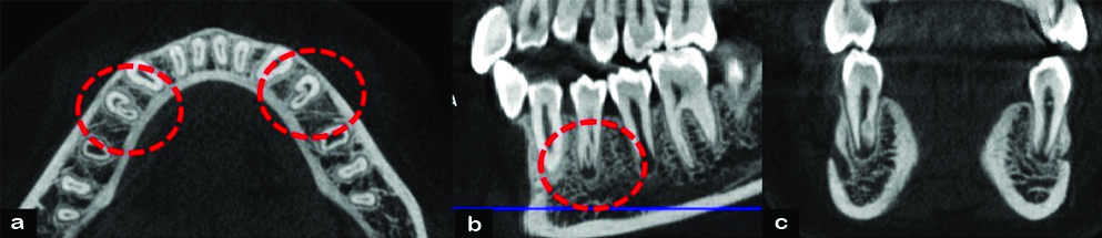
CBCT images demonstrating the presence of invagination #34 and #44: a) Axial view; b) Sagittal view; and c) Coronal view.
Case 2
A 12-year-old male patient reported to the Department of Pedodontics and Preventive Dentistry with the chief complaint of yellowish discolouration in lower left back tooth. There was history of sharp pain associated with the tooth since past 10 days, which aggravated on chewing. There was no significant past medical and dental history. Intra oral examination revealed a mixed dentition stage. The tooth #35 was partially erupted with a soft tissue bulge present around it. The tooth crown had a hypoplastic appearance with deep occlusal fissure. Additionally, a non-carious cervical defect was noted on the buccal aspect of the crown [Table/Fig-4]. On an intraoral periapical radiograph, no abnormality was noted with respect to #35. However incidentally the radicular portion of the tooth #34 revealed an open apex with a well-defined invagination in the apical third of the root canal, suggestive of Type II radicular dens invaginatus [1]. To rule out the bilateral involvement, IOPAR of the right mandibular premolar region was also taken [Table/Fig-5a,b]. The IOPAR revealed a similar finding suggesting Type II radicular dens invaginatus even in relation to tooth #44 [1]. Clinically, the tooth both #34 and #44 had normal appearance [Table/Fig-4]. The teeth were non tender on vertical and horizontal percussion. Mobility was absent. No soft tissue abnormalities were detected. Since the parents of the patient refused for further investigation, focal CBCT could not be taken in this case to arrive at the definitive diagnosis. However, through the medical history and a paediatric referral presence of associated syndromes were ruled out.
Intraoral image of the patient with Clinically sound #34 and #44.

Radicular dens invaginatus as seen on: a) IOPAR of #44; b) IOPAR of #34.

A conservative treatment plan in the form of regular follow-up once in every six months was formulated in order to prevent the likely complications from the dens invaginatus of the mandibular first premolars. The cervical defect of the tooth #35 was restored with Glass ionomer cement and the occlusal fissures were sealed using pit and fissure sealant.
Discussion
Radicular dens invaginatus is a rare dental anomaly which results due to the infolding of Hertwig’s epithelial root sheath [2]. Incisors and third molars are commonly affected than the other tooth types [3]. Maxillary teeth are commonly involved with extremely rare involvement of the mandible [4].
The exact aetiology of this condition is not clearly known. However, number of theories have been proposed which include retardation or acceleration of growth of internal enamel epithelium, increased pressure from the surrounding tissues during tooth formation, distortion of the enamel organ during tooth development or inadequate nutrition of a portion of a single tooth germ [5]. Different classifications of this condition were described by Hallett GE (1953), Ulmansky M and Hermel J (1964) and Vincent-Townend (1974) [6-8]. Oehlers FA classified dens invaginatus into four variants, based on clinical and radiographic criteria with a mention of the existence of this condition in the roots of teeth [9].
The radicular variant of dens invaginatus was also described by Bhatt AP and Dholakia HM [1]. The radicular dens invaginatus is further classified into Type I and Type II. The Type I present as an axial invagination in the wall of the root and is suggestive of an incomplete attempt at root bifurcation. The Type II form is extremely rare and is the true form of radicular dens invaginatus. It presents as an enamel-lined invagination within the root originating at an opening on the root itself [9]. Both the present cases are Type II radicular dens invaginatus involving the mandibular first premolars.
The presence of an invagination is likely to be associated with the risk of dental caries, pulpal involvement and periodontal inflammation, thus demanding the clinical attention. The various treatment options include conservative operative treatment, nonsurgical root canal treatment and surgical treatment like endodontic surgery, intentional re-implantation and extraction [4]. When root canal treatment of the involved tooth is indicated due to pulpal involvement, challenges while negotiating the canals should be kept in mind.
According to Hulsmann M et al., when there is no evidence of an entrance to the invagination and no clinical and radiographic signs of pathology are present, mere strict observation is recommended [10]. In the present cases, conservative management in the form of strict follow-up was undertaken with special emphasis on preventive strategies. Both the patients continue to be asymptomatic during the one year follow-up visit.
Conclusion
Radicular dens invaginatus is a rare occurrence when compared to coronal dens invaginatus. If it is asymptomatic, it can go undiagnosed. As the presence of an invagination is likely to be associated with the risk of dental caries, pulpal involvement, periodontal inflammation and complications during endodontic treatment, careful radiographic examination along with the knowledge of the variations in morphology of the internal anatomy of teeth are necessary for a successful treatment outcome.
[1]. Bhatt AP, Dholakia HM, Radicular variety of double dens invaginatusOral Surg Oral Med Oral Pathol 1975 39:284-87.10.1016/0030-4220(75)90230-3 [Google Scholar] [CrossRef]
[2]. Gannepalli A, Ayinampudi BK, Podduturi SR, Radicular dens invaginatus associated with radicular cyst in maxillary third molar-Rare case reportJournal of Pierre Fauchard Academy (India Section) 2014 28:133-36.10.1016/j.jpfa.2015.02.001 [Google Scholar] [CrossRef]
[3]. Stavrou E, Tosios KI, Stavrou IE, Globular radiopacity around the apex of an impacted maxillary third molarOral Surg Oral Med Oral Pathol Oral Radiol Endod 2007 103:594-98.10.1016/j.tripleo.2006.11.05017331757 [Google Scholar] [CrossRef] [PubMed]
[4]. Verma KG, Basavaraju S, Jindal S, Sachdeva S, Bilateral radicular dens in dente in mandibular premolarsJ Oral Maxillofac Radiol 2013 1:115-17.10.4103/2321-3841.126747 [Google Scholar] [CrossRef]
[5]. Beena VT, Sivakumar R, Heera R, Rajeev R, Choudhary K, Panda S, Radicular dens invaginatus: report of a rare caseCase Rep Dent 2012 2012:87193710.1155/2012/87193722900211 [Google Scholar] [CrossRef] [PubMed]
[6]. Hallet GE, The incidence, nature and clinical significance of palatal invagination in the maxillary incisors teethProceedings of the Royal Society of Medicine 1953 46:491-99.10.1177/003591575304600703 [Google Scholar] [CrossRef]
[7]. Ulmansky M, Hermel J, Double dens in dente in a single tooth-Report of a case And radiologic study of the incidence of small dens in denteOral Surg Oral Med Oral Pathol 1964 17:92-97.10.1016/0030-4220(64)90320-2 [Google Scholar] [CrossRef]
[8]. Vincent-Townend J, Dens invaginatusJ Dent 1974 2(6):234-38.10.1016/0300-5712(74)90024-4 [Google Scholar] [CrossRef]
[9]. Oehlers FA, The radicular variety of dens invaginatusOral Surg Oral Med Oral Pathol 1958 11:1251-60.10.1016/0030-4220(58)90278-0 [Google Scholar] [CrossRef]
[10]. Hulsmann M, Dens invaginatus: Aetiology, classification, prevalence, diagnosis, and treatment considerationsInt Endod J 1997 30:79-90.10.1111/j.1365-2591.1997.tb00679.x10332241 [Google Scholar] [CrossRef] [PubMed]