Systemic Lupus Erythematosus Presenting as Knee Joint Effusion
Basavaraj Baligar1, Uday S Bande2, BE Kalinga3, Y Chethan4
1 Assistant Professor, Department of General Medicine, Karnataka Institute of Medical Sciences, Hubballi, Karnataka, India.
2 Professor, Department of General Medicine, Karnataka Institute of Medical Sciences, Hubballi, Karnataka, India.
3 Assistant Professor, Department of General Medicine, Karnataka Institute of Medical Sciences, Hubballi, Karnataka, India.
4 Postgraduate Student, Department of General Medicine, Karnataka Institute of Medical Sciences, Hubballi, Karnataka, India.
NAME, ADDRESS, E-MAIL ID OF THE CORRESPONDING AUTHOR: Mr. Y Chethan, Postgraduate Student, Department of General Medicine, Karnataka Institute of Medical Sciences, Vivek Boys Hostel, No. 144, Vidyanagar, Hubballi-580022, Karnataka, India.
E-mail: chethany@ymail.com
Systemic Lupus Erythematosus (SLE) is a chronic, autoimmune, connective tissue disorder which has multi-organ involvement. Patient might rarely present with major joint sterile inflammatory effusion and evaluation of such patients for connective tissue disorders helps in the early diagnosis and treatment. We hereby, present the case of 35-year-old female with left knee joint effusion subsequently diagnosed to have SLE with lupus nephritis Class IIIa.
Connective tissue disorder, Diagnosis, Multi-organ involvement
Case Report
A 35-year-old female hailing from Hangal taluk (Hubballi district, Karnataka state) farmer by occupation presented to Orthopaedic casualty with history of left knee joint pain and swelling for one week duration. Pain was insidious in onset and progressive that restricted her movements and presented to emergency department. Swelling was insidious in onset and progression restricted to left knee joint only. There was no history of trauma, fever, other joint pains and swelling. Radiographic imaging of left knee joint showed joint effusion. She was suspected to have septic arthritis and was taken to minor Operation Theatre (OT) for aspiration of joint fluid. Aspirate was a clear fluid without pus or haemarthrosis. After aspiration patient was symptomatically better. She was referred to medicine department to evaluate the case further.
She had history of rashes all over the body for one month duration started with forearms then progressed to involve her abdomen, back and face. Rash was non itching, initially it was red in colour then over a period of time changed to black. She also had history of alopecia [Table/Fig-1], weight loss and easy fatigability for one month. There was no history of fever, chest pain, breathlessness, cough with expectoration, palpitations, haematuria, other joint pains, seizures, abnormal behavior, blurring of vision red eyes and repeated abortions in the past.
Photograph of the patient showing alopecia.
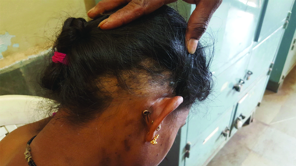
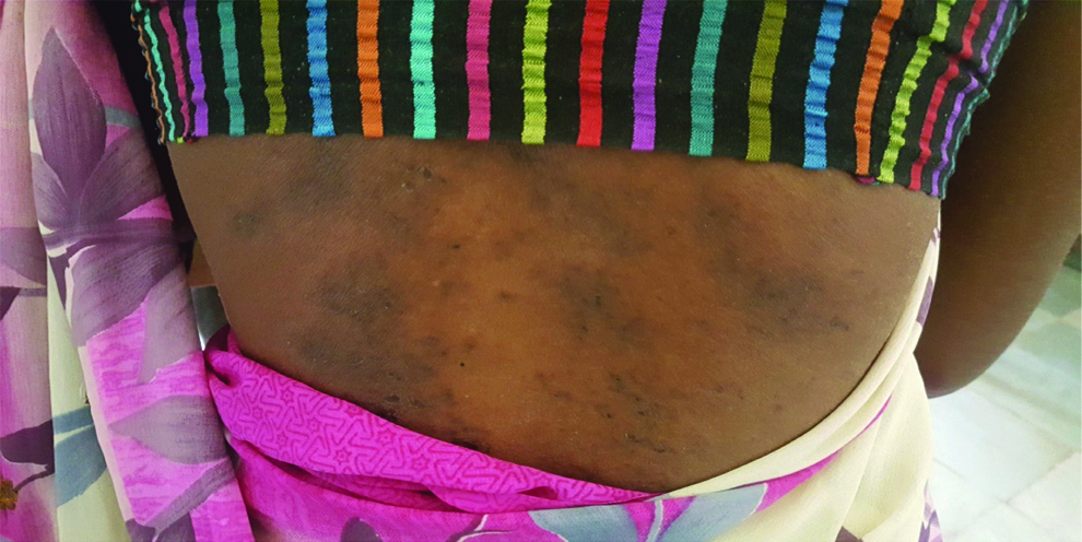
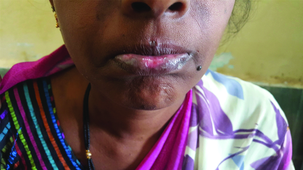
On examination, her vitals were stable. Rashes were maculopapular, blackish in colour, present over elbow, waist, lumbar region and over the bridge of the nose and cheek [Table/Fig-2,3,4]. Patient had pallor and oral ulceration [Table/Fig-5]. On systemic examination, there was no evidence of arthritis of other joints, pleural effusion, hepatosplenomegaly and focal neurological deficit.
Photograph of the patient showing malar rash.
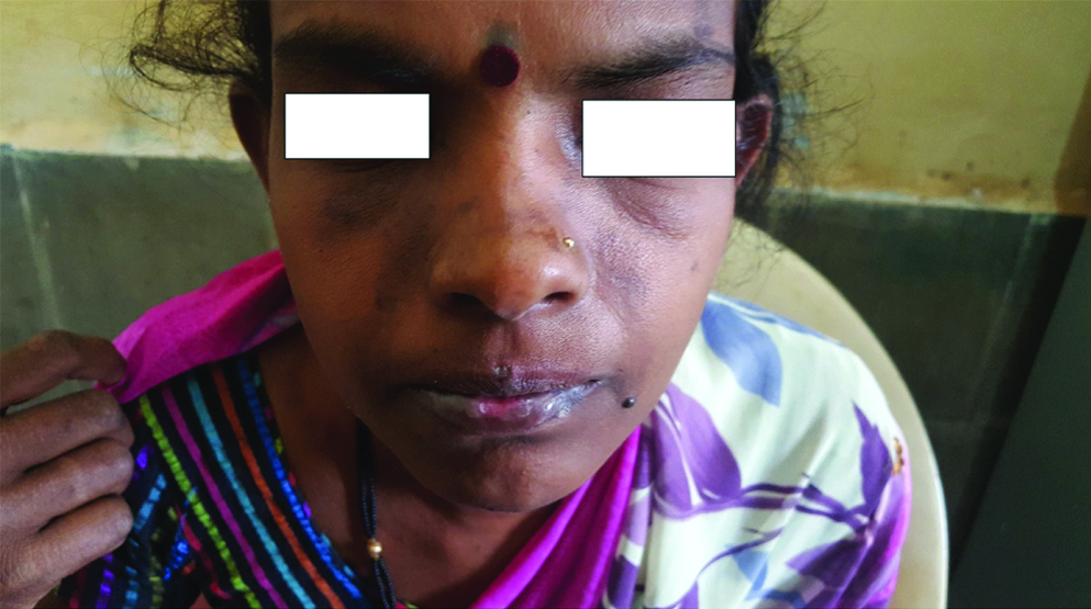
Photograph of the patient showing malar rash.
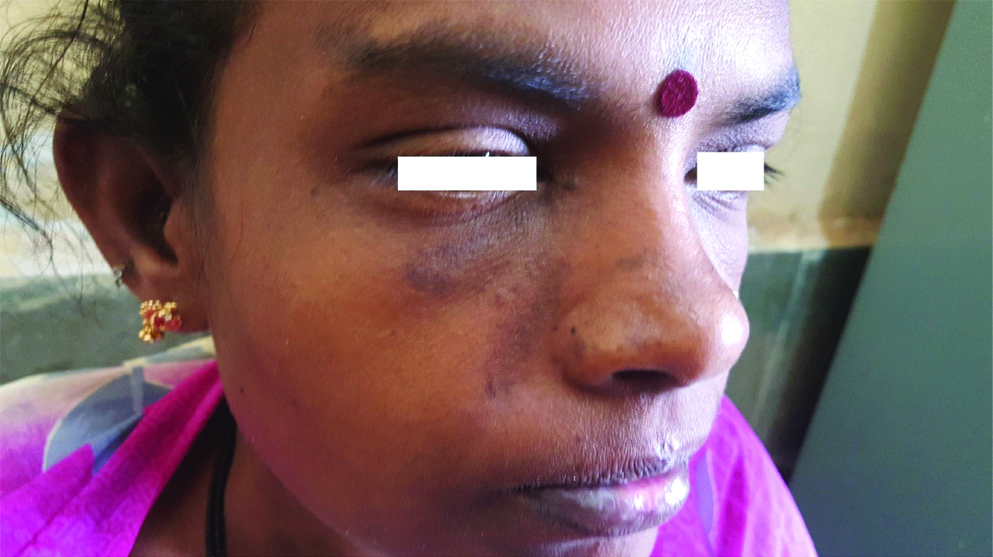
Photograph of the patient showing photosensitive rash.
Photograph of the patient showing ulcers over the lip.
With the above history and examination findings, her synovial fluid, blood and urine examination was sent [Table/Fig-6]. Patient was suspected to have connective tissue disorder as she had photosensitive and malar rashes, anaemia, thrombocytopenia and proteinuria along with her inflammatory knee joint effusion and evaluated accordingly. Her ANA screening test (ELISA) came strongly positive-13.1. ANA profile showed Anti Sm and Anti RNP positive. Patient’s haematological, renal, musculoskelatal and immunological criteria was positive (4 criteria) and hence diagnosis of SLE was confirmed. Nephrology opinion was sought and patient was subjected to renal biopsy for grading of nephritis after correcting her anaemia with three pints of whole blood. Biopsy report came as LUPUS NEPHRITIS FOCAL PROLIFERATIVE GLOMERULONEPHRITIS ISN/RPS CLASS III A with activity score 5/24 and chronicity score 0/12.
Left knee joint synovial fluid, blood and urine examination of the patient.
| Investigations | Values | Normal range |
|---|
| Synovial fluid |
| Colour | Pale yellow | Clear fluid |
| Proteins | 3.18 gm/dL | <2.5 gm/dL |
| Sugar | 46.36 mg/dL | 10 mg/dL lower than serum glucose (126 mg/dL) |
| Cell count | 1200 cells/cumm | 0-200 cells/cumm |
| Differential counts | N71% L20% synoviocytes 09% | Polymorphs <25% |
| Gram stain | No organism seen | negative |
| Adenosine Deaminase (ADA) | 16.2 IU/L | <40 IU |
| Blood investigations |
| HIV/HbSAg | Negative | - |
| Haemoglobin | 6.5 gm/dL | 11-16 gm/dL |
| White blood cell | 6500 cells/cumm | 4000-11000 cells/cumm |
| Platelets | 65000 lakhs/cumm | 1.5-4 lakhs/cumm |
| Blood urea | 35 mg/dL | 10-45 mg/dL |
| Serum creatinine | 0.8 mg/dL | 0.5-1.5 mg/dL |
| Erythrocyte sedimentation rate | 35 mm/hour | 0-20 mm/hour |
| C-reative protein | Positive | - |
| RA factor | Positive | - |
| Urine examination |
| Proteins | + | nil |
| Red blood cell | 2-3/HPF | 2-4/HPF |
| Pus cells | 3-4/HPF | 2-5/HPF |
| Crystals/casts | No casts/crystals | nil |
| 24 hour proteins | 820 mg/2 litres | <100 mg/day |
| Chest X-ray/2D ECHO | Within normal limit | - |
Meanwhile patient was started on methyl prednisolone 500 mg day one and 250 mg day two and day three. And Tab. Waysolone 1 mg/kg/body weight from day four. Patient was symptomatically better. No further effusion was noted. Patient’s consent was obtained before publishing the images.
Discussion
Systemic lupus erythematosus is a clinically heterogeneous autoimmune disease of complex aetiology, having a variable course and prognosis [1]. It effects primarily women in their childbearing years (20-40 years) [2]. Prevalence of SLE is 1:1000 [3]. The female to male ratio is 9:1 in adulthood and 4:1 in puberty [4]. Presentation of SLE is complex, as disease has multi-organ involvement [Table/Fig-7]. A minimum of four out of eleven criteria should be met in order to confirm the diagnosis of SLE patient with at least one in clinical and one in immunological category, well documented at any time during individual’s history [Table/Fig-8]. Some patients may have only one organ involved or only have some of the manifestations and will, therefore, not be diagnosed under the American College of Rheumatology (ACR) criteria. Such patients are classified as having “Incomplete” or “Latent” lupus [4]. The disease activity usually occurs in three phases i.e., flare, chronic, and long quiescence. A flare or relapsing remission is an exacerbation that occurs suddenly and unpredictably; patients are usually in good health between flares. Chronic SLE has persistent activity of some type such as chronic synovitis and chronic cytopenias. SLE is more aggressive in childhood with brain and kidney being the most affected organ [5]. Patients with long quiescence have a long remission period before having additional flare up [6,7]. Most common manifestations are fatigue, anorexia, weight loss followed by musculoskeletal symptoms. Polyarthralgia is most common.
Clinical features of systemic lupus erythematosus system features.
| Constitutional-Fatigue-Fever (in absence of infection)-Weight lossMusculoskeletal-Arthritis, Arthralgia, MyositisSkin-Butterfly rash, Photosensitivity-Mucous membrane lesion-Alopecia-Raynaud’s phenomenon-Purpura, Urticaria-VasculitisRenal-Hematuria, Proteinuria Casts-Nephrotic syndromeGastrointestinal-Nausea, Vomiting, Abdominal painPulmonary-Pleurisy, Pulmonary hypertensionCardiac-Pericarditis, Endocarditis, MyocarditisReticuloendothelial-Lymphadenopathy, Splenomegaly, HepatomegalyHaematologic-Anaemia, Thrombocytopenia, LeukopeniaNeuropsychiatric-Psychosis, Seizures, Organic brain syndrome-Transverse myelitis-Cranial neuropathies-Peripheral neuropathies |
Systemic lupus international collaborating clinic criteria for classification of systemic lupus erythematosus [1].
| Clinical ManifestationsSkin-Acute, subacute cutaneous LE-Chronic cutaneous LE-Oral ulcers-Alopecia-SynovitisNeurological-Seizures, Psychosis, Mononeuritis-Myelitis, Peripheral or cranial neuropathies-Acute confusional stateHematalogical-Leucopenia(<4000) or lymphopenia(<1000)-ThrombocytopeniaRenal-Prot/Cr .0.5-RBC casts-BiopsyImmunological manifestation-ANA > reference negative value-Anti-dsDNA-Anti-Sm-Antiphospholipid, Low serum complement and Positive direct Coombs test |
Renal biopsy is indicated in all patients of SLE. If renal biopsy is reported as systemic lupus and patient doesn’t have other clinical features mentioned above, he/she still qualifies for classification as SLE.
Most recent clinical studies of lupus nephritis have used the International Society of Nephrology (ISN) and the Renal Pathology Society (RPS) classification [8]. In the ISN/RPS classification, the addition of “a” for active and “c” for chronic changes gives physicians information regarding the potential reversibility of disease. In general, Class III and IV disease, as well as Class V accompanied by III or IV disease, should be treated with aggressive immunosuppression if possible, because there is a high risk for End-Stage Renal Disease (ESRD) if patients are untreated or undertreated.
The principle management of SLE is suppression of inflammation in an attempt to prevent organ damage [9-11]. The SLE management is mainly based on the disease manifestation and severity [12]. The EULAR also is referred to as “European League against Rheumatism” released recommendations for SLE treatments in 2007 [13].
Patients who meets the criteria for SLE (definite SLE 4 or more criteria, possible SLE <4 criteria) and without any potential end organ complication treated conservatively with drugs (NSAIDs, Hydroxychloroquine (HCQ), methotrexate, topical steroids, low dose glucocorticoids) and patients with potential life threatening complications started with high dose glucocorticoids along with mycophenolate mofetil or cyclophosphamide and to taper the drugs when the response is seen or to add on other drugs (Belimumab, rituximab and calcineurin inhibitors) if response is not seen [13-15].
Conclusion
Systemic lupus erythematosus is a very complex disease with multifactorial aetiology and multiple organ involvement making the diagnosis challenging. Clinical manifestations as well as immunological abnormalities assist in the diagnosis. Early diagnosis prevents patients ending in ESRD and have better quality of life.
[1]. Manson JJ, Rahman A, Systemic lupus erythematosusOrphanet J Rare Dis 2006 01:0610.1186/1750-1172-1-616722594 [Google Scholar] [CrossRef] [PubMed]
[2]. Maidhof W, Hilas O, Lupus: an overview of the disease and management optionsPharmacy and Therapeutics 2012 37(4):240-46.:249 [Google Scholar]
[3]. American College of Rheumatology Ad Hoc Committee on Systemic Lupus Erythematosus GuidelinesGuidelines for referral and management of SLE in adultsArthritis Rheumatol 1999 42(9):1785-96.10.1002/1529-0131(199909)42:9<1785::AID-ANR1>3.0.CO;2-# [Google Scholar] [CrossRef]
[4]. McAlindon T, Update on the epidemiology of systemic lupus erythematosus: new spins on old ideasCurr Opin Rheumatol 2000 12:104-12.10.1097/00002281-200003000-0000410751013 [Google Scholar] [CrossRef] [PubMed]
[5]. Pradhan V, Patwardhan M, Rajadhyaksha A, Ghosh K, Clinical and immunological profile of systemic lupus erythematosusIndian Pediatrics 2013 50(4):405-07.10.1007/s13312-013-0115-z23024105 [Google Scholar] [CrossRef] [PubMed]
[6]. Miah T, Haque MA, Mahmood T, Tarafder BK, Clinical profile, management and outcome of lupusMymensingh Med J 2008 17(2):S6-S11. [Google Scholar]
[7]. Gladman D, Urowitz M, American College of Rheumatology Ad Hoc Committee on Systemic Lupus Erythematosus Guidelines. Guidelines for referral and management of systemic lupus erythematosus in adultsArthritis Rheumtol 1999 42(9):1785-96.10.1002/1529-0131(199909)42:9<1785::AID-ANR1>3.0.CO;2-# [Google Scholar] [CrossRef]
[8]. Weening JJ, D’Agati VD, Schwartz MM, Seshan SV, Alpers CE, Appel GB, The classification of glomerulonephritis in systemic lupus erythematosus revisitedJ Am Soc Nephrol 2004 15:241-50.10.1097/01.ASN.0000108969.21691.5D14747370 [Google Scholar] [CrossRef] [PubMed]
[9]. Ginzler E, Sharon E, Diamond H, Kaplan D, Long-term maintenance therapy with azathioprine in systemic lupus erythematosusArthritis Rheum 1975 18:27-34.10.1002/art.17801801061115745 [Google Scholar] [CrossRef] [PubMed]
[10]. Lazaro D, Elderly-onset systemic lupus erythematosus: prevalence, clinical course and treatmentDrugs Aging 2007 24:701-15.10.2165/00002512-200724090-0000117727302 [Google Scholar] [CrossRef] [PubMed]
[11]. Amissah-Arthur MB, Gordon C, Contemporary treatment of systemic lupus erythematosus: an update for cliniciansTher Adv Chronic Dis 2010 1(4):163-75.10.1177/204062231038010023251736 [Google Scholar] [CrossRef] [PubMed]
[12]. Hahn BH, Management of Systemic Lupus Erythematosus. In: Harris ED, et al, edsKelley’s Textbook of Rheumatology 2005 7th edPhiladelphia, PaWB Saunders:1225-47. [Google Scholar]
[13]. Bertsias G, Ioannidis JP, Boletis J, Bombardieri S, Cervera R, Dostal C, EULAR recommendations for the management of systemic lupus erythematosus. Report of a Task Force of the EULAR Standing Committee for International Clinical Studies Including TherapeuticsAnn Rheum Dis 2008 67(2):195-205.10.1136/ard.2007.07036717504841 [Google Scholar] [CrossRef] [PubMed]
[14]. Hahn BH, McMahon MA, Wilkinson A, Wallace WD, Daikh DI, Fitzgerald JD, American College of rheumatology guidelines for screening, treatment, and management of lupus nephritisArthritis Care Res (Hoboken) 2012 64:797-808.10.1002/acr.2166422556106 [Google Scholar] [CrossRef] [PubMed]
[15]. Mosca M, Tani C, Aringer M, Bombardieri S, Boumpas D, Brey R, European League Against Rheumatism recommendations for monitoring patients with systemic lupus erythematosus in clinical practice and in observational studiesAnn Rheum Dis 2010 69(7):1269-74.10.1136/ard.2009.11720019892750 [Google Scholar] [CrossRef] [PubMed]