Postpartum Page Kidney Secondary to HELLP Syndrome
Sheshang Uday Kamath1, Bhushan Patil2, Sujata Kiran Patwardhan3
1 Senior Resident, Department of Urology, Seth G.S. Medical College and KEM Hospital, Mumbai, Maharashtra, India.
2 Associate Professor, Department of Urology, Seth G.S. Medical College and KEM Hospital, Mumbai, Maharashtra, India.
3 Head, Department of Urology, Seth G.S. Medical College and KEM Hospital, Mumbai, Maharashtra, India.
NAME, ADDRESS, E-MAIL ID OF THE CORRESPONDING AUTHOR: Sheshang Uday Kamath, 8th Floor, New MSB, Parel, Mumbai, Maharashtra, India.
E-mail: sheshangkamath@gmail.com
Page Kidney is a unique clinical entity causing hypertension secondary to external renal compression. We report the case of a female patient who was antenatally diagnosed to have HELLP syndrome and subsequently developed a spontaneous subcapsular haematoma causing Page kidney that was promptly diagnosed and managed in the postpartum period.
A 30-year-old female patient presented to the emergency department with chief complains of pain in the right lumbar region and intermittent high grade fever since 10 days. Patient was 20 days post-partum and presented with hypertension. An ultrasound of the abdomen and CECT abdomen was done to evaluate the cause for tenderness and to our surprise there was a subcapsular haematoma measuring 8×5×3 cm compressing the right kidney. An open exploration for evacuation of haematoma was done. Acute kidney injury with HELLP syndrome is a known entity however; renal subcapsular haematoma secondary to low platelets is a rare finding.
A high index of suspicion is essential to timely diagnose Page kidney. Page kidney as mentioned in previous literature and as observed in this case report is a reversible cause of hypertension and a low threshold for capsulotomy and drainage of the haematoma with optimal post-operative management of this condition would prevent long term sequela of hypertension among young patients.
Acute kidney injury, Haematoma, Hypertension
Case Report
A 30-year-old female patient presented to the emergency department with chief complaints of pain in the right lumbar region and intermittent high grade fever since 10 days. Patient was 20 days post-partum. She was diagnosed to have pregnancy induced hypertension for which she was on ACE inhibitors medication. Prior to delivery she was diagnosed to have HELLP syndrome and her blood pressure noted at the time of delivery was 190/100 mm of Hg and low platelets just prior to delivery. She received intravenous Labetalol and four units of platelet transfusion at the time of delivery and an uneventful forceps assisted delivery was performed. Patient was discharged on day 3 with a blood pressure of 100/80 mm of Hg.
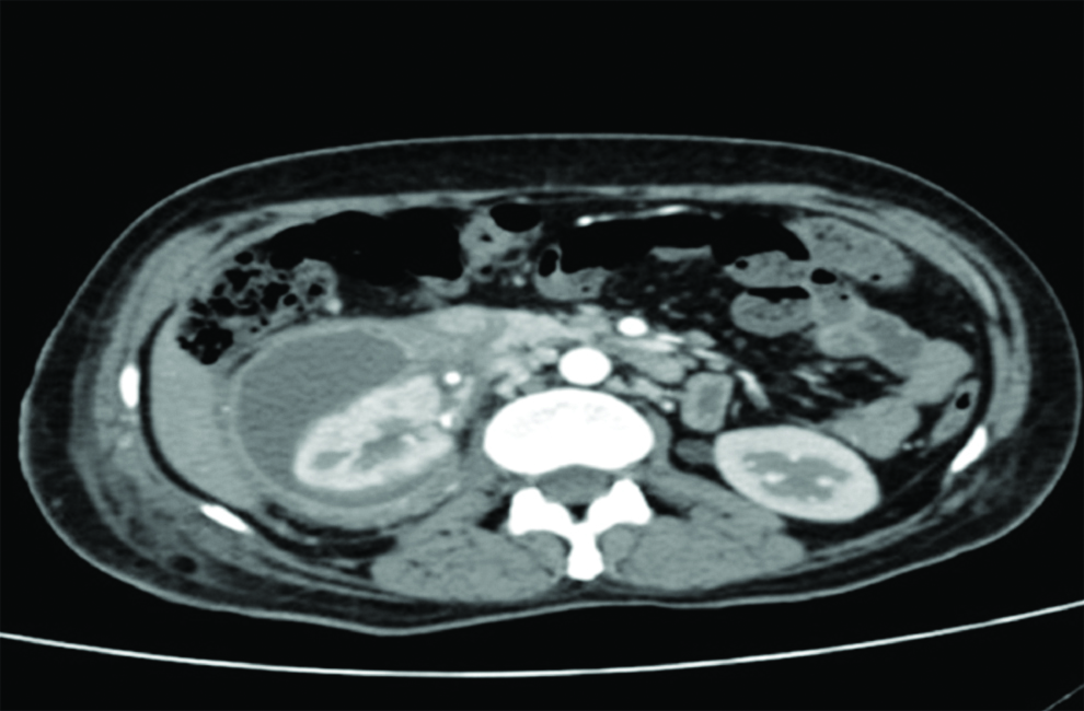
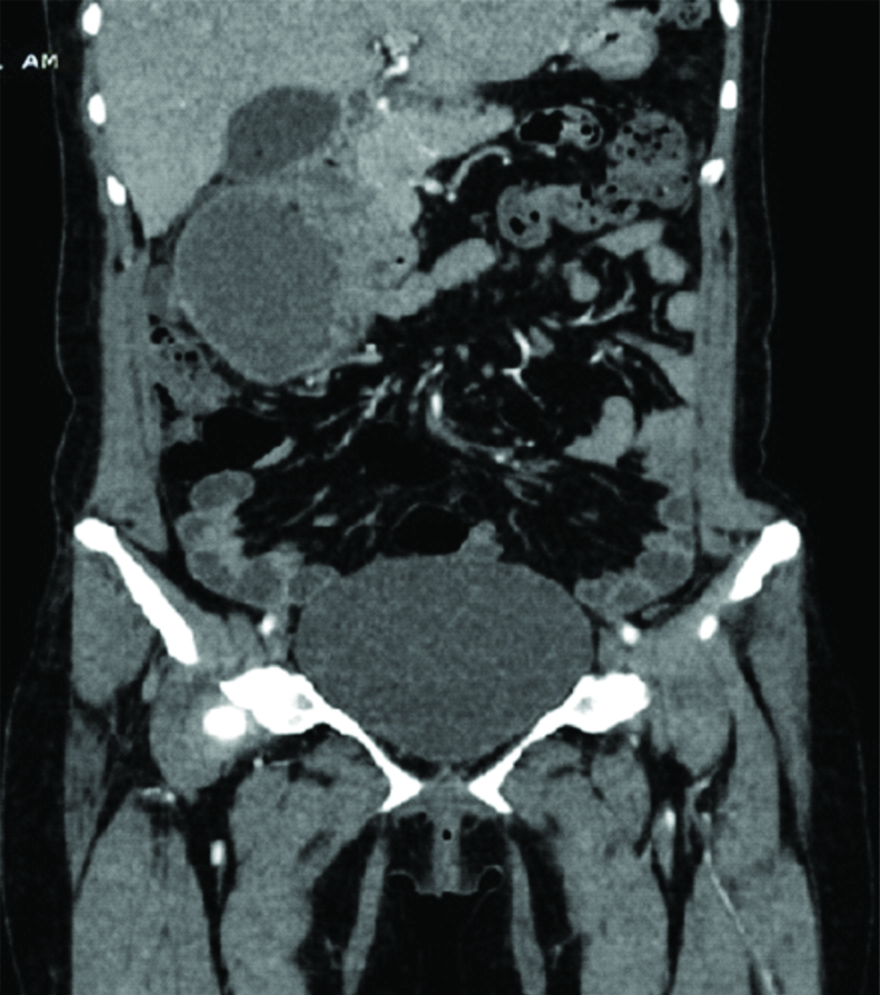
On having examined the patient in the emergency department patient had a blood pressure of 180/100 mm of Hg. Patient also had right flank tenderness. However, there was no local rise of temperature or skin changes over the right flank. A physician’s opinion was sought to control the blood pressure and intravenous labetalol stat dose was given and blood pressure was 140/100 mm of Hg. The laboratory investigation showed a fall in the hemoglobin from 9.5 to 8.2 gm/dL although the platelet counts (1.5 lac/cmm) and white blood cell counts (8500) were normal. The liver and renal function tests were within normal range as well. An ultrasound of the abdomen was done to evaluate the cause for tenderness and to our surprise there was a subcapsular haematoma measuring 8×5×3 cm compressing the right kidney. An ultrasound guided pigtail was inserted into the haematoma in the emergency department. Patient was started on ACE inihibitor for 48 hours yet the blood pressure was maintained at 140/100 mm of Hg. The pigtail drained only 20 mL over two days. A contrast enhanced CT scan was performed to evaluate the haematoma. As the haematoma persisted and was not adequately drained by the pigtail, an open exploration was planned. [Table/Fig-1,2] show images of the CT scan.
Cross sectional CT image showing right renal subcaspular haematoma
Coronal section of CT scan showing right renal subcaspular haematoma
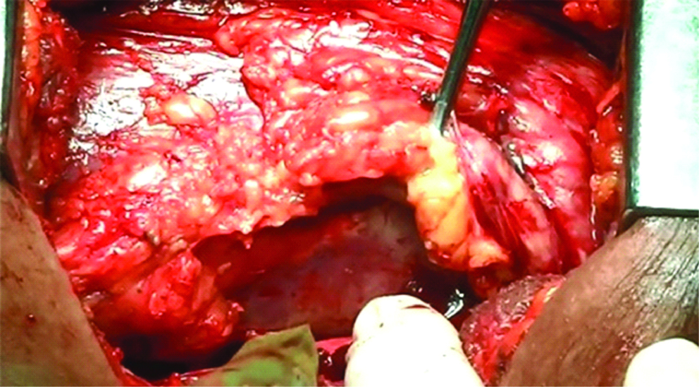
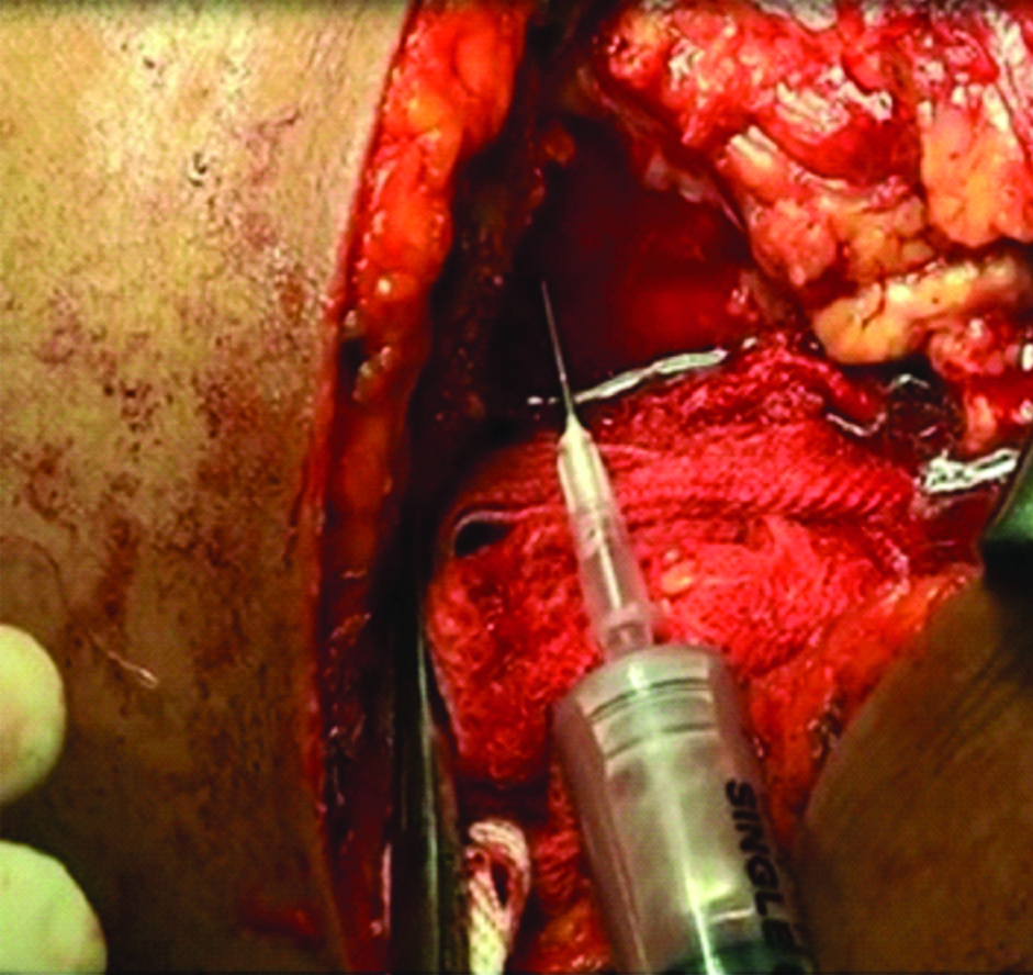
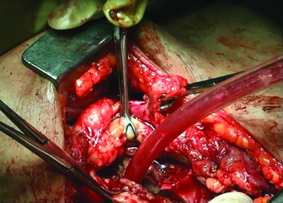
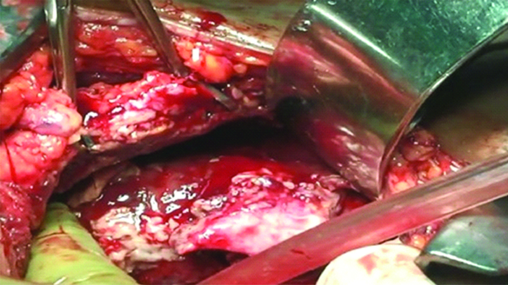
Right 12th rib bed incision was taken and Gerotas fascia dissected and capsule identified as shown in [Table/Fig-3]. Infected haematoma was aspirated as shown in [Table/Fig-4]. The capsule incised and infected haematoma drained as shown in [Table/Fig-5]. The underlying renal parenchyma is seen in [Table/Fig-6] after draining the haematoma. On postoperative day two the patient’s blood pressure dropped to 108/70 mm of Hg and subsequently the ACE inhibitor was stopped and she was discharged on post-operative day 5. On follow up after two months, the patient had a blood pressure of 106/70 mm of Hg and a renal scan showed a GFR of 56.5 mL/min for the right kidney.
Right renal capsule visualized after opening the Gerotas fascia.
Right renal subcapsular infected haematoma being aspirated
Capsulotomy done and haematoma drained
Renal parenchyma visualized after having drained the haematoma.
Discussion
Page Kidney is a unique clinical entity causing hypertension secondary to external renal compression. Experimentally, it was first described by Dr. Page in 1939 in a dog by wrapping the kidney with cellophane tape which induced hypertension in the dog [1]. Page kidney has been caused by either traumatic or spontaneous bleeding causing subcapsular haematoma. Few other conditions which have led to Page kidney include benign renal cyst and renal cell carcinomas [2]. The pathogenesis includes external compression of the kidney which leads to microvascular ischemia and activation of renin-angiotensin system and subsequent hypertension [3].
HELLP syndrome is a life threatening variant of pre-eclampsia which includes hemolysis, elevated liver enzymes and low platelets. Although, this condition is associated with antenatal hypertension, the raised blood pressure spontaneously resolves post-delivery. Subcapsular haematoma of the liver has been documented with HELLP syndrome [4]. Acute kidney injury with HELLP syndrome is a known clinical entity, however, a renal subcapsular haematoma secondary to low platelets is a rare finding.
In this particular case, although the HELLP syndrome and the blood pressure was managed intrapartum, the patient still developed post-partum hypertension which was unusual. We hypothesize that there may have been spontaneous gradual development of a right renal subcapsular haematoma in the immediate postpartum period which may have evolved over a period of 20 days. We believe this may have triggered renin-angiotensin axis which caused post-partum hypertension.
This is a peculiar case of Page kidney as the patient had been diagnosed to have hypertension in the antenatal period as well. This led to a diagnostic dilemma. However, there was normalcy of blood pressure in the immediate post natal period. Moreover, on ultrasound and CT scan there was an obvious subcapsular haematoma associated with the time of presentation which clinched the diagnosis. CT scan has been reported as the most sensitive and specific imaging modality in diagnosing Page kidney [5].
The initial management of non-traumatic Page kidney includes ACE inhibitors to control the hypertension. However, in case of Page kidney delay in management may lead to poor response [6]. Mathew et al., have reported successful ultrasound guided drainage of subcapsular haematoma in management of Page kidney [7]. Dopson SJ et al., has also shown good clinical response by either capsulotomy or nephrectomy in traumatic Page kidney [8,9].
We followed similar treatment guidelines and tried to manage the patient with ACE inhibitors followed by USG guided pigtailing. However, a surgical capsulotomy was finally performed and the patient responded in our case. Delay in exploration would have led to an organized haematoma and subsequent parenchymal injury and eventual nephrectomy.
In a review of case reports of page kidney, Smyth A et al., reported a low post intervention GFR among the non-traumatic group compared to the traumatic group [5]. In our case, GFR was 56.5 mL/min in the affected kidney which could again be attributed to early management and the age of our patient.
Conclusion
Page kidney is a reversible cause of hypertension and a timely capsulotomy and drainage of the haematoma with optimal postoperative management of this condition would prevent long term sequela of hypertension and loss of renal function among young patients.
[1]. Page I, The production of persistent arterial hypertension by cellophane perinephritisJAMA 1939 113:2046-48.10.1001/jama.1939.02800480032008 [Google Scholar] [CrossRef]
[2]. McCune TR, Stone WJ, Breyer JA, Page kidney: case report and review of the literatureAm J Kidney Dis 1991 18:593-99.10.1016/S0272-6386(12)80656-1 [Google Scholar] [CrossRef]
[3]. Massumi RA, Andrade A, Kramer N, Arterial hypertension in traumatic subcapsularperirenal hematoma (Page kidney). Evidence for renal ischemiaAm J Med 1969 46:635-39.10.1016/0002-9343(69)90082-5 [Google Scholar] [CrossRef]
[4]. Haram K, Svendsen E, Abildgaard U, The HELLP syndrome: clinical issues and management. A ReviewBMC Pregnancy Childbirth 2009 9:810.1186/1471-2393-9-819245695 [Google Scholar] [CrossRef] [PubMed]
[5]. Smyth A, Collins CS, Thorsteinsdottir B, Madsen BE, Oliveira GH, Kane G, Page kidney: etiology, renal function outcomes and risk for future hypertensionJ Clin Hypertens (Greenwich) 2012 14(4):216-21.10.1111/j.1751-7176.2012.00601.x22458742 [Google Scholar] [CrossRef] [PubMed]
[6]. Chung J, Caumartin Y, Warren J, Luke PP, Acute Page kidney following renal allograft biopsy: a complication requiring early recognition and treatmentAm J Transplant 2008 8:1323-28.10.1111/j.1600-6143.2008.02215.x18444936 [Google Scholar] [CrossRef] [PubMed]
[7]. Mathew A, Brahmbhatt B, Rajesh R, Kurian G, Unni VN, Page kidneyIndian Journal of Nephrology 2009 19(4):170-71.10.4103/0971-4065.5934220535256 [Google Scholar] [CrossRef] [PubMed]
[8]. Dopson SJ, Jayakumar S, Velez JC, Page kidney as a rare cause of hypertension: case report and review of the literatureAm J Kidney Dis 2009 54:334-39.10.1053/j.ajkd.2008.11.01419167799 [Google Scholar] [CrossRef] [PubMed]
[9]. Moriarty KP, Lipkowitz GS, Germain MJ, Capsulectomy: a cure for the Page kidneyJ Pediatr Surg 1997 32:831-33.10.1016/S0022-3468(97)90630-8 [Google Scholar] [CrossRef]