Breast cancer is the most common cancer among women worldwide and constitutes a significant burden even in under-developed countries with estimate suggesting that 60% of breast cancer deaths are from socioeconomically poor regions [1]. The stages of breast cancer can range from early, curable to metastatic breast cancer. Breast Conserving Surgery is becoming a preferred choice of treatment for breast cancer patients, especially in their early stages. It is less expensive and does not alter the social life of the woman as compared to Modified Radical Mastectomy. However, the outcome for survival and incidence of contralateral breast cancer is the same for both the surgeries [2]. Radiation Therapy finds its stand in the course of treatment and comes with a variety of treatment techniques due to the advancement in linear accelerators. The objective of radiotherapy is to provide a therapeutic dose to a distinct target while minimising the dose to adjacent normal tissues and critical organs. Achieving dose homogeneity in the breast is very important as it can help in reducing the late adverse effect. Changes in breast appearance have been found to be statistically higher in patients with more dose heterogeneity in the breast [3].
The 3DCRT is based on 3D anatomic information and uses dose coverage that will conform to the target volume as much as possible with minimum possible dose to normal tissue [4]. Often wedges or compensators are used to alter the intensity profile to offset contour irregularities. The 3DCRT method is simplistic and superior in terms of low-dose volume, integral dose and treatment time [5]. In IMRT, non-uniform fluence is delivered to optimise the composite dose distribution [4]. Optimal fluence profiles for a given set of beam directions are determined through inverse planning. This technique reduces maximum dose and improves conformity and homogeneity of the target volumes [6,7]. But, there is an increased risk almost by double for the secondary malignancies by the multiple field IMRT [5,8]. As there is an upturn in the survivors of breast cancer patients in the recent past, secondary malignancies and the long-term radiation induced toxicities in the heart and lungs are of concern [9]. This study aimed to compare three different radiotherapy techniques – 3DCRT, two field IMRT and multiple field IMRT in the treatment of Carcinoma Breast.
Materials and Methods
This retrospective, dosimetric study was done in the Department of Radiotherapy and Oncology during the period January-August 2016. Ten female breast cancer patients who were previously treated with whole breast radiation after Breast Conservative Surgery (BCS) were included in this study. After getting the ethical committee approval and written informed consent of the patients, the planning Computed Tomography (CT) images of these patients were used to generate three different radiotherapy plans for the purpose of comparison retrospectively. Out of 10 cases selected 80% were left-sided, and 20% were right-sided breast cancer cases. The CT images were acquired on Philips Brilliance 16-Big Bore CT machine. The slice thickness was 5 mm and acquired with the patients positioned supine, head first, flat on their back on a stable board. The mid sternal line was set parallel to the table. The arm on the side to be treated was placed perpendicular to the body. Their face and head were turned a little upward and contralateral to the treatment side. The patients were immobilised using four clamp thermoplastic moulds.
Structure definition: The target and Organ At Risk (OAR) delineation were done as per Radiation Therapy Oncology Group (RTOG) breast cancer guidelines [10] by the radiation oncologists on Tomo Con 3.0 workstation (Tatra Med software s.r.o, Slovak Republic). The Planning Target Volume (PTV) was generated from the Clinical Target Volume (CTV) with 3 mm margin from the skin. Structure PTV 2 cm with additional 2 cm anterior margin was created to account for patient motion uncertainities. The OAR contoured were the heart, contralateral breast, ipsilateral lung, contralateral lung and spinal cord. A no dose region was created using the external contour excluding the target by 1 cm. This structure was created to reduce the spread of the dose to the normal surrounding tissues and organs.
Treatment planning: The software used was Oncentra Treatment Planning system, version 4.3 (Nucletron B.V., Veenendaal, The Netherlands). Direct Step and Shoot (DSS) algorithm and Collapsed cone (GPU) algorithm were used for dose optimisation and dose calculation respectively. The DSS optimisation algorithm optimises the Multi Leaf Collimator (MLC) settings and monitor units directly to achieve the objectives. At the end of the optimisation process, the MLC segments are ready for delivery without further processing. This is also known as direct machine parameter optimisation [11].
A dose of 50Gy in 25 fractions was prescribed to PTV. A 6MV and 15MV coplanar photon beams from Elekta Precise Digital LINAC model 1500 which accommodates 40 pairs of MLCs was used in treatment planning. For all the 10 patients, three plans, namely 3DCRT, two field IMRT and multiple field IMRT were generated. The planning objective was to ensure at least 95% of the target volume receives 95% of the prescribed dose. While planning, the conformance of the MLC was made to the PTV 2 cm.
3DCRT: Two tangential beams with beam angles between 310°-320° for medial field and 130°-140° for lateral field were set for left-sided cases as shown in [Table/Fig-1]. The beam angles set were 50°-56° for medial field and 230°-232° for lateral for right-sided cases. The dose was prescribed to a normalisation point depending on the need to meet the dose coverage. Beam weights were adjusted, and appropriate wedge angles were chosen to achieve desired coverage and reduce the hotspots to an acceptable level. Besides, field in field was created to lessen hotspot and to improve homogenous dose distribution.
a) Medial and Lateral Tangential wedge beams in 3DCRT; b) Multiple Field IMRT using 7 Beams; c) Two Field (Medial and Lateral Tangential beams) IMRT.
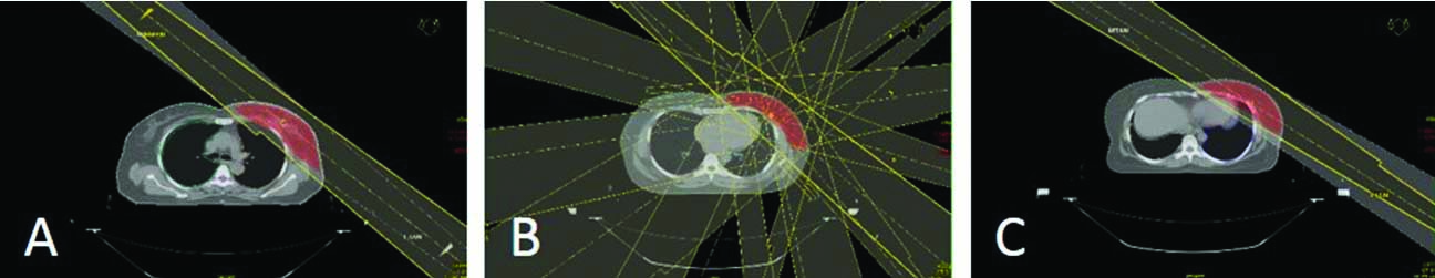
Multiple field IMRT: Seven beams were used where each beam had nearly ten segments or sub-field. The beam angles given for left-sided cases were 315°, 350°, 20°, 50°, 80°, 110° and 145° as shown in [Table/Fig-1]. The beam angles set for right-sided cases were 45°, 10°, 340°, 310°, 280°, 250° and 215°. The dose objectives and constraints mentioned in [Table/Fig-2] were given according to Memorial Sloan Kettering Cancer Centre (MSKCC) guidelines for the IMRT of breast [12].
Dose volume constraints given to the target and critical structures.
| Structure | Specification |
|---|
| PTV | DO* : Uniform dose of 50Gy |
| DC† : Min dose of 45Gy to 95% volume |
| Ipsilateral lung | V20Gy≤30% |
| Dmean≤22Gy |
| Heart | V25Gy≤25% |
| Dmean≤20Gy |
| Dmax≤53Gy |
| Contralateral breast | Dmean<5Gy |
| Contralateral lung | V20Gy≤8% |
*DO: Dose Objective, †DC: Dose Constraint
Two field IMRT: Two tangential beams were set with beam angles same as that of the 3DCRT plan. These beams were without wedges as shown in [Table/Fig-1], segmented into 10 sub-field. Optimisation was performed with dose objectives and constraints as given in [Table/Fig-2].
Dosimetric parameters: With respect to the target, data regarding tumour volume (cc), D2 (dose received by 2% of the PTV), D5 (dose received by 5% of the PTV), D95 (dose received by 95% of the PTV), Dmean (mean dose received by the PTV), V49.5Gy (volume of the PTV that receives a dose of 49.5Gy) and V53.5Gy (volume of the PTV that receives a dose of 53.5Gy) were collected. The Dmean was noted for ipsilateral lung, contralateral lung, heart and contralateral breast. Additionally, V20Gy and V10Gy (volume of the organ that receives a dose of 20 Gy and 10Gy respectively) for ipsilateral lung and V25Gy (volume of the organ that receives a dose of 25Gy) for heart were used for the study. The conformity and homogeneity indices were calculated for each plan.
The conformity index was found using the Conformation Number (CN) formula proposed by Van’t Riet et al., as below [13].
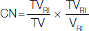
where, CN=Conformation Number (ranges between 0 and 1, 1 implies the ideal case and 0 or close to 0 implies absence of conformity); TVRI =Target Volume covered by the Reference Isodose; TV=Target Volume; VRI =Volume of the Reference Isodose.
The formula chosen for homogeneity index [14] was,

where, HI=Homogeneity Index; D5 =minimum dose in 5% of PTV indicating the maximum dose; D95 =minimum dose in 95% of PTV indicating the minimum dose.
The lower the index value, i.e., closer to 1, better the dose homogeneity. The index value, however, increases with lesser homogeneous plans.
Statistical Analysis
The paired t-test was performed for the parameters to check if there are any differences between the planning techniques. The analysis was done by comparing each planning technique individually against the other. A significance level, p=5% or 0.05 was chosen.
Results
A total of 10 female patients who underwent whole breast radiation were included in this study. Mean age of patients was 47 years and age ranged from 36 to 66 years. Also, 80% of the cases were left-sided breast [Table/Fig-3]. Treatment plans were generated using three different techniques viz., 3DCRT, Two Field IMRT and Multiple Field IMRT. Plan quality parameters such as Conformity index (CN), Homogeneity Index (HI), D2, Dmean, Monitor Units (MU), V47.5Gy and V53.5Gy were taken from Dose Volume Hisogram (DVH) was tabulated and are compared [Table/Fig-4]. The dose to critical organs such as Ipsilateral lung (Dmean,V20 and V10), Heart (V25Gy and Dmean), Dmean of Contralateral lung and Contralateral breast was also reported and compared. [Table/Fig-5]. All results were statistically analysed using paired t-test for p-value of 5% or 0.05.
Demographic details of the 10 breast cancer patients.
| Characteristics | Value |
|---|
| Mean age, years | 47 (range 36-66) |
| Laterality |
| Left breast | 8/10 (80%) |
| Right breast | 2/10 (20%) |
| Stage |
| I | 2/10 (20%) |
| IIA | 5/10 (50%) |
| IIB | 3/10 (30%) |
| Pathological node status |
| Positive | 1/10 (10%) |
| Negative | 9/10 (90%) |
| Hormone receptor status |
| Positive | 5/10 (50%) |
| Negative | 5/10 (50%) |
| HER2/neu status |
| Positive | 3/10 (30%) |
| Negative | 7/10 (70%) |
Comparison between the three techniques (two at a time) for the PTV.
| Parameter | 3DCRT (A) | Two field IMRT (B) | Multiple field IMRT (C) | p-value* (A&B) | p-value* (B&C) | p-value* (C&A) |
|---|
| CN | 0.59±0.08 | 0.61±0.07 | 0.78±0.05 | 0.105 | 0.0001 | 0.0001 |
| HI | 1.11±0.01 | 1.09±0.01 | 1.08±0.01 | 0.001 | 0.141 | 0.0001 |
| D2 (Gy) | 53.5±0.44 | 52.95±0.49 | 52.72±0.41 | 0.008 | 0.175 | 0.002 |
| Dmean (Gy) | 50.73±0.27 | 50.65±0.27 | 50.56±0.3 | 0.543 | 0.551 | 0.09 |
| V47.5Gy (%) | 95.2±0.86 | 95.87±0.71 | 96.86±1.10 | 0.149 | 0.040 | 0.0004 |
| V53.5Gy (%) | 2.16±1.44 | 0.98±0.75 | 0.60±0.63 | 0.029 | 0.181 | 0.005 |
| MU/# | 408.2±22.59 | 346.5±67.91 | 815±180.17 | 0.013 | 0.0001 | 0.0001 |
*Paired t-test was used for identifying statistical differences between the plans.
Comparison between the three techniques (two at a time) for the OAR.
| Parameter | 3DCRT (A) | Two field IMRT (B) | Multiple field IMRT (C) | p-value* (A&B) | p-value* (B&C) | p-value* (C&A) |
|---|
| Ipsi-lateral lung | V20Gy (%) | 18.45±4.37 | 16.55±2.95 | 20.27±2.11 | 0.151 | 0.002 | 0.218 |
| V10Gy (%) | 24.08±4.88 | 22.64±3.26 | 41.54±7.70 | 0.314 | 0.0001 | 0.0001 |
| Dmean (Gy) | 9.77±2.10 | 8.77±1.40 | 13.12±1.19 | 0.09 | 0.0001 | 0.0002 |
| Contra-lateral lung | Dmean (Gy) | 0.39±0.10 | 0.39±0.08 | 4.60±1.38 | 0.889 | 0.0001 | 0.0001 |
| Heart | V25Gy (%) | 6.61±5.51 | 4.53±3.58 | 6.35±3.60 | 0.044 | 0.004 | 0.465 |
| Dmean (Gy) | 5.15±3.02 | 4.01±2.00 | 12.21±3.04 | 0.03 | 0.0001 | 0.0001 |
| Contra-lateral breast | Dmean (Gy) | 0.77±0.28 | 0.69±0.32 | 2.31±0.25 | 0.363 | 0.0001 | 0.0001 |
*Paired t-test was used for identifying statistical differences between the plans.
Discussion
Therapeutic radiation has to tread the path of adequately treating the volume of interest to planned doses while sparing the surrounding normal tissues. Recent developments in the field of radiation delivery have progressively enabled increasing conformity of the dose to the tumour while progressively reducing dose to normal tissues. IMRT is the standard-of-care in several tumours sites such as cancers of the head and neck and prostate cancers. However, its role in the treatment of breast cancer has been questioned [15]. Irrespective of the site of treatment, theoretical benefits of utilising a highly conformal form of radiotherapy cannot be denied. This dosimetric study attempted to identify if IMRT would offer significant advantages in terms of both coverage of tumour volume and organ sparing in patients with breast cancer.
Conformal radiotherapy techniques stress at delivering target dose as if to form an envelope around the tumour alone. Closer the value of Conformation number (CN) to 1 [13], better is the conformity of the plan. In our study cohort, best conformity was achieved by multiple field IMRT technique. Conventional radiotherapy planning techniques frequently result in missing of the tumour volume at the edges due to presence of surrounding normal tissues such as contralateral breast. IMRT intuitively has an advantage of superior conformity, and this has been reported in other dosimetric studies [16-18]. For instance, in a dosimetric study by Beckham et al, IMRT was found to significantly improve the conformity when compared to conventional technique (0.91 vs 0.48, respectively) [16]. Both the IMRT techniques provided homogeneous dose distribution with reduced areas of breast receiving doses >107% compared to 3DCRT. These results are in agreement with other reports on variation in dose-homogeneity with treatment technique [17,18]. Better the dose homogeneity, greater the chance to reduce skin and subcutaneous tissue toxicity and potentially improved cosmetic outcome [19]. Additionally, maximum dose to the target (D2 and V53.5Gy) was on the higher side for 3DCRT. By these illustrations, it was found that target requirements were better achieved by the IMRT techniques. Cases of left-sided breast tumour need more attention and regard since the heart is in proximity while for right-sided cases cardiac dose is due to scattered dose [20]. Multiple field IMRT resulted in higher lung and heart doses and also highest doses in the contralateral breast compared to the other two techniques. The use of tangential parallel opposed beams in whole breast radiotherapy helps in reducing the dose to lung tissue. This beam arrangement in combination with two field IMRT technique resulted in a better reduction of dose to the lungs and heart and was in accordance with Li JS et al. [21]. Li JS et al., compared intensity-modulated tangential beam irradiation with conventional tangential photon beam irradiation for breast treatment. According to them, dose comparisons showed that the percentage volume of the lung receiving more than 20 Gy dose (V20Gy) during the entire treatment was reduced by about 10%. The percentage volume of the heart receiving more than 30 Gy dose (V30Gy) is reduced from 3.3% to 0.3% [21].
The MUs for multiple field IMRT was more than double for seven cases and almost double for the remaining three cases compared to that of two field IMRT. The MU for two field IMRT was also significantly lower when compared to 3DCRT technique. The higher MU in 3DCRT plans is because of the use of wedge. In this study, a universal motorised wedge located in the head of the Linear Accelerator is used. The presence of a metal wedge can increase the scatter dose outside the field [22]. Since the cancer survival rate is increasing [9,23], the quality of life after treatment necessitates interest. Furthermore, dose to the contralateral breast can increase the risk of contralateral breast cancer, especially in those under 45 years of age [24]. The use of MLC instead of a physical wedge to achieve homogenous dose distribution inside the target volume in two field IMRT reduced the mean dose to contralateral breast. On the whole, it was found that sparing of critical organs was well achieved by the two field IMRT technique.
Limitation
It is limited by a small number of patients that were used for comparisons between the different plans. However, since the different treatment techniques were planned and compared on the same patient image-sets, and challenges in treatment planning vary little in between patients, we believe that a larger number of patient image-sets would lead to a similar conclusion.
Conclusion
In conclusion, the two field IMRT technique was clearly found to have better homogeneity when compared to 3DCRT while delivering lower doses to critical organs compared to multiple field IMRT. Though the multiple field IMRT plan helped in achieving better dose homogeneity and conformity, the dose to critical organs was on the higher side compared to two field IMRT. Hence, two field IMRT plan technique can be adopted in routine clinical practice for treatment of breast cancer patients post breast conserving surgery. This study can be further extended to Volumetric Modulated Arc Therapy (VMAT), latest advanced treatment planning and delivery technique in Indian scenario for comparison and analysis.
*DO: Dose Objective, †DC: Dose Constraint
*Paired t-test was used for identifying statistical differences between the plans.
*Paired t-test was used for identifying statistical differences between the plans.