Fungal Pericarditis due to Aspergillus nidulans: A Rare Case Report
Sarika P Kombade1, Kumar S Abhishek2, Vijaya Lakshmi Nag3
1 Assistant Professor, Department of Microbiology, AIIMS, Jodhpur, Rajasthan, India.
2 Senior Resident, Department of Microbiology, AIIMS, Jodhpur, Rajasthan, India.
3 Professor and Head, Department of Microbiology, AIIMS, Jodhpur, Rajasthan, India.
NAME, ADDRESS, E-MAIL ID OF THE CORRESPONDING AUTHOR: Dr. Kumar S Abhishek, AIIMS Resident’s Hostel, Jodhpur-342005, Rajasthan, India.
E-mail: get.abhi1904@gmail.com
Disseminated Aspergillosis though rare is usually seen among immunocompromised and neutropenic patients. Aspergillus spp are usually considered to be a laboratory contaminants until its association with the disease is established on repeated isolation. The present is of Aspergillus nidulans associated with pericarditis post Anti-Tuberculous Treatment (ATT) initiation in a 67-year-old male patient with known case of Pulmonary Tuberculosis (PTB). Aspergillus nidulans was identified on the basis of culture on Sabouraud Dextrose Agar (SDA) and Lacto-Phenol Cotton Blue (LPCB) wet mount preparation.
Anti-tuberculous treatment, Disseminated aspergillosis, Pulmonary tuberculosis
Case Report
A 67-year-old male patient presented to the TB Chest OPD with the history of chest pain, breathlessness and cough since one month. For which he was admitted in the SMS Medical College where he was diagnosed for pulmonary tuberculosis with pleuro-pericardial effusion, pleural tapping was done and Anti-Tuberculous Treatment (ATT) Cat. I was started. Following ATT, patient developed acute renal infection and ATT induced Hepatitis, with supportive treatment patient recovered and was discharged. After 7-8 days patient complained of aggravated chest pain and loose motion.
Patient was admitted to AIIMS Jodhpur on 26-7-2017 with provisional diagnosis of Pleurisy; pneumothorax with pulmonary effusion. Treatment started with Inj. Metrogyl TDS (Metronidazole 400mg thrice daily) and all needful investigations were performed.
Biochemical analysis revealed raised level of Procalcitonin (0.96 ng/mL). Serum Urea and creatinine level was also raised to 243 mg/dL and 1.67 mg/dL respectively. His direct, indirect and total bilirubin level was 1.43, 1.17 and 2.60 mg/dl respectively which were above normal range for the age and sex of the patient. Haemogram suggested neutrophilic leucocytosis with leucoerythroblastic blood picture associated with mild anaemia.
Two-Dimensional Echocardiography (2D ECHO) was suggestive of pericardial thickening. On Contrast Enhanced Computed Tomography (CECT) thorax, significant pericardial effusion with pericardial thickening with moderate bilateral pleural effusion was observed. On the ground of CECT findings, Computed Tomography (CT) guided pericardial tapping was done and pericardial fluid was sent for aerobic and fungal culture and sensitivity.
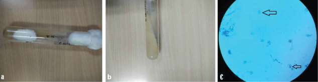
As soon as the sample was received in Microbiology laboratory, it was inoculated on routine aerobic culture media and on a set of SDA with Cycloheximide and gentamicin and incubated at 25oC and 37oC. KOH mount was also performed where no fungal elements were demonstrated. Aerobic culture was sterile after 48 hours of incubation while after three weeks of incubation, fungal culture appeared on both the tubes of SDA with gentamicin. Velvety, white coloured growth was obtained on obverse side [Table/Fig-1a,b] with no pigmentation. LPCB mount was prepared and viewed under 40X objective magnification. LPCB microscopy revealed abundant round Hulle cells [Table/Fig-1c] with few cleistotheicium and occasional bi-serrated short phialide developed on vesicle directly with hyaline septate hyphae arising from unbranched conidophore. Presence of Hulle cells narrowed down the diagnosis to Aspergillus nidulans, Aspergillus ustus or Aspergillus versicolor which are known to produce abundant Hulle cells. Absence of diffusing pigment and round regular Hulle cells differentiated it from Aspergillus ustus which produces irregular to elongated Hulle cells, while the presence of cleistotheicium ruled out the possibility of Aspergillus versicolor. On the basis of above findings, fungus was identified as Aspergillus nidulans.
(a) Growth on SDA (Obverse): White velvety growth. (b) Growth on SDA (Reverse): No diffusible pigment. (c) LPCB Mount (Arrow): Hulle Cells.
We decided to repeat fungal culture if possible to look for the same isolate on repeated sample. Aspergillus being the most common laboratory contaminant, to establish isolate as the cause of this condition only when the repeated sample yields the same isolate. But, as the patient left the hospital against medical advice, it was not possible for us to re-confirm the isolate and also to look for response to anti-fungal treatment.
Discussion
Fungal infections of the heart and pericardium are rare and usually associated with disseminated fungemia. Prolonged use of antibiotics, immunosuppressive agents, and parenteral nutrition is becoming more frequent, and these factors may be contributing to the increased incidence of fungemia. Invasive aspergillosis has also become an increasingly prevalent disease in immunocompromised patients [1]. Our patient was on ATT following diagnosis of pulmonary TB with pleuro-pericardial effusion. Gradually patient developed chest pain, hepatitis and diarrhoea, probably ATT induced. Further investigations revealed cardiac tamponade with effusion and Aspergillus nidulans was isolated from the pericardial aspirate. Though rare fungemia and invasive aspergillosis are also reported to cause endocarditis, myocarditis or pericarditis. So, we chose to present it as a case report. The classic clinical manifestation is sudden onset of fever, and a pleural rub, suggesting pulmonary thromboembolism. If left untreated, the aspergillus may extend into neighbouring anatomical structures and organs, e.g., the mediastinum, ribs, vertebrae, oesophagus, and pericardium [1].
Aspergillus species are usually ubiquitous saprophytes. Aspergillus fumigatus is so by far the most frequently isolated pathogen but other species like Aspergillus nidulans has also been recognised to be human pathogen. A.nidulans is known to cause invasive infections in Chronic-Granulomatous Diseases (CGD). Unlike A. fumigatus, A.nidulans rarely causes aspergillosis in neutropenic and immunocompromised patients.
Cardiac tamponade and overwhelming sepsis are the usual fatal complications of purulent pericarditis, rare complications includes myocardial necrosis and infarction. Aspergillus pericarditis with associated myocardial involvement in form of myocardial abscess has been described in few case reports [2]. Prosthetic valve endocarditis rarely presents with pericarditis or myocardial abscess. Most of such cases occur in settings of disseminated invasive aspergillosis [3].
Lungs are the commonest organ to be affected by the Aspergillus spores via inhalation, with rapid haematogenous dissemination to vital organs like heart and CNS forming micro abscesses. In myocarditis symptoms progresses insidiously and rapidly, and we usually loose the patients. Isolation of the fungus from blood carries extremely low sensitivity rate of 6.4%-11% and thus, tissue biopsy is usually required [4]. In immunocompromised patients presenting with Aspergillus pericarditis or myocarditis without any sign of transmural myocardial infarction, extension of myocardial abscess into the pericardial space stands to be the most common mechanism of dissemination throughout the pericardium. Serum antigen studies have shown good result using galactomannan and 1,3-beta-D-glucan and are sufficient to make a presumptive diagnosis and initiate antifungal treatment [5,6]. The major limitation of this case study was lack of follow up and confirmation of the isolate on repeated culture, as the patient left the medical service against any medical advice. So, it was difficult for us to establish direct connection of the isolate with the present condition of the patient.
Conclusion
Our case highlights Aspergillus nidulans though rare but has potential to cause pericarditis and pericardial effusion in immunocompetent patients with other underlying medical conditions.
[1]. Balows Topley WWC, Collier L, Sussman Wilson SG, Microbiology & Microbial Infections 2006 vol. 5:10e [Google Scholar]
[2]. Kupsky DF, Alaswad K, Rabbani BT, A rare case of Aspergillus pericarditis with associated myocardial abcess and echocardiographic response to therapyEchocardiography 2016 33(7):1085-88.10.1111/echo.1321427009593 [Google Scholar] [CrossRef] [PubMed]
[3]. Le Moing V, Lortholary O, Timsit JF, Couvelard A, Bouges-Michel C, Wolff M, Aspergillus pericarditis with tamponade: Report of a successfully treated case and reviewClin Infect Dis 1998 26:451-60.10.1086/5163269502470 [Google Scholar] [CrossRef] [PubMed]
[4]. El-Hamamsy I, Durrleman N, Stevens LM, Perrault LP, Carrier M, Aspergillus endocarditis after cardiac surgeryAnn Thorac Surg 2005 80:359-64.10.1016/j.athoracsur.2004.08.07015975413 [Google Scholar] [CrossRef] [PubMed]
[5]. Pfeiffer CD, Fine JP, Safdar N, Diagnosis of invasive aspergillosis using a galactomannan assay: A meta-analysisClin Infect Dis 2006 42:1417-27.10.1086/50342716619154 [Google Scholar] [CrossRef] [PubMed]
[6]. Pickering JW, Sant HW, Bowles CA, Roberts WL, Woods GL, Evaluation of a (1-3)-beta-D-glucan assay for diagnosis of invasive fungal infectionsJ Clin Microbiol 2005 43:5957-62.10.1128/JCM.43.12.5957-5962.200516333082 [Google Scholar] [CrossRef] [PubMed]