Is Preoperative Anthropometric Data Assessment Reliable Indicator for Estimating Femoral Tunnel Length in Anterior Cruciate Ligament Reconstruction?
Nagamanickam Rajendran Sutharsen1, Prem Kotian2, Rajendra Annappa3, Srikanth Mudiganty4, Sharan Mallya5, Nabeel Mohammed6
1 Postgraduate, Department of Orthopaedics, Kasturba Medical College, Manipal Academy of Higher Education, Mangalore, Karnataka, India.
2 Professor, Department of Orthopaedics, Kasturba Medical College, Manipal Academy of Higher Education, Mangalore, Karnataka, India.
3 Assistant Professor, Department of Orthopaedics, Kasturba Medical College, Manipal Academy of Higher Education, Mangalore, Karnataka, India.
4 Assistant Professor, Department of Orthopaedics, Kasturba Medical College, Manipal Academy of Higher Education, Mangalore, Karnataka, India.
5 Senior Resident, Department of Orthopaedics, Kasturba Medical College, Manipal Academy of Higher Education, Mangalore, Karnataka, India.
6 Senior Resident, Department of Orthopaedics, Kasturba Medical College, Manipal Academy of Higher Education, Mangalore, Karnataka, India.
NAME, ADDRESS, E-MAIL ID OF THE CORRESPONDING AUTHOR: Dr. Nagamanickam Rajendran Sutharsen, Postgraduate, Department of Orthopaedics, Kasturba Medical College, Manipal Academy of Higher Education, Mangalore-576104, Karnataka, India.
E-mail: n.r.s349@gmail.com
Introduction
Correct length of the femoral tunnel is required for adequate length of graft inside the femoral canal. Length of femoral tunnel depends on mass, height, dimensions of lateral femoral condyle and amount of flexion of knee intraoperatively. Increasing weight and BMI affects the length of femoral canal since they affect positioning of limb in maximal knee flexion.
Aim
To measure tunnel length in independent femoral tunnel drilling using the medial portal and to correlate the tunnel length with anthropometric data (height, weight, BMI of patient) and with width and depth of Lateral Femoral Condyle (LFC) based on MRI.
Materials and Methods
The present prospective study including 40 patients were conducted. Participants were selected by a non-random convenience sampling methodology. Complete details of patients were collected by verbal communication with the patients and their attendees, clinical examination, baseline investigations, radiological investigations and surgical findings. Radiological investigations included preoperative assessment of LFC in MRI. Surgical findings were determined by measuring the femoral tunnel length after initial drilling through the medial portal using a depth gauge placed through the medial portal. SPSS version 17.0 was used for data analysis.
Results
A total of 40 patients, 30 males and 10 females were included. Mean BMI was 24.22 kg/m2 with the range of 18 to 30. Mean width of LFC was 32.9 mm with range of 26.2 mm to 37.3 mm and depth of LFC was 58.79 with range of 50.7 mm to 66.2 mm. Mean femoral tunnel length was 38.4 mm with range from 32 mm to 44 mm.
Conclusion
Average femoral tunnel length in Anterior Cruciate Ligament (ACL) reconstruction was 38.4 mm for assessed 40 patients. This correlated positively with height and weight of the patient. It also correlated positively with the width (p=0.027) and depth (p=0.029) of lateral femoral condyle.
Anthropometry, Lateral femoral condyle, Orthopaedic surgery
Introduction
Recent advances in orthopaedic surgery have improved surgical outcome, particularly in the field of ACL. Anatomical reconstruction of ACL surgery has become the cornerstone in recent years. The correct tunnel placement is specifically crucial for successful arthroscopic ACL reconstruction surgery and it requires good tensile, stretchable, anatomical placement of the autograft [1-3].
ACL injury is encountered routinely in sports medicine. Literature has reported poor outcomes for ACL injuries when managed on a conservative basis; hence, reconstructive surgery is the established treatment of choice particularly in young patients with an active lifestyle, including sportsmen. The most common mode of injury resulting in ACL ligament injury is the external rotational force (valgus) on knee bending [4].
The main purpose of ACL reconstruction is not only to restore knee stability but also to normalise knee kinematics after reconstructive surgery. ACL injury produces functional impairment and provokes the early onset of degenerative osteoarthritic changes at the knee joint due to predisposing ACL ligament injury and unopposed activity of anterior translation of tibia over the femur. Earlier, ACL reconstruction focused primarily on non-anatomical approach using single bundle reconstruction by transtibial technique. However, this technique provides only anterior stability in knee flexion [5-7].
Anatomical placement of femoral tunnel is important irrespective of method of reconstruction whether single-bundle or double-bundle method. The ideal position of the tibial and femoral placement of the canals in an anatomical location is the central part of native ACL footprints. Recent advances in imaging modalities and ultimately better understanding of anatomy have led to improvements in surgical arthroscopic techniques. Furthermore, availability of advanced fixation devices has resulted in better clinical outcomes even when using the single-bundle ACL reconstructive surgery. In spite of this, some studies have reported that 10% to 30% of the patients that have undergone single-bundle ACL reconstruction had experienced rotational instability and later progression to osteoarthritis. It has been proposed that such issues result due to lack of the Postero-Lateral (PL) bundle in the single-bundle ACL reconstructed knee [6-8].
Femoral canal for placement of the graft in a central position of native ACL is drilled in the lower part of the lateral femoral condyle. Canal has to be more horizontal which gives better rotational control in addition to translational stability anteroposteriorly.
Correct length of the femoral tunnel is required for adequate length of graft inside femoral canal. Length of femoral tunnel depends on mass, height, dimensions of lateral femoral condyle and amount of flexion of knee intraoperatively. Increasing weight and BMI affects length of femoral canal since they affect the positioning of limb in maximal knee flexion.
The present study aimed to measure the tunnel length in independent femoral tunnel drilling using medial portal and to correlate the tunnel length with anthropometric data (height, weight, BMI of patient) and with width and depth of LFC based on MRI.
Materials and Methods
A 2-year prospective study from June 2015 to June 2017 was conducted in hospitals affiliated with Kasturba Medical College, Mangalore. The study population was composed of 40 patients between the age of 20 to 50 years attending the Department of Orthopedics in Kasturba Medical College and its allied hospitals in Mangalore, Karnataka, India. Study approval was taken from the Institutional Ethics Committee (IEC) of Kasturba Medical College, Mangalore, Karnataka, India. The objectives were explained to the participants involved in the study following which written informed consent was taken. Confidentiality of the information was strictly adhered to, by assuring the patients that no details of their status will be revealed and data will be used only for research purposes.
Inclusion criteria consisted of patients undergoing primary arthroscopic ACL reconstruction using anatomic femoral tunnel drilling and graft size ≥8 mm in thickness with no posterior wall fracture. All patients were fixed with the suspensory fixation on femoral side. Patients with revision ACL reconstruction, ACL reconstruction as part of multi-ligamentous reconstruction, associated fractures or bony injuries, chronic ACL insufficiency with osteoarthritis, avulsion injury of the ACL, improper drilling in femoral tunnel were excluded from the study. Participants were selected by a non-random convenience sampling methodology.
Data was collected by the verbal communication with the patients and their attendees, clinical examination, baseline investigations, radiological investigations and surgical findings. Radiological investigations included preoperative assessment of Lateral femoral condyle LFC in MRI using Radi-Ant Dicom viewer done by two Senior Radiologists (to avoid intra-observer variations). Mean of these two readings was taken. Surgical findings were determined by measuring the femoral tunnel length after initial drilling through the medial portal using a depth gauge placed through the medial portal.
All patients underwent the same technique for independent femoral tunnel drilling, through the anteromedial portal on the ACL footprint [Table/Fig-1]. The ACL was first visualised using an arthroscope in a viewing (anterolateral) portal. The shaver was then used to remove the torn ACL stump using an instrumental (anteromedial) portal; however, a small cuff of tissue was preserved at the femoral ACL footprint, to look over the size, location and shape of the footprint.
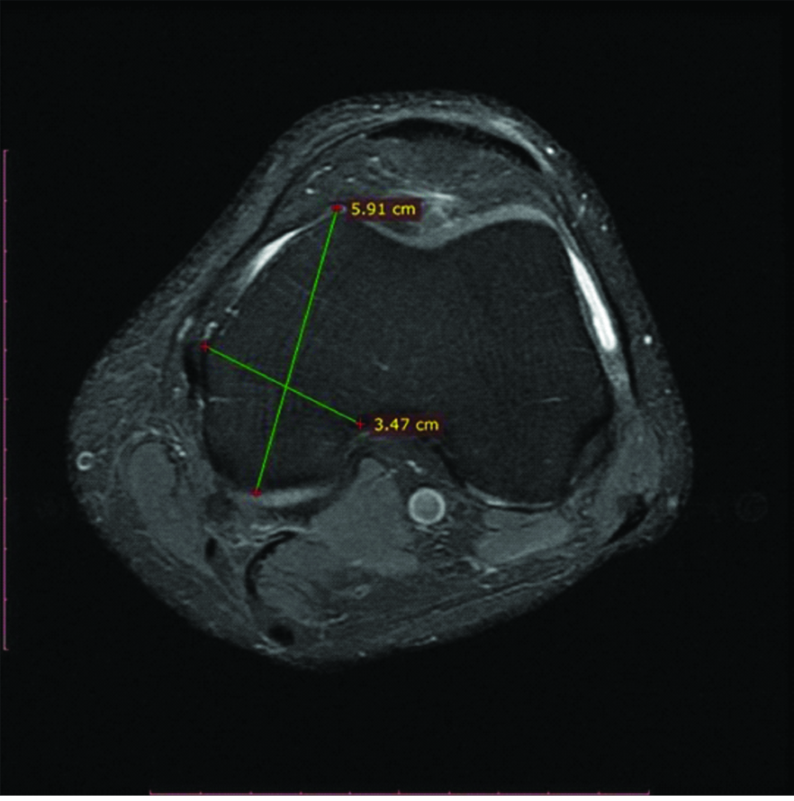
Measurement of width and depth in MRI.

A secure database was maintained with all patient information. Tunnel length was recorded at the time of drilling intraoperatively. In addition, height, weight, and BMI were recorded to correlate patient demographics with tunnel length. The width and anteroposterior length of the LFC, as measured on magnetic resonance images or plain radiographs, were also correlated with tunnel length. On magnetic resonance images, the width and depth of the LFC were measured between 15 mm and 20 mm proximal to the joint line at the point of greatest width of the LFC; these were always measured at a right angle to one another. On plain radiographs, the depth of the LFC was measured on the lateral view and the width was measured on the anteroposterior view, again approximately 15 mm to 20 mm proximal to the joint line; magnification error was taken into account by the PACS software used in the measurements.
Statistical Analysis
SPSS version 17.0, Pearson’s correlation and t-test were used for data analysis.
Results
In the present study a total of 40 patients, 30 males and 10 females were included. Mean BMI was 24.22 with range of 18-31. Mean width of LFC was 32.9 mm with range of 26.2 mm to 37.4 mm and depth of LFC was 58.79 mm with range of 50.7 mm to 66.2 mm. Mean femoral tunnel length was 38.4 mm with range from 32 mm to 44 mm [Table/Fig-2].
Summary of data collection.
| Variables | n | Mean | Std. Deviation | 95% Confidence Interval for Mean | Minimum | Maximum |
|---|
| Lower Bound | Upper Bound |
|---|
| Height | 40 | 166.40 | 8.04 | 163.83 | 168.97 | 148.00 | 181.00 |
| Weight | 40 | 67.63 | 11.24 | 64.03 | 71.22 | 42.00 | 97.00 |
| BMI | 40 | 24.23 | 3.44 | 23.13 | 25.32 | 18.00 | 31.00 |
| Width of LFC | 40 | 32.93 | 3.43 | 31.84 | 34.03 | 26.00 | 37.40 |
| Depth of LFC | 40 | 58.79 | 3.78 | 57.58 | 60.00 | 50.70 | 66.20 |
Femoral tunnel length in ACL reconstruction correlated positively with width (p=0.027) and depth (p=0.029) of lateral femoral condyle [Table/Fig-3]. Correlation analysis for various variables is shown in [Table/Fig-4].
Correlation of preoperative MRI with lateral condyle depth and width measurements.
| Variables | Sex | n | Mean | SD | t-test p-value |
|---|
| Width of LFC | Male | 30 | 33.62 | 2.731 | 0.027* |
| Female | 10 | 30.88 | 4.555 |
| Depth of LFC | Male | 30 | 59.53 | 3.027 | 0.029* |
| Female | 10 | 56.55 | 5.004 |
Significant *
| Variables | Correlations |
|---|
| Height | Weight | BMI | Width of LFC | Depth of LFC |
|---|
| Age | Pears | -.106 | 0.058 | 0.236 | -.074 | -.023 |
| p-value | .515 | 0.722 | 0.142 | 0.649 | 0.889 |
| Height | Pearson Correlation | | 0.423** | -.074 | 0.629** | 0.615** |
| p-value | | 0.006 | 0.651 | <0.001 | <0.001 |
| Weight | Pearson Correlation | | | 0.800** | 0.525** | 0.545** |
| p-value | | | <0.001 | 0.001 | <0.001 |
| BMI | Pearson Correlation | | | | 0.173 | 0.205 |
| p-value | | | | 0.285 | 0.206 |
| Width of LFC | Pearson Correlation | | | | | 0.830** |
| p-value | | | | | <0.001 |
**Correlation is significant at the 0.01 level
Discussion
The anatomical positioning of the femoral and tibial tunnels in the central part of native ACL is of utmost importance in ACL reconstruction. We used Antero Medial portal in independent femoral tunnel drilling which produced average tunnel length of 38.4 mm. Ideal or minimal tunnel length is unknown; tunnel length was long to hold adequate graft and promotes healing of graft, least possibility of posterior wall fracture.
Accessory Antero Medial portal has been advised by many in literature wherein the location of the tunnel is more anatomical and horizontal with very fewer chances of a posterior blow out [1,4,5]. Authors did not have any posterior blowout in the present study and tunnel lengths were comparable to other studies available in the literature. When accessory Antero Medial portal was used many authors believe that the angle of femoral canal was more anterior in axial plane in lateral femoral condyle. During drilling placing the knee in maximal flexion allows better length and a fewer chance for the posterior blowout.
The correlations authors have obtained in the present study are similar to results in the previous study by Tompkins M et al., which is understandable since larger individuals have larger condyles. Tompkins M et al., also included knee flexion angle in their study which is correlated negatively with weight and BMI which can be assumed due to interference of soft tissues for knee flexion but tunnel lengths did not correlate with knee flexion angles [1]. We placed the knee in maximal flexion in the present study, however authors have not measured it intraoperatively. Authors have found none of these variables causing short femoral tunnels.
Several studies have described post-operative tunnel positions and compared the results with known characteristics in the literature including its influence on clinical outcome [1,7,8]. There is a gap in literature with regards to patient-specific optimal tunnel orientation preoperatively, although correct femoral and tibial tunnel positioning a major factor in the overall success of ACL reconstructions.
Iyyampilla G et al., in their study concluded that a sagittal inclination of greater than 45° and an axial angle of 30°-45° resulted in a tunnel length of more than 35 mm [7]. This is achieved by flexing the knee maximally so that the hip flexion increases with increase in the sagittal inclination. Hence, theoretically weight and increased BMI may reduce knee flexion angle and affect femoral tunnel length. However, these did not affect femoral tunnel length in the present study. Tompkins M et al in their study recorded knee flexion angles from 122°-147° which was small to affect tunnel length.
Limitation
The main limitation of the present study was that sample size was small, there was no control group and the long-term clinical effects was not followed up.
Conclusion
Average femoral tunnel length in ACL reconstruction was 38.4 mm for assessed 40 patients. This correlated positively with height and weight of the patient. It is also proportional to the width and depth of LFC, however there was no correlation with BMI. Therefore, in ACL reconstruction tunnel lengths tend to be greater with increasing patient height, weight and larger LFC dimension.
Significant *
**Correlation is significant at the 0.01 level
[1]. Tompkins M, Milewski MD, Carson EW, Brockmeier SF, Hamann JC, Hart JM, Femoral tunnel length in primary anterior cruciate ligament reconstruction using an accessory medial portalArthroscopy 2013 29(2):238-43.10.1016/j.arthro.2012.08.01923270787 [Google Scholar] [CrossRef] [PubMed]
[2]. Strobel MJ, Manual of arthroscopic surgery 2002 1st edBerlinSpringer-Verlag:46010.1007/978-3-540-87410-2 [Google Scholar] [CrossRef]
[3]. Bedi A, Raphael B, Maderazo A, Pavlov H, Williams RJ 3rd, Transtibial versus anteromedial portal drilling for anterior cruciate ligament reconstruction: a cadaveric study of femoral tunnel length and obliquityArthroscopy 2010 26(3):342-50.10.1016/j.arthro.2009.12.00620206044 [Google Scholar] [CrossRef] [PubMed]
[4]. Basdekis G, Abisafi C, Christel P, Influence of knee flexion angle on femoral tunnel characteristics when drilled through the anteromedial portal during anterior cruciate ligament reconstructionArthroscopy 2008 24(4):459-64.10.1016/j.arthro.2007.10.01218375279 [Google Scholar] [CrossRef] [PubMed]
[5]. Pastrone A, Ferro A, Bruzzone M, Bonasia DE, Pellegrino P, Elicio DD, Anterior cruciate ligament reconstruction creating the femoral tunnel through the anteromedial portal. Surgical techniqueCurrent Reviews in Musculoskeletal Medicine 2011 4(2):52-56.10.1007/s12178-011-9078-721541700 [Google Scholar] [CrossRef] [PubMed]
[6]. Celiktas M, Kose O, Sarpel Y, Gulsen M, Can we use intraoperative femoral tunnel length measurement as a clue for proper femoral tunnel placement on coronal plane during ACL reconstruction?Arch Orthop Trauma Surg 2015 135(4):523-28.10.1007/s00402-015-2173-225701457 [Google Scholar] [CrossRef] [PubMed]
[7]. Iyyampillai G, Raman ET, Rajan DV, Krishnamoorthy A, Sahanand S, Determinants of femoral tunnel length in anterior cruciate ligament reconstruction: ct analysis of the influence of tunnel orientation on the lengthKnee Surg Relat Res 2013 25(4):207-14.10.5792/ksrr.2013.25.4.20724368999 [Google Scholar] [CrossRef] [PubMed]
[8]. Guglielmetti LGB, Shimba LG, do Santos LC, Severino FR, Severino NR, de Moraes Barros Fucs PM, The influence of femoral tunnel length on graft rupture after anterior cruciate ligament reconstructionJournal of Orthopaedics and Traumatology: Official Journal of the Italian Society of Orthopaedics and Traumatology 2017 18(3):243-50.10.1007/s10195-017-0448-928213787 [Google Scholar] [CrossRef] [PubMed]