Anatomical and Morphometric Study of the Pes Anserine Tendons in the Knee
Marcelode Azevedoe Souza Munhoz1, Fernando Bento Cunha2, Gliuliano Mestriner3, Fauzi Carvalho Ferreira4, Rafael Vicentino Leme5, Ewerton Alexandre Galdeano6, Arthur Marques Alcaraz7, Marcelo Rodrigues Cunha8
1 Faculty, Department of Orthopaedics, Instituto Jundiaiense de Ortopediae Traumatologia, Hospital de Caridade São Vicente de Paulo HSV, Jundiai, São Paulo, Brazil.
2 Faculty, Department of Orthopaedics, Instituto Jundiaiense de Ortopediae Traumatologia, Hospital de Caridade São Vicente de Paulo HSV, Jundiai, São Paulo, Brazil.
3 Faculty, Department of Orthopaedics, Instituto Jundiaiense de Ortopediae Traumatologia, Hospital de Caridade São Vicente de Paulo HSV, Jundiai, São Paulo, Brazil.
4 Faculty, Department of Orthopaedics, Instituto Jundiaiense de Ortopediae Traumatologia, Hospital de Caridade São Vicente de Paulo HSV, Jundiai, São Paulo, Brazil.
5 Faculty, Department of Orthopaedics, Instituto Jundiaiense de Ortopediae Traumatologia, Hospital de Caridade São Vicente de Paulo HSV, Jundiai, São Paulo, Brazil.
6 Faculty, Department of Morphology and Pathology, School of Medicine of Jundiaí (FMJ), Jundiaí, São Paulo, Brazil.
7 Faculty, Department of Morphology and Pathology, School of Medicine of Jundiaí (FMJ), Jundiaí, São Paulo, Brazil.
8 Faculty, Department of Morphology and Pathology, School of Medicine of Jundiaí (FMJ), Jundiaí, São Paulo, Brazil.
NAME, ADDRESS, E-MAIL ID OF THE CORRESPONDING AUTHOR: Dr. Marcelode Azevedoe Souza Munhoz, Rua Francisco Telles, 250, Jundiai, São Paulo, Brazil.
E-mail: masmunhoz@me.com
Introduction
The anatomical and topographical study of insertion of the pes anserinus is of fundamental importance for obtaining autogenous grafts in knee ligament reconstruction.
Aim
To evaluate the topographical parameters of insertion of the pes anserinus in the medial region of the knee.
Materials and Methods
Seven cadavers were selected for bilateral analysis of the knees. The following measurements were obtained with a digital caliper: 1) bilateral distance from the articular surface of the medial tibial plateau to the superior border of the pes anserinus tendons; 2) bilateral distance from the anterior tuberosity of the tibia to the insertion of the pes anserinus; 3) bilateral distance from the insertion of the pes anserinus tendons to their respective vínculas tendíneas. The data were analysed statistically by ANOVA and Tukey’s test using the BioEstat 5.3 software.
Results
There was no significant difference in the measures of the pes anserinus tendons in relation to the medial joint line. Bilateral analysis showed variation in the distance between the anterior tuberosity of the tibia and insertion of the pes anserinus (p=0.2923), as well as in the distance between the insertion of the tendons and their respective vínculas (p<0.01).
Conclusion
The distance between the flexor tendons and medial tibial articular surface is a safe anatomical parameter. In this respect, the tendon of the semitendinous muscle exhibits less anatomical variation and greater reliability for providing autogenous grafts in knee ligament repair.
Autogenous grafts, Knee, Knee ligament repair, Tendon
Introduction
In orthopaedics, knowledge of anatomical structures is fundamental for surgical treatment in order to minimise the time of surgery, to optimize outcomes, and to prevent complications [1-3]. At present, the semitendinosus and gracilis tendons are one of the main graft sources for knee ligament reconstruction and are also used for the reconstruction of other joints [1,4-6]. The advantages of these grafts include the absence of antigenicity because they are autologous grafts, lower donor site morbidity when compared to the central third of the patellar ligament, and preservation of the integrity of the knee extensor mechanism [3-9].
The lack of knowledge of the anatomy of the pes anserinus tendons that comprise the sartorius, gracilis and semitendinosus muscles may lead to technical problems during their removal, such as damage to the saphenous nerve and tibial collateral ligament, difficulty in identifying the specific anatomical site of their insertion [6,7,10], amputation of the graft, an inadequately short size, and equivocal removal of the sartorius tendon [2-4]. Loss of one of the tendons has been reported in the literature and requires the removal of another graft from a second donor site [2-4,8,9]. These technical difficulties can be overcome by increasing the knowledge of the anatomical references of the donor site [3,4,10,11].
The objective of this study was to evaluate the topographical anatomical parameters of insertion of the pes anserinus in the medial region of the knee.
Materials and Methods
The study was approved by the Ethics Committee of the School of Medicine of Jundiaí (Protocol 1.726.813). Seven formalin-preserved adult cadavers (4 female and 3 male) from the Laboratory of Anatomy, School of Medicine of Jundiaí, were selected. The cadavers were submitted to dissection of the anteromedial region of the right and left knees for exposure of the pes anserinus, anterior tuberosity of the tibia, and joint line of the knee [12,13]. The following measurements of the anatomical structures were obtained with a 150-mm digital caliper (Western PRO):
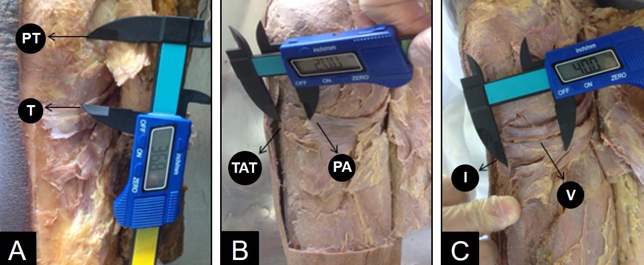
Distance (mm) from the articular surface of the medial Tibial Plateau (PT) to the superior border of the Tendons (T) of the sartorius, gracilis and semitendinosus muscles [Table/Fig-1a]. For PT, a medial distance of 15 mm in relation to the intercondylar area of the tibia was standardised.
Distance (mm) from the anterior Tuberosity of the Tibia (TAT) to the insertion of the Pes Anserinus (PA) [Table/Fig-1b].
Distance (mm) from the insertion (I) of the gracilis and semitendinosus tendons to the their respective vínculas (V) [Table/Fig-1c].
Measurement of the millimetric distances of anatomical structures with digital caliper 150 mm Western® PRO.
For statistical analysis, the data were tabulated and bilateral differences in the morphometric measurements were analysed statistically by ANOVA using the BioEstat 5.3 software.
Results
Anatomy Analysis: No anatomical variations were observed in the insertion sites of the tendons that compose the pes anserinus. In addition, there was no second víncula tendinea for the same muscle or variations in the trajectory of the tendons. The tendons of the semitendinosus and gracilis muscles originated in the distal third of the medial region of the thigh and projected to the tendon of the semimembranosus muscle. Both tendons were arranged in parallel to the tendon of the sartorius muscle in the posteromedial region of the medial epicondyle. At the height of the sulcus of insertion of the tendon of the semimembranosus muscle, the sartorius, gracilis and semitendinosus tendons composed the pes anserinus, which was facing the anteromedial aspect of the tibia. Projecting on the subtendinous bursa of the sartorius muscle and anserine bursa, the tendons of the sartorius, gracilis and semitendinosus muscles were inserted on the anteromedial side of the tibia.
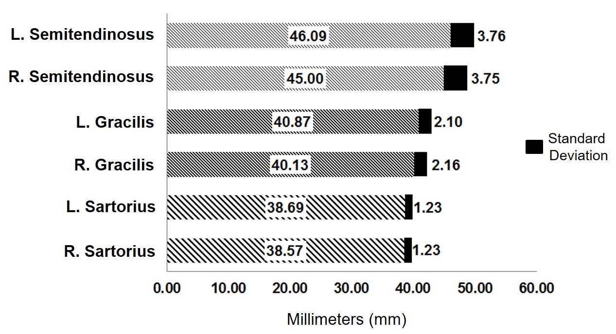
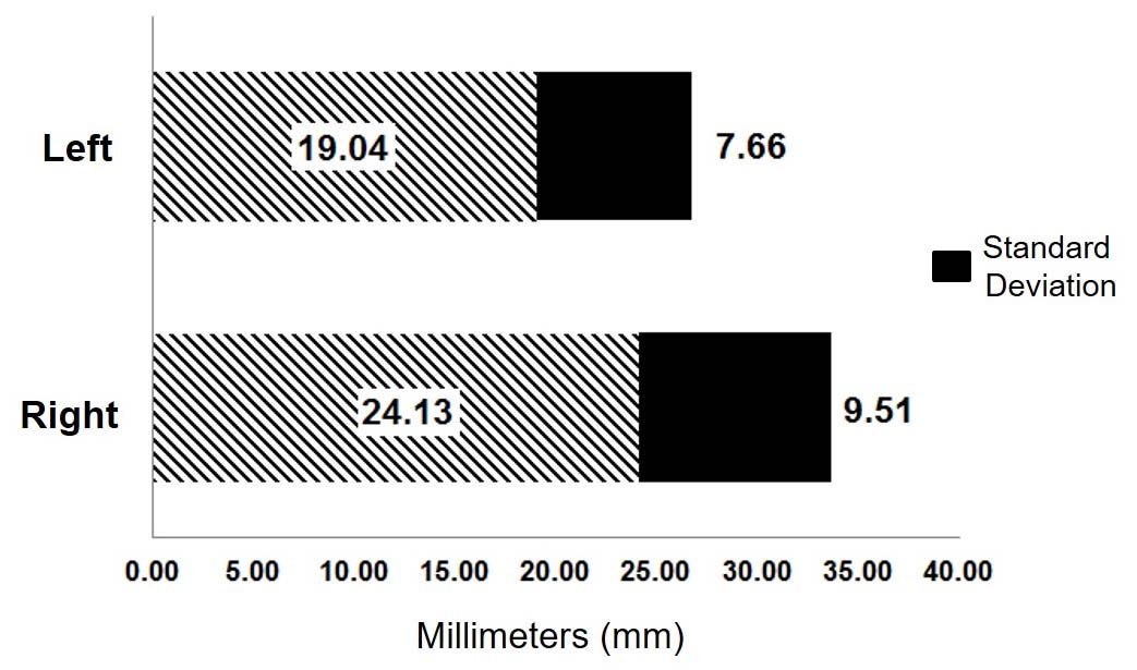
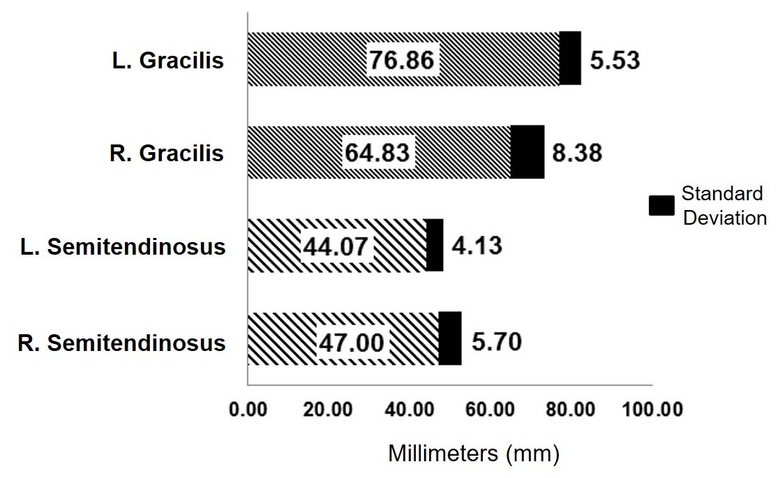
Morphometric and Statistical Analysis (ANOVA): Statistical analysis did not reveal any significant difference in the bilateral comparison of of the distance between the tendons that compose the pes anserinus and anteromedial surface of the tibial plateau [Table/Fig-2] as well as in comparison of the distance of the anterior tuberosity of the tibia between the left and right side [Table/Fig-3]. Bilateral statistical analysis of the distance between insertion of the tendons that compose the pes anserinus and their respective vínculas tendíneas showed a significant difference (p<0.01) between the right and left gracilis tendon and between the right and left semitendinosus tendon. The same difference was observed between the homolateral gracilis and semitendinosus tendons [Table/Fig-4].
Distances between the tendons of the muscles that make up the pes anserinus (T) and the antero-medial surface of the tibial plateau (PT). (L) Left; (R) Right.
Distances between the anterior Tuberosity of the Tibia (TAT) and the insertion of the Pes Anserinus (PA).
Distances between the insertion of the tendons of the muscles that compose the pes anserinus (I) and their respective tendinous vincula (V). (L) Left; (R) Right.
Discussion
With the advances in surgical techniques and the increase in the incidence of traumatic injuries, knee ligament reconstruction has become a common procedure in orthopedics, with the knee flexors being the main graft sources. Greater emphasis on the anatomical knowledge of this donor site can prevent undesired complications, such as the incorrect identification of the gracilis and semitendinosus muscles and amputation of the graft at the time of its extraction [4,8,9,12].
Comparison of the flexor tendon measures in relation to the joint showed similar lateral measures and little variance in the distance between each flexor tendon and joint. In this respect, the mean distance from the insertion of the pes anserinus to the medial tibial plateau was 41.55 mm, which is an auxiliary reference for identification and surgical planing. Similarly, Grassi CA et al., reported a mean difference of 41 mm (range: 37 to 60 mm) between the articular surface of the medial tibial condyle [9].
Analysis of the mean distance from the insertion of the pes anserinus to the TAT revealed no significant difference in laterality. Thus, this anatomical reference might be useful for identifying the insertion of the pes anserinus during surgical procedures. In the same study of Grassi CA et al., the mean distance was 6.88±1 mm [9]. However, these authors did not compare the right and left sides of the same cadaver as done in our study, a fact impairing comparison in relation to laterality.
Comparison of the measures to the insertion of the pes anserinus, showed a greater distance for the vincula of the gracilis compared to the semitendinosus. When the distance of each vincula alone was analysed and compared between the right and left knee, the distance of the vincula of the semitendinosus was found to be more symmetric than that of the gracilis in terms of laterality. These results suggest that during dissection it is easier to first look for and identify the vincula of the semitendinosus since it is closer to the insertion and little variation is observed between the right and left side.
Conclusion
The distance between the flexor tendons and medial articular surface of the tibia is a safe anatomical parameter. The distance between the insertion of the pes anserinus and anterior tuberosity of the tibia was variable and differed significantly between the right and left knees. The víncula tendínea of the semitendinosus muscle was closer to the anterior tuberosity of the tibia, while the víncula tendínea of the gracilis muscle was not a reliable anatomical parameter because of the greater variability in the results.
[1]. Ivey M, Prud’homme J, Anatomic variations of the pes anserinus: a cadaver studyOrthopedics 1993 16(5):601-06.10.3928/0147-7447-19930501-148327387 [Google Scholar] [CrossRef] [PubMed]
[2]. Pagnani MJ, Warner JJ, O’Brien SJ, Warren RF, Anatomic consideration in harvesting the semitendinosus and gracilis tendons and a technique of harvestAm J Sports Med 1993 21:565-71.10.1177/0363546593021004148368418 [Google Scholar] [CrossRef] [PubMed]
[3]. Lopes CL, Arantes G, De Oliveira RL, Pinto DM, Gonçalves MC, Gonçalves RC, Anatomical reference point for harvesting a flexor graft during arthroscopic reconstruction of the anterior cruciate ligamentRev Bras Ortop 2015 5:64-67. [Google Scholar]
[4]. Oliveira VMD, Tatsuo A, Cury RDPL, Avakian RJrAD, De Camargo OPA, Anatomical study of the gracile and semitendinous muscles insertionActa Ortop Bras 2006 14(1):7-10.10.1590/S1413-78522006000100001 [Google Scholar] [CrossRef]
[5]. Noyes F, Noyes’s Knee Disorders: Surgery, Rehabilitation, Clinical Outcomes E-Book 2009 1st EditionSaunders [Google Scholar]
[6]. Thompson JC, Netter. Atlas de anatomia ortopédica 2011 2nd EditionElsevier [Google Scholar]
[7]. Ferrari JD, Ferrari DA, The semitendinosus: anatomic considerations in tendon harvestingOrthop Rev 1991 20:1085-88. [Google Scholar]
[8]. Charalambous CP, Kwaees TA, Anatomical considerations in hamstring tendon harvesting for anterior cruciate ligament reconstructionMuscles Ligaments Tendons J 2012 2(4):253-57. [Google Scholar]
[9]. Grassi CA, Fruheling M, Abdo JC, de Moura MFA, Silva JLV, Hamstring tendons insertion – an anatomical studyRev Bras Ortop 2013 48(5):417-20.10.1016/j.rbo.2012.07.01131304145 [Google Scholar] [CrossRef] [PubMed]
[10]. Gray H, Mayo GC, Gray Anatomia 1988 29th EditionRio de JaneiroGuanabara Koogan [Google Scholar]
[11]. Putz R, Pabst R, Sobotta Atlas of Human Anatomy, Package 2008 15th EditionGuanabara Koogan [Google Scholar]
[12]. Scott WN, Insall & Scott Surgery of the Knee 2011 5th EditionChurchill Livingstone [Google Scholar]
[13]. Hoppenfeld S, Thomas H, Hutton R, Physical examination of the spine and extremities 1976 1st EditionBookseller Rating [Google Scholar]