Radius is the bone which is present on the lateral aspect of forearm. The word radius in Latin means ray [1]. In the context of the radius bone, a ray rotates around its axis line extending diagonally from the centre of the capitulum to the centre of the distal ulna. Radius is usually ossified from three centres one primary centre for the body, and two secondary centres, one for the upper end and one for the lower end [1]. The primary centre for the body appears around the eighth week of intrauterine life, ossification commences in the lower end at second year and in the upper end at fifth year and sometimes a new secondary A2 centre may arise for radial tuberosity at fourteen or fifteen years [1]. The proximal and distal end of radius are commonly involved in trauma, about 2-6% of fractures occur in the proximal end and about 8-20% occurs in distal end which is more commonly seen in geriatric population [2,3]. People between the age of 30-40 years have 20% of elbow injuries especially radial head fracture and it is more common in women than in men [4]. If the fracture is severe then the person should undergo radial head prosthesis [5]. The dimensions of the bicipital tuberosity, radial head, and radial styloid process will be useful in various surgical procedures such as radial head prosthesis, reconstruction of the bicipital tendon and reconstruction of proximal trauma [6]. Distal end of the radius is common for malignant tumour and it is treated by endo prosthesis [7]. Reduction in the radius length along with the altered palmar tilt, has substantial loss of movements in the forearm and wrist joint and grip strength [8,9]. In order to treat these kinds of injuries we should know the normal anatomical morphometry of proximal and distal radius. Radius also plays a major role in sex determination of an unknown decomposed body or bone [10]. By using the range of the length, sensitivity and specificity of the bone it is easy to determine the sex and age [11]. This study was aimed to study the morphometric parameters of proximal and distal end of the radius which correlates for treating the injuries that could happen in that region. However, the availability of new intra-and extra-medullary implants and an increased incidence of iatrogenic injuries, call for a more detailed study of the proximal and distal end of the radius.
Materials and Methods
The study was designed as an observational study, size of the sample is 160 bones in which 80 are right side bone and 80 are left side bones. Both right and left side bones are without damage. The study setting was done in the Department of Anatomy, Saveetha Medical College, Thandalam, Chennai, Tamil Nadu, India. The following parameters were observed and measured using digital vernier caliper, divider, normal ruler and inelastic thread.
The parameters measured and observed were:
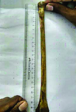
Length of the radius on the medial and lateral aspect: It was measured by using a normal 30 cm ruler, for measuring the length at the medial aspect the ruler was kept at the medial aspect of the radial head above and lower border of the ulnar notch below as shown in [Table/Fig-1]. For measuring the length of lateral aspect, the ruler was kept at the lateral aspect of the head above and edge of the styloid process below.
Measuring the length of the radius on the medial aspect of the radius bone.
Circumference of radius head: It was measured by using an inelastic thread, the radial head was surrounded by the thread and the end was marked, by using a normal ruler, the thread was measured, and the circumference was noted.
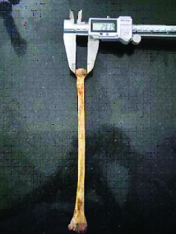
Diameter of the radius: The jaws of the vernier calliper were kept in the middle of the radial head and the diameter was measured as shown in the [Table/Fig-2].
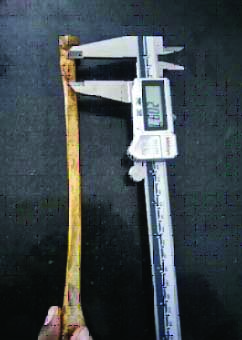
Measuring the length of the radial head on the medial aspect.
Length of the radial head on the medial and lateral aspect: It was measured with the help of digital vernier caliper, the length at the medial aspect was measured from the upper border of the medial aspect of the radial above and lower border of medial aspect of head below. For measuring the lateral aspect, the lower jaw of digital vernier caliper was kept at the upper border of lateral aspect of the head above and lower border of lateral aspect of head below as shown in [Table/Fig-3].
Measuring the diameter of the radial head.
Distance between the radial head and the radial tuberosity: The area between the lower border of the radial head and upper surface of the radial tuberosity was measured by using the divider.
Circumference of the radial neck: The inelastic thread was surrounded around the neck of the radius and the end of the thread was marked. The marked area of the thread was measured with the help of the ruler.
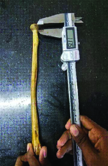
Length of the radial tuberosity: The lower jaw of the vernier caliper was kept on the upper surface of the radial tuberosity above and the point of junction between the tip of the radial tuberosity and the readings were noted as shown in the [Table/Fig-4].
Measuring the length of radial tuberosity.
Length of styloid process: It was not measured specifically, by subtracting the value of the length of the bone on medial and lateral aspect gives the value of styloid process.
Length of ulnar notch on the anterior and posterior aspect: The lower jaw of the vernier caliper was kept on the upper border of the ulnar notch above and lower border of the ulnar notch below and measured on the anterior and the posterior end of the notch and the readings were noted.
Width of the ulnar notch: The lower jaw of the vernier caliper was kept between the anterior and posterior end the ulnar notch and the readings were noted.
Inferior articular facet of radius in anterior and posterior aspect: The lower jaw of vernier caliper was measured between the edges of the anterior, posterior and base of the inferior articular facet.
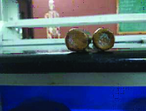
Shapes of radial head: With the help of paper and pencil the head of the radius was traced drawn on a paper and the shape was observed. In [Table/Fig-5], the first radial head was elliptical whereas the second head was circular.
Shapes of the radial head (the first radial head is elliptical in shape and second is circular in shape).
Shape of radial tuberosity: The shape of the radial tuberosity was observed by shading it.
Prevalence of types of curvature at the head and neck zone: Types of curvature were classified as flat and low concave curvature.
Prevalence of morphological variants of radial tuberosity: Morphological variants of bicipital tuberosity were classified as smooth, a single ridge or bifid ridge.
The determined measurements and photographs were taken.
Results
The mean value was determined from 160 bones in which 80 bones were right side bones and 80 bones were left side bones are:
The mean length of radius bone in medial side (22.7 cm) and lateral side (23.7 cm), circumference of the head (6.3 cm), length of the head from medial side (0.67 cm) and lateral side (0.32 cm), distance between the radial head and the radial tuberosity (0.75 cm), length of the radial tuberosity (3.36 cm), circumference of the neck (4.64 cm).
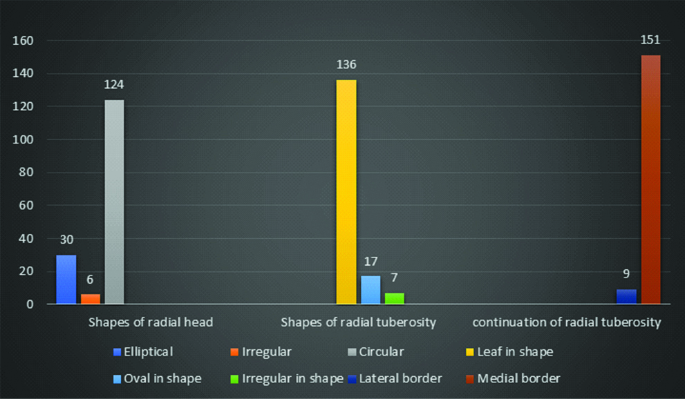
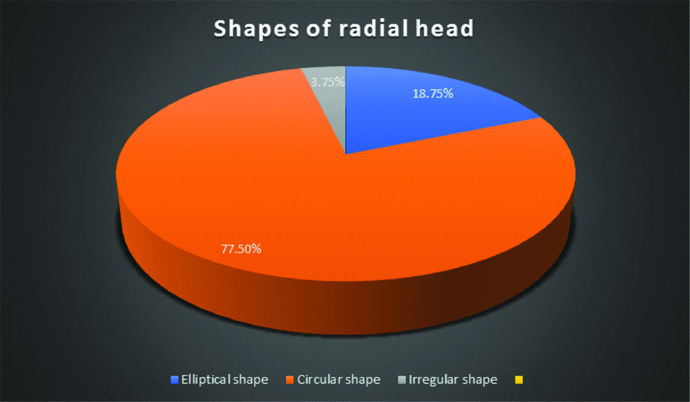
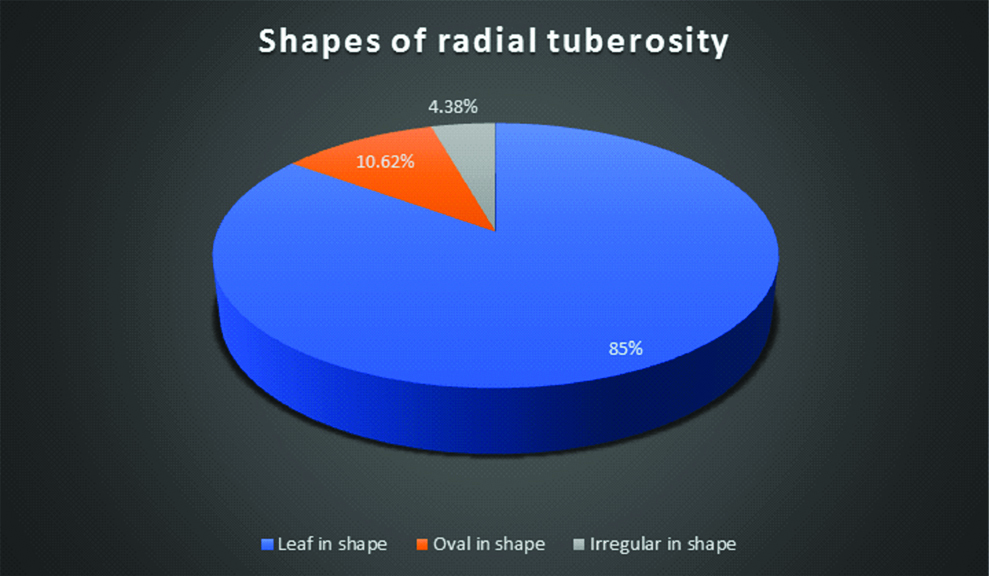
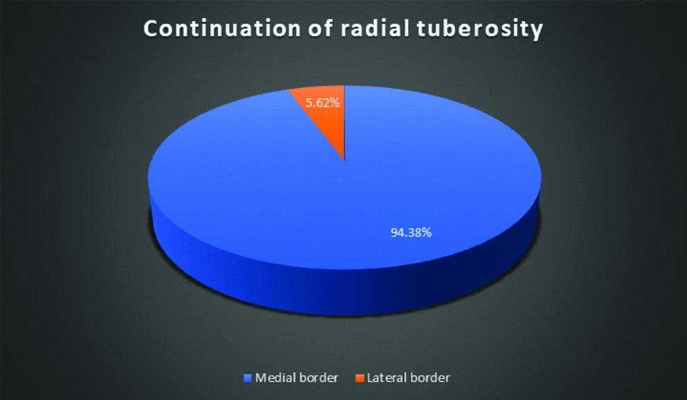
The mean width of the ulnar notch (1.41 cm), length of the ulnar notch from anterior aspect (0.30 cm) and posterior aspect (0.62 cm), length of the styloid process (1.01 cm), circumference of the distal end of the bone (8.40 cm), circumference of the distal end of the shaft (5.63 cm), scaphoid and lunate fossa from anterior (2.79 cm), posterior (2.50 cm), and base (1.32 cm). The readings are tabulated in [Table/Fig-6]. It was observed that radial head of 124 bones i.e., 77.5% appear in circular shape and 30 bones i.e., 18.75% were elliptical in shape and six bones i.e., 3.75% are irregular in shape as shown in [Table/Fig-7,8]. It was also observed that the shape of the radial tuberosity was leaf in shape for 136 bones i.e., 85% and 17 bones i.e., 10.62% were oval in shape and 7 bones i.e., 4.38% are irregular as shown in [Table/Fig-7,9]. In 151 bones i.e., 94.38% the radial tuberosity continues as medial border but in 9 bones i.e., 5.62% bones continue as lateral border as shown in [Table/Fig-7,10].
The mean value is determined from 160 bones in which 80 bones are right side bones and 80 bones are left side bones.
| Parameters | Mean | Standard Deviation |
|---|
| Right | Left | Right | Left |
|---|
| Length of the Radius bone | Medial | 22.7 cm | 22.5 cm | 1.26 cm | 1.24 cm |
| Lateral | 23.7 cm | 23.4 cm | 1.30 cm | 1.27 cm |
| Length of the Radial head | Medial | 0.6 cm | 0.6 cm | 0.07 cm | 0.06 cm |
| Lateral | 0.32 cm | 0.30 cm | 0.05 cm | 0.04 cm |
| Circumference of distal end | Distal | 8.40 cm | 8.38 cm | 0.81 cm | 0.79 cm |
| Shaft | 5.65 cm | 5.63 cm | 0.65 cm | 0.63 cm |
| Circumference of Radial head | 6.3 cm | 6.1 cm | 0.60 cm | 0.58 cm |
| Circumference of Radial neck | 4.64 cm | 4.62 cm | 0.53 cm | 0.51 cm |
| Length of ulnar notch | Anterior | 0.30 cm | 0.28 cm | 0.06 cm | 0.05 cm |
| Posterior | 0.62 cm | 0.60 cm | 0.07 cm | 0.06 cm |
| Width of ulnar notch | 1.41 cm | 1.40 cm | 0.08 cm | 0.07 cm |
| Length of styloid process | 1.01 cm | 1.00 cm | 0.04 cm | 0.03 cm |
| Length of Radial tuberosity | 3.36 cm | 3.34 cm | 0.24 cm | 0.23 cm |
| Distance between radial tuberosity and head | 0.75 cm | 0.73 cm | 0.11 cm | 0.09 cm |
| Scaphoid and lunate fossa | Anterior | 2.79 cm | 2.77 cm | 0.31 cm | 0.29 cm |
| Posterior | 2.50 cm | 2.48 cm | 0.22 cm | 0.20 cm |
| Base | 1.32 cm | 1.30 cm | 0.11 cm | 0.09 cm |
| Diameter of radial head | 4.11 cm | 4.10 cm | 0.03 cm | 0.02 cm |
Variations seen in the shape of radial head, radial tuberosity and its continuation.
Shapes of radial tuberosity.
Continuation of radial tuberosity.
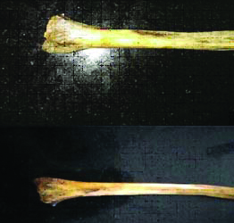
The variations seen in the distal aspect of the radius were the number of tubercles present at the dorsal aspect of radius which were more on all right-side bones than on the left side as shown in [Table/Fig-11].
Comparing the dorsal tubercles on the right and left side bone (the above image is right side and below is left side).
Discussion
The radius is the lateral side bone of the forearm. Its upper end articulates with the lower end of humerus at the elbow joint and with the ulna at the superior radioulnar joint. Its lower end articulates with the scaphoid and lunate bones of the hand at the wrist joint and with the ulna at the inferior radioulnar joint. Gupta C et al., Rajasree G et al., and Van Riet RP et al., have found that, the mean length of the radius on the right and left side is 23.12 cm±2.11 cm and 23.29 cm respectively [12-14] while in present study, the length of the radius is measured separately on the length of the radius in medial side is 22.7 cm±1.26 cm in right side bone and 22.5 cm±1.24 cm on the left side. The length of the radius on lateral side was 23.7 cm±1.30 cm on the right side and 23.4 cm±1.27 cm on the left side.
The mean length of the right radius on the medial side was 22.7±1.26 cm. Similarly, the mean length of the left radius on the lateral side was 22.5±1.25 cm. This knowledge has clinical application in fracture healing and hand prosthesis. As far as we know, this unique way of measuring the length of the radius in present study is not found in previous studies.
Van Riet RP et al., has determined the mean length of radial neck as 1.3 cm [14], while in present study the length of the radial neck was measured as distance between the radial head and the radial tuberosity and the mean length we determined was 0.75 cm±0.11 cm and 0.73±0.9 cm on the right side and left side respectively.
Rajsree G et al., has determined that the mean length of the lateral side of the radial head is 5.71 mm on the left side and on the right side is 6.5 mm. The mean anteroposterior diameter of the radial head on the right side is 15.41 mm and on the left side is 15.45 mm [13], however in present study, we determined that the mean length of the lateral side of the radial head was 0.6 cm on the left side and on the right side was 0.6 cm. The mean anteroposterior or radial head diameter on the right side is 4.11 cm and on the left side was 4.10 cm. The length of the radial head and diameter of the radial head was greater in present study results.
Captier G et al., found that the radial head was elliptical in 57% of cases and in 43% of cases it was circular [15] however, Gupta C et al., has determined that the 64% of cases are circular and 26% of cases are oval but, in present study, we have determined that the 77.5% of cases are circular and 18.75% are elliptical in shape [12]. As the parameters are mainly significant for the radial head prosthesis, it is must to have a clear determination about the shape of the head.
Gupta C et al., also found the length of the radial head at medial and lateral ends, diameter of the head, head thickness at the anterior, posterior and lateral sides, and depth of articular facet in total radius were 0.9, 0.75, 1.91, 0.42, 0.32, 0.30, and 0.19 cm, respectively [12]. In present study, we found that the mean length of the lateral side of the radial head was 0.30 cm±0.04 cm on the left side and on the right side was 0.32 cm±0.05 cm. The mean diameter of the radial head on the right side was 4.11±0.03 cm and on the left side was 4.10 cm±0.02 cm. The mean circumference of the head of radius on the left side was 6.1 cm±0.58 cm and on the right side is 6.3±0.60 cm. The measurement of length of radial head is less when compared to their study. Since in both study, vernier caliper was used, this differences in the values may be a geographical characteristic of the population.
Gupta C et al., determined the mean length and width of bicipital tuberosity on right side are 2.02 and 1.25 cm, respectively, in the left side are 1.92 cm and 1.21 cm respectively [12]. In present study, it was found the mean length of bicipital tuberosity in right side was 3.36±0.24 cm and in the left side was 1.23±0.23 cm, respectively. The result values of present study were greater than the values of their study. In present study also, we found mainly distance between radial head and bicipital tuberosity which is 0.75±0.11 cm on right side and 0.73±0.09 cm on the left side.
Gupta C et al., determined the length of styloid process as 1±0.13 cm and 0.97±0.14 cm on the right and left side however in present study [12], we have determined 1.01±0.04 cm and 1.00±0.03 cm on right and left side respectively. It shows that the mean length of styloid process is lesser in present study, however James I et al., has determined the mean length of styloid process as 11.0±1.4 and 10.8±1.5 mm [16] which is more or less similar to present study.
Koslowsky C et al., determined there is no statistical difference between the right and left side bone [17] but in our study, we have determined there is a statistical difference between the right and left side bone.
The knowledge of shape and size of the head of radius will be useful for the radial head prosthesis [18]. The size of the radial head plays the major role in influencing radial head replacement [19]. The knowledge about the parameters of distal end of the radius will be useful for the distal end prothesis due to Colles’ fracture [20]. The knowledge of size and shape of radial tuberosity will be useful in bicipital tendon reconstruction.
Limitation
The limitation of the study was that incompletely ossified bones, broken bone, bones that are damaged at the region of head and the distal end were not taken.
Conclusion
The morphometric parameters of proximal and distal ends of radius are useful in the designing of prosthesis, for the head and distal end of the radius and for the reconstruction of bicipital tendon. The distal end prosthesis is used in Colles’ fracture and in surgical removal of tumour in the distal end, which is useful for orthopaedic surgeons and oncologist.