Sex determination of an anonymous individual is one of the main step when human skeletal remains are found, both in forensic investigation and archaeological studies. Hence, evaluation of sexual dimorphism of bones in the human population is interesting for both forensic experts as well as anthropologists [1].
The hip bone is a perfect bone for sex determination; since, it reveals the dissimilarities between the two sexes and likewise displays a specific adaptation of women hip bone for childbearing [2]. It is believed that for the sex determination of human skeleton the hip bone shows the highest accuracy levels [3]. The sexual dimorphism in the shape and size of the pelvis is very great since women giving birth to infants [4]. The parts of the pelvis which are more resilient to damage can be used for sex determination, including the GSN and the auricular surface of the ilium [5]. In pelvis, the GSN has an advantage because it is recognisable early in fetal development. Studies have shown a statistically significant level of sexual dimorphism in GSN [6]. The form and size of the GSN are associated with the size of the pelvic inlet. Therefore, multiple studies have demonstrated that the GSN is highly accurate for estimating sex when used alone [7-9].
There are different methods for determination of sex in human skeletal remains including visual examination, bones anthropometric measurements, anthropometric measurements with use of statistics, X-ray examination, and Microscopic examination [10]. CT is a high speed method and can capture high level details of bones’ features and there is no need to remove soft tissue. Therefore, it is a perfect instrument to save time to protect remains from physical manipulation [11]. 3D-CT images of the pelvis reproduce complex curved features, and it stored data format facilitates computerised geometrical analysis which helps to archive the data and use them in the future [11,12]. There are many skeletal or collapse bodies in which determination of sex is the first step. No study has been performed previously in Iran, using GSN 3D-CT scan for sex determination.
The present study was done to evaluate the role of GSN parameters in sex determination in the Iranian population by means of 3D images, reconstructed by multi-slice CT.
Materials and Methods
Study Design and Participants
In the present cross-sectional study, 237 cases (121 females and 116 males) who had undergone pelvic CT for various reasons, in Rasoul-e-Akram Hospital between September 2016 and February 2017 were included in the study. All the patients who met the inclusion criteria were enrolled in this six month study. Sample size was calculated using α = 0.05, β = 0.2, μ1 = 27.06, μ2 = 25.41, SD1 = 3.53, SD2 = 3.37 (20) and was estimated to be minimum sample of 140 cases.
Inclusion and Exclusion Criteria
Patients who underwent pelvic CT according to their physician’s advise were included in the study. Individuals who had severe pelvic injuries due to a fall or accident, those who had deformed or malformed bones and also with congenital abnormalities as well as non-Iranians, were excluded from the study.
Ethical Considerations
The study was performed according to the Ethical principles of declarations of Helsinki. The study is approved by Ethical Committee of Iran University of Medical Sciences. (Ethical code: IR.IUMS.FMD.REC 1396.9411223010) All patients were aware of the study and a written informed consent was obtained from each participant.
Data Collection
The GSN parameters including the width, depth and posterior segment were applied to measure the 3D-CT radiographs of participants’ hip bone using digital instruments with an accuracy of 0.01° and 0.01 mm [Table/Fig-1]. The measured data with sex information were then recorded in a checklist. At first, the GSN landmarks (Pyriformis tubercle, Ischial Spine and the deepest point of the GSN) were determined by Ruler Syngo software in the division then the following parameters were measured.
Dimension of GSN from 3D images of the pelvis reconstructed from CT scan imaging system.
A: Ischial Spine; B: Pyriformis tubercle; C: Deepest point of greater sciatic notch; AB: Width of greater sciatic notch; AB: Width of greater sciatic notch; OB: Posterior segment of sciatic notch; OC: Depth of greater sciatic notch; ACB: Total angle of greater sciatic notch; OCB: Posterior angle of greater sciatic notch
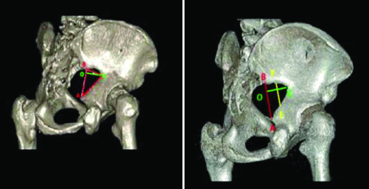
Maximum width (AB): Measured as the distance between the pyriformis tubercle (B) and the point of the Ischial spine (A).
Mid width (EF): GSN width at midpoint of OC line.
Maximum depth (OC): Considered as the perpendicular distance between the deepest points of the GSN (C) to the maximum width.
Posterior segment (OB): Measured as the distance between pyriformis tubercle (B) and the point of O (Vertical cross-sectional width and the maximum depth of GSN)
Index I=Depth OC/Width AB×100
Index II=Posterior segment OB/Width AB×100
Index III (EF/AB)
Index IV (EF/CO)
Total angle=ACB: Measured as an angular distance between the point of pyriformis tubercle (B), the deepest point of the GSN (C) and the point of Ischial spine (A).
Posterior angle=BCO: was determined as an angular distance between the point of pyriformis tubercle (B), deepest point of the GSN (C) and the point of O.
Construction of 3D images from pelvic CT scan
All 3D-CT graphs of 237 individuals were taken by imaging device (Aquilion-advance, Toshiba, 120 KV, 100 MA, 16 slices, 5 mm-thick layers). Untitled measurement data of adult pelvic CT scans were converted to field data using a data conversion and visualisation program.
Statistical Analysis
The collected data were analysed using the SPSS statistical software package version 21.0 (SPSS Inc, Chicago, IL, USA). To compare the mean of variables which causes difference, an independent sample t-test was used and a p-value of <0.05 was considered to be statistically significant. The qualitative variables were evaluated using chi-square test. Correlation coefficient (r) was evaluated by Pearson’s correlation test to analyse the significance of relation between quantitative variables. Roc curve was used to determine the sensitivity and specificity of the parameters.
Results
A total of 237 cases (121 females and 116 males) with the mean age of 50.53 years (20- 89 years) participated in this study.
Equality of means was evaluated by the independent Student’s t-test and p-values were calculated. Statistically significant differences between means related to sex were found for parameters including Max width (p<0.001), mid-width (0.001), Total angle (<0.001), Post angle (<0.001), Post Segment (<0.001), index 1,2,3 and 4 (<0.001). While there was no difference between Max depth and means related to sex in right (p=0.767) and left side (p=0.561) [Table/Fig-2].
Comparison of different GSN parameters in left and right sides with regard to sex.
| Parameter | Sides | Gender | Mean | Standard deviation | Minimum | Maximum | Range | p-value |
|---|
| Width | Right | Male | 44.93 | 5.94 | 32.10 | 64.40 | 32.30 | <0.001 |
| Female | 52.93 | 8.07 | 34.90 | 82.70 | 47.80 |
| Left | Male | 45.06 | 5.79 | 30.30 | 63.40 | 33.10 | <0.001 |
| Female | 55.06 | 7.48 | 38.70 | 71.40 | 32.70 |
| Midwidth | Right | Male | 34.27 | 4.85 | 23.10 | 50.40 | 27.30 | <0.001 |
| Female | 36.68 | 4.56 | 24.50 | 49.70 | 25.20 |
| Left | Male | 34.21 | 4.26 | 24.60 | 43.60 | 19.00 | <0.001 |
| Female | 37.98 | 4.78 | 21.60 | 49.90 | 28.30 |
| Depth | Right | Male | 30.18 | 4.44 | 17.80 | 40.70 | 22.90 | 0.767 |
| Female | 30.02 | 4.07 | 21.10 | 40.20 | 19.10 |
| Left | Male | 31.34 | 4.32 | 21.70 | 39.80 | 18.10 | 0.561 |
| Female | 31.00 | 4.51 | 21.20 | 41.70 | 20.50 |
| Total angle | Right | Male | 66.31 | 9.50 | 48.37 | 105.92 | 57.55 | <0.001 |
| Female | 80.91 | 10.38 | 50.25 | 112.01 | 61.76 |
| Left | Male | 65.71 | 9.23 | 45.66 | 108.00 | 62.34 | <0.001 |
| Female | 82.59 | 9.60 | 45.15 | 106.73 | 61.58 |
| Post angle | Right | Male | 16.64 | 9.72 | 1.90 | 61.00 | 59.10 | <0.001 |
| Female | 32.95 | 9.66 | 8.83 | 51.04 | 42.21 |
| Left | Male | 17.25 | 8.34 | 1.55 | 48.69 | 47.14 | <0.001 |
| Female | 35.97 | 9.62 | 8.90 | 55.98 | 47.08 |
| Post segment | Right | Male | 9.40 | 4.87 | 0.45 | 46.90 | 46.45 | <0.001 |
| Female | 20.39 | 7.88 | 5.00 | 41.10 | 36.10 |
| Left | Male | 10.56 | 5.56 | 0.33 | 29.00 | 28.67 | <0.001 |
| Female | 23.15 | 7.89 | 6.50 | 41.60 | 35.10 |
| Index 1 | Right | Male | 68.22 | 12.83 | 34.76 | 98.97 | 64.21 | <0.001 |
| Female | 57.66 | 10.22 | 36.66 | 102.20 | 65.54 |
| Left | Male | 70.46 | 12.21 | 43.39 | 112.21 | 68.82 | <0.001 |
| Female | 56.91 | 8.89 | 36.60 | 89.54 | 52.94 |
| Index 2 | Right | Male | 20.79 | 10.31 | 1.00 | 60.31 | 59.31 | <0.001 |
| Female | 37.80 | 12.15 | 11.85 | 71.52 | 59.67 |
| Left | Male | 23.29 | 11.59 | 0.78 | 60.30 | 59.52 | <0.001 |
| Female | 41.77 | 11.53 | 13.80 | 73.54 | 59.74 |
| Index 3 | Right | Male | 76.44 | 8.71 | 40.05 | 103.56 | 63.51 | <0.001 |
| Female | 69.93 | 7.34 | 66.59 | 95.41 | 28.82 |
| Left | Male | 76.34 | 7.81 | 58.60 | 92.61 | 34.01 | <0.001 |
| Female | 69.52 | 8.30 | 51.55 | 93.97 | 42.42 |
| Index 4 | Right | Male | 115.64 | 26.47 | 68.09 | 198.72 | 130.63 | <0.001 |
| Female | 123.84 | 18.98 | 66.21 | 193.19 | 126.98 |
| Left | Male | 111.24 | 19.92 | 67.59 | 175.45 | 107.86 | <0.001 |
| Female | 124.52 | 20.47 | 58.69 | 180.87 | 122.18 |
The parameters Depth (0.008), Post segment (0.017), and Index 2 (0.015) showed statistically significant differences in two sides of the body; however, the other parameters showed no differences in right and left sides and can be used for sex determination [Table/Fig-3].
Comparison of different GSN parameters in right and left sides.
| Parameters | Side | Mean | Standard deviation | p-value |
|---|
| Width | Right | 49.01785 | 8.14991 | 0.130 |
| Left | 50.16924 | 8.3642 |
| Midwidth | Right | 35.50802 | 4.85053 | 0.159 |
| Left | 36.14076 | 4.90429 |
| Depth | Right | 30.10253 | 4.25224 | 0.008 |
| Left | 31.16962 | 4.41814 |
| Total Angle | Right | 73.76911 | 12.3442 | 0.624 |
| Left | 74.33211 | 12.64706 |
| Post angle | Right | 24.97097 | 12.66153 | 0.119 |
| Left | 26.81084 | 12.99795 |
| Post segment | Right | 15.01582 | 8.57668 | 0.017 |
| Left | 16.98975 | 9.30489 |
| Index 1 | Right | 62.83059 | 12.70805 | 0.540 |
| Left | 63.54447 | 12.60939 |
| Index 2 | Right | 29.47916 | 14.12715 | 0.015 |
| Left | 32.7303 | 14.79632 |
| Index 3 | Right | 73.12207 | 8.66776 | 0.749 |
| Left | 72.8657 | 8.74616 |
| Index 4 | Right | 119.8336 | 23.27739 | 0.377 |
| Left | 118.0258 | 21.23027 |
Correlations between all parameters and age were analysed using Pearson’s correlation coefficient test. There was no statistical difference between age and most variables of the GSN in men (p>0.05). All of the parameters except right AB (r= -0.195, p=0.036), mid-width right (r= -0.328, p<0.001) and left (r= -0.352, p<0.001), index 3 right (r= -0.201, p=0.031) and left (r= -0.246, p=0.008) and index 4 right (r= -0.196, p=0.035) had no correlation with age. In comparison with men, significant correlation was found between age and right mid-width (r= -0.268, p=0.003), left mid-width (r= -0.290, p<0.001), left OB (r= 0.198, p=0.029), left index 2 (r=0.221, p=0.015), index 3 right (r= -0.381, p<0.001) and left (r= -0.483, p<0.001) and 4 right (r= -0.320, p<0.001) and left (r= -0.285, p=0.002) (p<0.001) in women [Table/Fig-4].
The correlation of GSN parameters and age.
| Parameters | Sides | Male | Female |
|---|
| Pearson correlation | p-value | Pearson correlation | p-value |
|---|
| Width | Right | -0.195 | 0.036 | 0.048 | 0.601 |
| Left | -0.157 | 0.093 | 0.139 | 0.129 |
| Midwidth | Right | -0.328 | 0.001 | -0.268 | 0.003 |
| Left | -0.352 | 0.001 | -0.290 | 0.001 |
| Depth | Right | 0.003 | 0.975 | 0.134 | 0.143 |
| Left | -0.140 | 0.134 | 0.078 | 0.408 |
| Total Angle | Right | -0.138 | 0.140 | -0.047 | 0.606 |
| Left | 0.108 | 0.250 | 0.075 | 0.414 |
| Post angle | Right | -0.007 | 0.938 | 0.108 | 0.237 |
| Left | 0.116 | 0.215 | 0.169 | 0.064 |
| Post segment | Right | 0.038 | 0.687 | 0.153 | 0.094 |
| Left | -0.009 | 0.927 | 0.198 | 0.029 |
| Index 1 | Right | 0.154 | 0.099 | 0.077 | 0.401 |
| Left | 0.008 | 0.929 | -0.059 | 0.520 |
| Index 2 | Right | 0.094 | 0.316 | 0.165 | 0.070 |
| Left | 0.040 | 0.672 | 0.221 | 0.015 |
| Index 3 | Right | -0.201 | 0.031 | -0.381 | 0.001 |
| Left | -0.246 | 0.008 | -0.483 | 0.001 |
| Index 4 | Right | -0.196 | 0.035 | -0.320 | 0.001 |
| Left | -0.142 | 0.127 | -0.285 | 0.002 |
Roc curve was used to determine the sensitivity and specificity of the parameters. The most accuracy in sex determination was seen in post angle and post segment 90.3% and 89.5% respectively. The most sensitivity belongs to Total angle. Post angle and post segment have the most specificity (91.1%) [Table/Fig-5,6]. The most accuracy in right side belongs to post segment (88.7%) and post angle (88.6%). The most specificity belongs to post angle (87.9%). The most sensitivity in the right side is belong to Width (81%) and after that total angle, Index 1, Index 2, and Index 4 have the similar sensitivity (80.2%). The most accuracy for left side was related to post angle (92.3%) and then total angle and post segment (90.8%). The most specificity belongs to post angle (91.4%). Post segment has the most sensitivity (82.6%) and the parameters width, total angle, post angle, and Index 3 has the second rank in sensitivity (80.2%).
Roc curve analysis of the Post segment, Post angle, and Total angle.
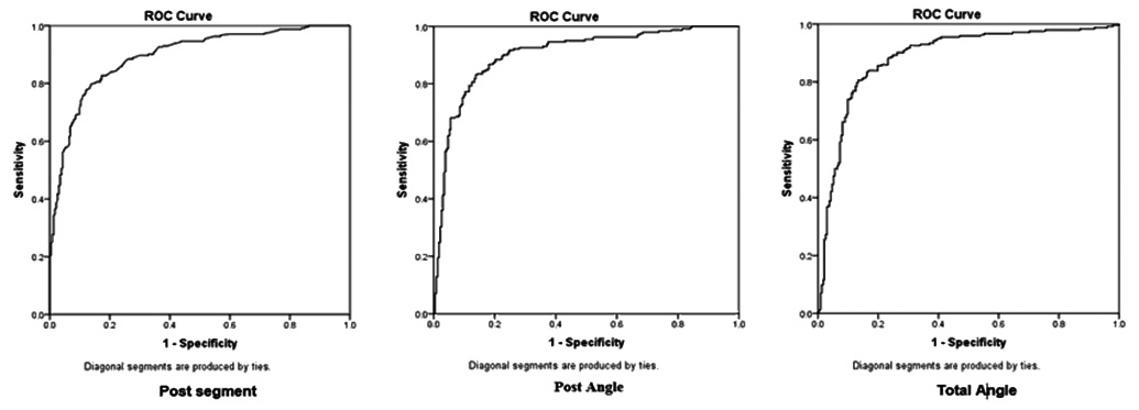
Sensitivity, specificity, accuracy, and cut off point.
| Parameter | Cut-off | Sensitivity | Specificity | AUC |
|---|
| Width | 49.75 | 70.7% | 80.6% | 0.826 |
| Midwidth | 36.65 | 53.3% | 72.8% | 0.680 |
| Depth | 30.05 | 50% | 47.4% | 0.480 |
| Total angle | 69.55 | 90.9% | 72.4% | 0.886 |
| Post angle | 28.12 | 76% | 91.1% | 0.903 |
| Post segment | 16.25 | 71.5% | 91.1% | 0.895 |
| Index 1 | 60.02 | 35.5% | 23.3% | 0.207 |
| Index 2 | 29.29 | 80.2% | 88.4% | 0.865 |
| Index 3 | 71.90 | 36% | 31% | 0.266 |
| Index 4 | 131.40 | 35.1% | 80.2% | 0.658 |
Discussion
The hip bone is usually considered as an important sexually dimorphic region of the human skeleton and the GSN is one of the main features that is normally used as a reliable source for sex determination [8]. Given the sexual dimorphism in different patterns and levels, the standards of specific populations and species cannot be applicable for all human beings. By measuring several variables simultaneously through the digital tools and electronic software, it is possible to increase accuracy and reduce error in sex determination analyses. Among different tools, the GSN parameters are a reliable indicator of sex determination. The present study compared different patterns of GSN parameters involved in sex determination of Iranian population using CT micrographs.
In the present study, the GSN was wider in female than male (respectively p<0.001). The result of the study is consistent with other previous studies. In studies of Dnyanesh S et al., (p<0.05), Kalsey G et al., (p=0.02) and Devadas P et al., (p=0.0003), the width is greater in female than in male [10,13,14]. The width is significantly different in female (p<0.001) and male (p<0.001) in both sides. Jain SK and Choudhary AK, showed that the width is larger in left side in male (p<0.05) and larger in right side in female (p>0.05) [15]. In another study by Kalsey G et al., the width was larger on the left side for female and on the right side in male although the difference was not significant (respectively p=0.533 and p=0.551) [13]. This difference in results can be due to racial differences. In addition, the Midwidth is greater in female than male in the present study (p<0.001). This parameter is slightly larger in left side (p=0.159) both in male and female. The Midwidth is significantly different in female (p<0.001) and male (p<0.001) in both sides. In previous studies this parameter of GSN was not evaluated.
The results of the current study revealed that the depth parameter was not significantly different in male and female on right (p=0.767) and left sides (p=0.561). This means that the depth parameter cannot be used for sex determination. The result of the present study is consistent with the studies of Dnyanesh S et al., right (p>0.05) and left sides (p=0.212) that showed that the depth is not significantly different between male and female [10]. In the study by Kalsey G et al., the result was the same and there was no significant difference in depth in two sexes (p=0.06) [13]. However, in the Raut R et al., and Naqshi BF et al., study the maximum depth showed a significant different between the two sexes (respectively p<0.05 and p=0.006) [16,17].
Total angle is significantly different in male and female in both right and left sides (p<0.001). Moreover, total angle is not significantly different in two sides (p=0.624). These findings are similar to Dnyanesh S et al., Kalsey G et al., Raut R et al., study. They showed that the total angel is significantly larger in female in the both sides but it is not different on two sides (respectively p<0.001) [10,13,16].
The result of the present study shows that the post angle is significantly different in male and female in right and left sides (p<0.001) and it is not different in left and right sides (p=0.119). In Raut R et al., and Singh S et al., study it is significantly greater in female than male (respectively p<0.001) in both sides [16,18]. The result is also similar in Dnyanesh S et al., and Kalesy et al., study [10,13].
The result of the current study revealed that the post segment is significantly different in male and female an both sides (p<0.001). With regard to the side, post segment shows significant difference between the right and left sides (p=0.017). Therefore, post segment cannot be used for sex determination without considering the side. In Shah S et al., (p<0.001) and Dnyanesh S et al., (p<0.001) Raut R et al., (p<0.001), Naqsh BF et al., (p=0.03) and Alizadeh Z et al., (p<0.001) study the post segment is significantly different in male and female and in female it is larger than male [2,10,16,17,19].
Index 1 is significantly different between male and female an both sides and it is greater in male (p<0.001). In addition, there is no different in Index 1 in two sides (p=0.540). The female had larger width they had smaller Index 1. The result are consistent with the result of Shah S et al., Dnyanesh S et al., and Kalsey G et al., studies [2,10,13].
Index 2 showed a significant different between male and female in both sides (p<0.001). Index 2 was significantly different in right and left sides (p=0.015). Index 2 is calculated by dividing OB/AB and since the post segment is meaningfully higher in female, the index 2 parameter is higher in female. Hence, it cannot be used for sex determination without considering the side. The result are consistent with the results of Shah S et al., Dnyanesh S et al., Kalesy et al., and Akpan T et al., studies [2,10,13,20].
Right Width, Right and Left Midwidth, Right and Left Index 3, and Right Index 4 have correlation with age in male and Right and Left Midwidth, Left Post segment, Left Index 2, and Right and Left Index 3 and 4 are correlated with age in female. It is obvious that age has more effect on GSN parameters in female. This difference may be due to obesity, nutritional problems, frequent childbirth and life style in women.
In the present study, the most accurate parameters for sex determination were posterior angle and posterior segment width (90.3% and 89.5% respectively). In a study by Takahashi H et al., it was shown that the most accuracy rate was related to Post Angle with accuracy of 91% which is similar to the result of present study [21].
Limitation
Since, the parameters were measured manually, there was a possibility of mistake in measurement.
Conclusion
Except for depth, the other GSN parameters are different between male and female can be used for sex determination. Except for Depth, post segment and Index 2, most of the parameters are not statistically different between right and left sides, so they can be used for sex determination irrespective of their sides. Moreover, post angle and post segment have the most accuracy in sex determination. Application of 3D-CT micrographs in present study helped us to easily quantify sexual dimorphism in the GSN, suggesting 3D-CT can be considered as one of the valuable tools in practical forensic Osteology investigation due to the great accuracy to measure the sex differences.