Two Trunked Brachial Plexus: Multiple Variations in Formation, Course and Distribution
Pooja Singh1, Manisha B. Sinha2
1 Senior Resident, Department of Anatomy, All India Institute of Medical Sciences, Raipur, Chhattisgarh, India.
2 Associate Professor, Department of Anatomy, All India Institute of Medical Sciences, Raipur, Chhattisgarh, India.
NAME, ADDRESS, E-MAIL ID OF THE CORRESPONDING AUTHOR: Dr. Pooja Singh, Flat No. 128, Kalpkriti Parisar, Awadhpuri, Risali, Bhilai, Diatrict- Durg, Chhattisgarh - 490006, India.
E-mail: poojaanilsingh@gmail.com
Brachial plexus is a very complicated nerve network of upper limb. Many anatomical variations have been documented regarding its formation, course and distribution. Understanding of variations in brachial plexus is very important for clinicians, especially during surgical approach of axilla and neck region. Multiple variations in the left brachial plexus of about 70-year-old male cadaver, during routine dissection classes of upper extremities was noted in this variation of brachial plexus in which the middle trunk was absent. Other associated anomalies of same plexus were- all the three cords and their branches lie lateral to the axillary artery. Coracobrachialis was supplied by more than one branch of lateral cord. A loop was formed by joining of medial as well as lateral pectoral nerve around superior thoracic artery and there was also a connection present between lateral and medial root of median nerve. Therefore, the purpose of this paper was to unfold the complexity of brachial plexus. These changes may or may not affect the functioning of upper extremity of this person, but the knowledge of these variations are necessary during the surgical procedures as well as to evaluate unexplained sensory or motor loss after trauma. It also helps the clinicians for proper understanding of some previously unexplained clinical symptoms.
Axillary Artery, Brachial Plexus, Coracobrachialis, Median Nerve, Single Cord, Superior Thoracic Artery
Case Report
We observed an anomaly in the formation of brachial plexus with additional variations observed in the left upper extremity of 70-year-old male cadaver at the Department of Anatomy, AIIMS, Raipur, Chhattisgarh, India.
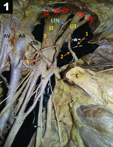
Brachial plexus on left side was formed by two trunks only. The upper trunk was formed by the fusion of C5 and C6 root, While C7 root was fused with C8 and T1 to form a single trunk as a lower trunk. Anterior division of upper trunk (C5 and C6) joined with few fibres of lower trunk to form the lateral cord [Table/Fig-1]. Lateral pectoral nerve, emerged from anterior division of upper trunk (C5 and C6) and formed a loop with medial pectoral nerve (that aroused from anterior division of lower trunk). This loop encircled the superior thoracic artery and then branches from this loop were supplied to pectoralis muscles [Table/Fig-1]. Medial cord was formed by anterior division of lower trunk while posterior cord was formed by posterior division of both trunks. A connection between lateral root and medial root of median nerve was also found. Coracobrachialis muscle got innervated by three nerves, the first nerve directly arose from lateral cord, second one was musculocutaneous nerve, also arose from lateral cord, and third nerve was branch of musculocutaneous nerve [Table/Fig-2]. The explanation behind these variations was that during development, there was continuous splitting and joining of fascicles that took place. These rearrangements and regrouping of axons/fascicles would decide the final innervations (sensory/motor) of targeted regions [1].
Two trunked brachial plexus, encircling the superior thoracic artery
C – Clavicle (cut); AA- Axillary Artery; AV- Axillary Vein; STA- Superior Thoracic Artery; C5, C6, C7, C8 and T1 – Ventral Rami of corresponding nerves; UT- Upper Trunk of Brachial Plexus; LT- Lower Trunk of Brachial Plexus
1. Nerve to Subclavius; 2. Suprascapular Nerve; 3. Lateral Pectoral Nerve; 4. Medial Pectoral Nerve;
Relation of brachial plexus with axillary artery, innervation of coracobrachialis and connection between two roots of median nerve
ICBN- Intercostobrachial Nerve; MCNA- Medial Cutaneous Nerve of Arm; MCNFA- Medial Cutaneous Nerve of Forearm; MN (red arrow) - Median Nerve; CB- Coracobrachialis
1. Axillary Nerve; 2. Radial Nerve; 3. Musculocutaneous Nerve;
Yellow arrows- Other nerves that are supplied to Coracobrachialis
Green arrows- Connection between Lateral and Medial Cord of Brachial Plexus
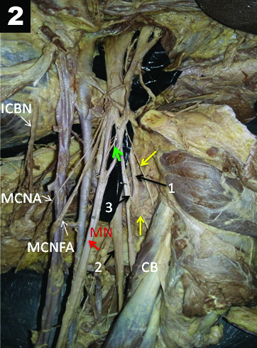
Apart from that, all cords and their branches were present on lateral aspect of all the three parts of axillary artery. Only medial cutaneous nerve of arm and forearm crossed it from lateral to medial aspect on upper half of arm [Table/Fig-2]. Further course and distribution of all nerves were normal. On right side, there was normal pattern of brachial plexus.
Discussion
Ontogeny Repeats Phylogeny
Rao PVVP and Chaudary SC stated that during development, the human brachial plexus appears as a single radicular cone in the upper limb bud [2]. Formation of any plexus is due to anastomosis of certain spinal nerves, which then develops into a solid plate. After that due to separation of solid plate, they get divided into trunks and divisions. The posterior division supplies extensor muscles and their surfaces and the anterior division supplies flexor muscles and their surfaces [3].
Tropic substances like brain-derived neurotropic growth factor- c-kit ligand, neutrin-1, neutrin-2 etc., attract the correct growth cone that is essential to take the right path [4]. Changes in the signal pathway of mesenchymal cells and neural growth factors, at the time of fission of brachial plexus can lead to variations. Ontogenically, the variations of brachial plexus in the present paper could be due to improper separation of solid plate or due to a failure of proper division of the radicular cone occurred [5]. Tracing the phylogeny, no trunk formation is seen in amphibians, reptilians and dogs. In gorillas, lemurs and marsupials, two trunked brachial plexus is found. In these animals C4, C5 and C6 roots join to form upper trunk while C7, C8 and T1 roots join to form lower trunk [6].
Nayak BS et al., reported an absence of middle trunk and stated that C5, C6 and C7 formed upper trunk whereas C8 and T1 formed lower trunk [7]. Other case that was reported by Singla RK et al., was similar to our case, he also reported the absence of middle trunk, while C7 root blended with C8 and T1 to form lower trunk [8]. This case was different from previous studies due to presence of other associated anomaly along with the two trunked brachial plexus.
Clinical Implication
It is very important to understand the variations of formation as well as distribution of brachial plexus. The knowledge about these variations in the brachial plexus would be helpful to carry out various surgical procedures and post operative management of tumors and traumas of brachial plexus successfully. It is also important for anesthetists, to give brachial plexus block at different levels- interscalene, infraclavicular and supraclavicular etc. for various limb surgeries [9].
Conclusion
The understanding of variations of brachial plexus is important for analyzing clinical signs/symptoms as well as for proper diagnosis. It is prone to damage in open or close external injury and in obstetric injuries. It may be pressed by cervical rib or regional tumor. Apart from the clinicians, these variations are also important for radiologists, anesthesiologists, neurosurgeons, neurologists, vascular surgeons and orthopaedic surgeons.
[1]. Singh R, Tubbs RS, Kumar R, Abnormal cord formation of brachial plexusBasic Sciences of Medicine 2016 5(1):1-4. [Google Scholar]
[2]. Rao PVVP, Chaudary SC, Communication of the musculocutaneous nerve with the median nerveEast African Med J 2000 77(9):498-503. [Google Scholar]
[3]. Hamilton WJ, Boyd JD, Mossman HW, Peripheral nervous systemIn: Human Embryology (pre-natal development of form and function) 1962 3rd editionCambridge, EnglandW. Heffer and Sons Limited:358-60. [Google Scholar]
[4]. Larson WJ, Development of peripheral nervous systemIn: Human Embryology 2001 3rd edPennsylvaniaChurchill Livingstone:115-16. [Google Scholar]
[5]. Sannes HD, Reh TA, Harris WA, Axon growth and guidanceIn: Development of nervous system 2000 New YorkAcademic Press:189-97. [Google Scholar]
[6]. Uzel AP, Bulla A, Steinmann G, LaurentJoye M, Caix P, Absence of the musculocutaneous nerve and its distribution from median nerve: about two cases and literature reviewMorphology 2011 95(311):146-50.10.1016/j.morpho.2011.10.00122079600 [Google Scholar] [CrossRef] [PubMed]
[7]. Nayak BS, Nagabhooshana S, Venkatramanal V, A rare variation in the formation of the upper trunk of brachial plexus: a case reportJ Neuro Anat 2005 4:37-38. [Google Scholar]
[8]. Singla RK, Sharma RK, Shree B, A two trunked brachial plexus: A case reportJ Clin Diag Res 2013 7(4):704-05.10.7860/JCDR/2013/4580.288623730651 [Google Scholar] [CrossRef] [PubMed]
[9]. Bala A, Sinha P, Tamang BK, Sarda RK, Anatomical variation: median nerve formation - a case vignetteJ Clin Diagn Res 2014 8(6):AD03-04.10.7860/JCDR/2014/7620.445525120965 [Google Scholar] [CrossRef] [PubMed]