Double Perineal Ani with Duplication of External Genitalia in a Septicemic Neonate: A Case Report
Bismay Das1, C Mohanty2, Rajniti Prasad3, Braj Kishore Das4
1 Senior Resident, Department of Pathology, AIIMS, Bhubaneswar, Odisha, India.
2 Professor, Department of Anatomy, Institute of Medical Sciences, Varanasi, Uttar Pardesh, India.
3 Professor, Department of Paediatrics, Institute of Medical Sciences, Varanasi, Uttar Pardesh, India.
4 Professor, Department of Paediatrics, Institute of Medical Sciences, Varanasi, Uttar Pardesh, India.
NAME, ADDRESS, E-MAIL ID OF THE CORRESPONDING AUTHOR: Dr. Rajniti Prasad, Professor, Department of Paediatrics, Institute of Medical Sciences, B.H.U, Varanasi, Uttar Pardesh, India.
E-mail: rajnitip@gmail.com
Double perineal ani, a rare congenital anomaly is due to persistence of urogenital septum with widening of hind gut. Only few cases have been reported in literature. We report a rare case of double perineal ani with duplication of external genitalia and ectopia vesicae in a septicaemic male child. Ultrasonography showed no duplication of internal organs. The treatment was started with intravenous cefotaxime and amikacin but died within 2 hours of admission. Autopsy could not be done. Early presentation to hospital and multidisciplinary approach is required for management of such cases.
Ectopia vesicae, Hind gut, Septicaemia
Case Report
A two-month-old male delivered from 26 years primigravida mother vaginally presented in emergency for not accepting feed for three days and fast breathing for one day. Antenatal and perinatal period were uneventful. There was no history of maternal drug or radiation exposure.
On examination, child was lethargic and tachypnic (Respiratory Rate: 62/minute). His capillary refill time was less than three seconds. All primitive reflexes were depressed. The examination of cardiovascular system and chest was unremarkable. He had no hepatosplenomegaly.
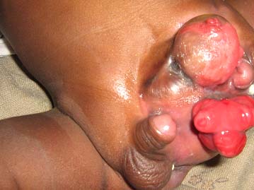
On local examination, he had multiple anomalies i.e., two functional anal opening with simultaneous passage of stool during defecation, double penis and scrotal sac. There was a defect in lower abdominal wall with two irregular fleshy mass over hypogastric region. The right anal opening was below the scrotum and left anal opening in the area between penis and scrotum. He had bilateral hemiscrotum and undescended testis. The right penis was well developed and appeared clinically normal but partially buried, whereas left penis was smaller and almost buried under a fleshy mass, rotated and only glans could be visualized. Meatus could be seen in both penis but stenosed. There was leakage of urine from under surface of right penis and from the area between fleshy mass and left penis (hypospadius). Anal orifices were separated on either side of midline with corresponding external genitalia and were quite far apart [Table/Fig-1].
Showing double anal opening, penis and scrotum with ectopia vesicae.
His complete blood count showed neutrophilic leucocytosis, with a haemoglobin value of 120 gm/L (haematocrit 36.8%), total leucocyte count 13.2x10⁹/L (neutrophils 8.8x10⁹/l, lymphocytes 3.8x10⁹/L) and platelet count 175x109/L. His C-reactive protein was 42 mg/L and blood culture showed growth of E.Coli; report collected after death), which was sensitive to cefotaxime, amikacin and netromycin. Chest radiograph showed infiltrates in both lung fields. Ultrasonography of abdomen showed bilaterally normal kidneys and ureter but bladder could not be differentiated. Abdominal Computed Tomography (CT), Magnetic Resonance Imaging (MRI) and cystogram was planned but could not be done as patient had died two hours after admission. Parent had not given consent for autopsy.
At admission, patient was given Inj. Cefotaxime and amikacin with oxygen inhalation considering respiratory distress but within 15 minutes shifted to mechanical ventilation as per his arterial blood gas analysis and SPO2 (PaO2-46, PaCo2, pH-7.234, HCO3-12 mEq/L, SPO2-82%).
Discussion
Double perineal ani with duplication of external genitalia and ectopia vesicae is extremely rare in male. The two ani may be separated by a thin septum or may lie wide apart from each other for about 1.5 cm. Both ani function simultaneously and the continence is normal as they pass through sphincter complex (puborectalis and levator ani muscle) [1]. It is nine times more common in females [1]. Banu T et al., reported two well formed ani, total colon duplication and two vagina in a 42-day-old female baby from Bangladesh [2]. Choi SK and Park WH, observed anal canal duplication is the most distal and the least frequent digestive duplication and recommended early surgical removal to avoid delayed presentation of infection such as perianal abscess or fistula formation as in our case [3]. Kratz JR et al., reported anal canal duplication associated with presacral cyst in a 16-year-old girl [4]. Samuk I et al., reported associated lipomyelomeningocele and urinary tract abnormalities and Mukunda R et al., diphallus with anorectal malformation [5,6].
Swaika S et al., reported caudal duplication syndrome and diverse combination of gastrointestinal, genitourinary, spinal and limb anomalies (two hemiphalluses with two patent external genital orifices, complete duplication of colon and two stenotic anal orifices [1]. The prognosis of previously reported cases is good with early surgical interventions.
The embryological basis of this anomaly [7] is probably due to persistence of urogenital septum with widening of hind gut, thereby dividing the endodermal elements into two halves to form two urachuses, bladders and urogenital sinuses. The two urogenital sinus opening could lead to widening of ectodermal urethral plate and canalization will result in paired external urethral orifices. Another possibility could be splitting at the stage of development of the primitive streak, when cranial ends fuse but not the caudal end. The third possibility could be the presence of human homologue of mouse mutant disorganization gene, which disrupts the orderly process of organogenesis and induces a variety of developmental anomalies in structures derived from all germ layers.
Treatment of this condition includes early medical and surgical interventions involving paediatric surgeon, paediatric urologist, and a plastic surgeon, to maximize management effectiveness.
Conclusion
This case was being reported because of late presentation, which had led to death due to septicaemia and associated duplication of external genitalia and ectopia vesicae. Early and planned medical and surgical interventions using a multidisciplinary team approach is required in such cases.
[1]. Swaika S, Basu S, Baddra RC, Sarkar R, Maitra SK, Caudal duplication syndrome-report of a case and review of literatureIndian J Surg 2013 75:S484-87.10.1007/s12262-013-0838-z24426655 [Google Scholar] [CrossRef] [PubMed]
[2]. Banu T, Chowdhury TK, Hoque M, Hannan MJ, Congenital double anus with total colon duplication: a case reportJ Pediatr Surg 2007 42:E1-E2.10.1016/j.jpedsurg.2006.11.00117208529 [Google Scholar] [CrossRef] [PubMed]
[3]. Choi SK, Park Wh, Anal canal duplication in InfantsJ pediatr Surg 2003 38:758-62.10.1016/jpsu.2003.5016112720188 [Google Scholar] [CrossRef] [PubMed]
[4]. Kratz JR, Deshpande V, Ryan DP, Goldstein AM, Anal canal duplication associated with presacral cystJ Pediatr Surg 2008 43:1749-52.10.1016/j.jpedsurg.2008.05.02618779021 [Google Scholar] [CrossRef] [PubMed]
[5]. Samuk I, Levitt M, Dlugy E, Kravarusic D, Ben-Meir D, Raiz G, Caudal duplication syndrome: the vital role of multidisciplinary approach and staged correctionJ Pediatr Surg Rep 2016 4:01-05.10.1055/s-0035-157037028018799 [Google Scholar] [CrossRef] [PubMed]
[6]. Mukunda R, Bendre PS, Redkar RG, Hambarde S, Diphallus with anorectal malformation-case reportJ Pediatr Surg 2010 45:632-34.10.1016/j.jpedsurg.2010.01.00320223333 [Google Scholar] [CrossRef] [PubMed]
[7]. Taneja AK, Zaffani G, Amato-Filho AC, Queiroz Lde S, Zanardi Vde A, Menezes-Netto JR, Caudal duplication syndromeArq Neuropsiquiatr 2009 67:695-96.10.1590/S0004-282X200900040002319722052 [Google Scholar] [CrossRef] [PubMed]