Nematodes are probably the most widespread and abundant group of animal often occurring in huge numbers in very diverse environments. The largest number of helminthic parasites of humans belongs to the phylum nematoda and Ascaris lumbricoides is the most common of human helminthic parasites. Commonly known as roundworm, Ascaris lumbricoides is cosmopolitan, especially prevalent in the tropical countries like India. The adult roundworm commonly lives in small intestine, retaining its position by the virtue of its own muscle tone. Eggs of roundworm are bile stained, appearing yellowish brown in colour. Roundworm is a restless wanderer, showing great inquisitiveness as it tends to move itself from the original site, and get into any aperture it may find on its way. So it’s quite possible to encounter this commonest and largest intestinal nematode of humans at some odd sites in the body. Here, we are presenting a very rare case of finding colourless (i.e., non bile stained) eggs with a female roundworm isolated from an ectopic site.
Case Report
A 31-year-old female was admitted in the Surgical Intensive Care Unit (SICU) of the institute since about a month after she developed a complication following Lower Segment Caesarian Section (LSCS). During her stay in SICU, she suffered from multiple febrile episodes. She was in delirium throughout her stay in there. On 27th day in SICU, patient’s relatives noticed ‘something’ coming out of patient’s left nasal opening. They quickly informed about the same to the hospital staff and, then the duty doctor removed this single worm; whole body intact; by gloved hands. The worm was immediately sent to the microbiology laboratory of this institute.
As the patient was shifted to another hospital for further management on the same day; no further investigations regarding worm infection were possible.
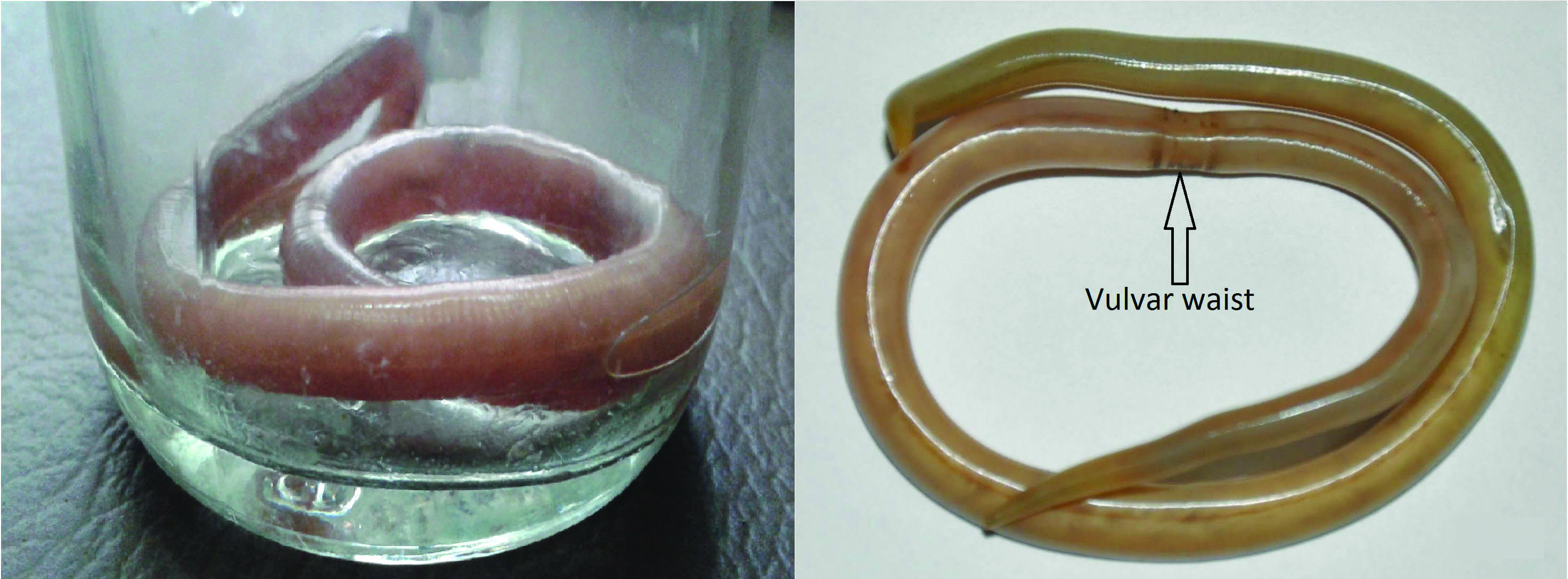
The worm received in the microbiology laboratory was studied carefully. The body of the worm was elongated, un-segmented, cylindrical, tapering gradually at one end, somewhat less so on the other end. Colour of the worm was pinkish cream. A lateral line was seen as a whitish streak along the entire length of the body. Total length was 23 cm, and diameter of the body was about 4-5 mm at the widest point. The posterior end was straight. Vulvar waist was seen, well developed, situated at junction of anterior and middle one thirds [Table/Fig-1].
Showing freshly isolated worm on the left side and worm preserved in formalin on the right side.
Depending on these observational findings, the recovered worm was confirmed to be an adult female of Ascaris lumbricoides [1-4].
The worm was washed in normal saline immediately, and the same fluid used for worm washing was observed under microscope. Microscopy (40X, high power) revealed plenty colourless eggs of sizes around 80-90 μm by 50-55 μm, oval or elliptical in shape; and having a thin outer shell irregularly coated with protein. All of these appeared to contain small atrophied ovum along with refractile granular material [Table/Fig-2].
Showing comparison of eggs of roundworm found normally (left: upper and lower. Source-www.cdc.gov/dpdx/ascariasis/index.html) and those found in this case (right, 40X).
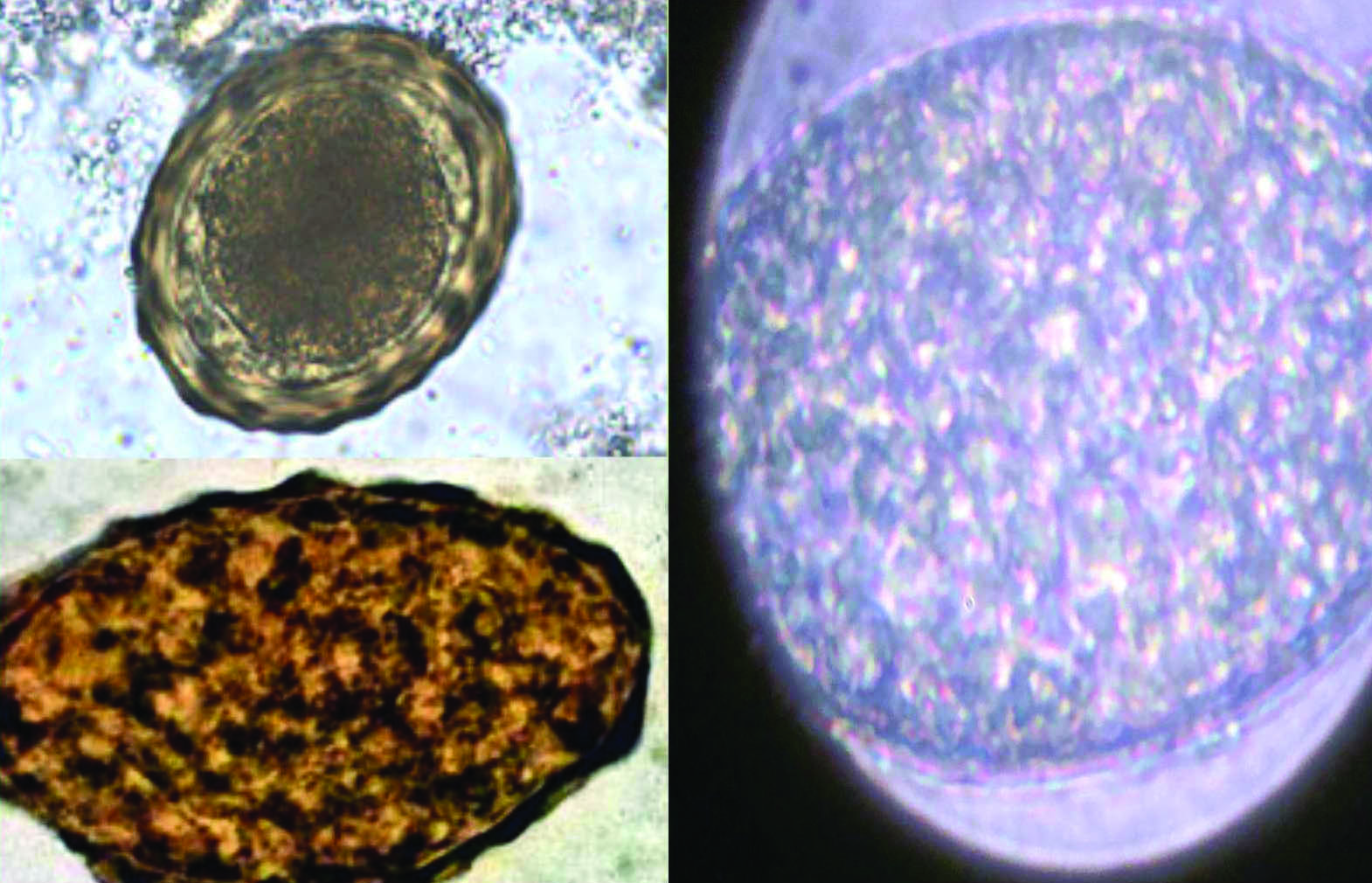
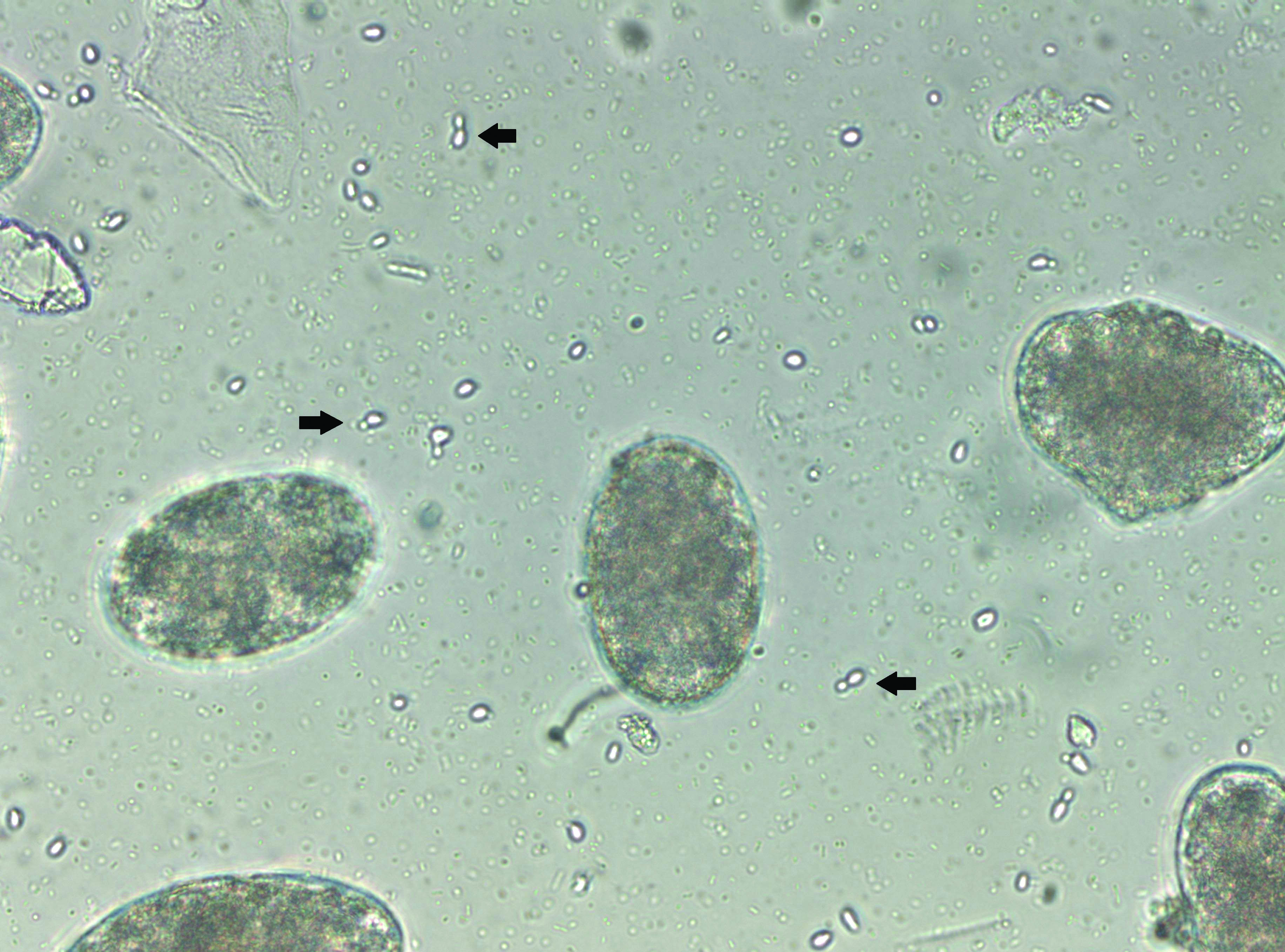
With the help of these findings, it was inferred that all these structures seen in the wash fluid were eggs of Ascaris lumbricoides [3]; most of the eggs were unfertilized and all of them were colourless hence, non-bile stained. We also noticed few budding yeast cells attached to many eggs [Table/Fig-3].
Showing presence of budding yeast cells along with colourless round orm eggs (40X).
Discussion
Ascariasis is at the top in the list of neglected tropical diseases, having global prevalence of 807 million. India is among the regions of highest prevalence [3,5].
According to a Lancet editorial, the Ascaris burden worldwide is so enormous that if placed head to tail, the worms would encircle the world 50 times [6]. But recovery of adult roundworms from ectopic sites is either rare or rather seems under reported. Also, identification of the worms as male or female; and observation of eggs remains questionable whenever and wherever a microbiologist is not available. Although, there are a few incidences of roundworm isolation from ectopic sites [7-12].
A similar case is reported from Senegalese in 2011, when an adult female Ascaris lumbricoides was observed to be coming out of oropharynx of an adult male patient who was a victim of road traffic accident. Nothing is mentioned in the article about whether any eggs were observed along with the female worm [8].
A unique case of scrotal ascariasis is reported in 2012 from Kolkata. Migration of roundworm was suspected through a surgical fistula resulting in unusual ectopic ascariasis. Whether any attempts were made to look for eggs, remains unrevealed [9].
Three cases of hepatic abscess due to ascaris are reported from Kangra, Himachal Pradesh in 2015. One patient vomited out the worm while in other two cases; ascaris worm was found in the pus bags in which draining fluid from the abscesses was being collected. Report mentions nothing about sex of the worm or about the eggs [10]. Another case of pancreatic ascariasis is reported from Tamilnadu in 2015 without any details about eggs [11].
In the present case, the diseased was a female with average nutritional status and poor personal hygiene. Patient was bed ridden and was in delirium since about a month in SICU, hence general body movements of the patient were restricted, that’s why; the worm might have got chance to come to this unusual site (i.e., nasopharynx) so easily. The patient was from lower social economical group, working as laborer on farm fields. As roundworm eggs are present in soil, patient must have acquired the infection due to poor personal sanitation habits.
Although, the exact reasons behind such wandering of roundworms are not clearly understood; yet, fever (generally above 102°F) and inflammatory responses by the host tissue where the worm is dwelling; probably explain the migration. Although, we encountered a female worm in this case; male roundworms are more responsive to such stimuli than the female worms [4].
What intrigued us more was the peculiar finding about the eggs. Most of the eggs were unfertilized and more importantly, all were colourless i.e. unstained by bile [Table/Fig-3].
Unfertilized eggs are fairly common in humans, most commonly produced as result of the host harbouring only female worms [13]. But eggs of the roundworm are always bile stained and brownish (golden brown) in colour [2]. The unfertilized egg too, is brownish in colour and rather darker than the fertilized egg [14].
The reason behind this finding might be in the migrating tendency of this worm. As the diseased was bed ridden, a small immature worm that was in intestines might have moved upwards in stomach and then oesophagus and while its course towards nasopharynx; it matured and started laying eggs. Incidentally, the worm itself was at ectopic site, so there were very few chances of it getting fertilized, hence the unfertilized eggs. Also, there was no chance for the eggs to get exposed to bile, hence the eggs were colourless.
We also observed budding yeast cells along with the roundworm eggs [Table/Fig-3]. The yeast cells might be from the nasopharyngeal mucosa of the patient. Or is it possible that there is an association of roundworm eggs and yeast cells? Such kind of association is well known between bacterium Wolbachia pipientis and dengue virus [15]. In one of the recent studies, it has been shown that this endosymbiont bacterium can interfere with microbial and parasite infection in insects, including viruses in mosquitoes [15]. On the same line, we may have to explore the chances of association between roundworms and other micro-organisms like candida.
The infective form of ascariasis for human beings is the embryonated eggs and the mode of transmission is faeco-oral [16]. Eggs survive in the soil for longer periods. Thick outer wall enables them to sustain for months or years in cold and dry conditions [17]. No practical means of killing ascaris eggs in the soil has been discovered [4].
Although ascariasis is one of the most common of the human parasitic infections, it is also generally asymptomatic, disease being the exception rather than the rule [18]. Still, the World Health organization (WHO) has laid emphasis on Neglected Tropical diseases (NTDs) in their 2010 report and parasitic diseases constitute a majority of these NTDs, ascariasis being included under the heading, Soil-transmitted helminthiasis [3]. NTDs affect the poor; and hence, eliminating or reducing these diseases will certainly improve the quality of lives of people living in the tropics [3].
Prophylactic use of anti-helminthic drugs is effective in reducing the morbidity due to helminthiasis, mainly due to ascaris; simultaneously improving the digestion as well [19,20]. But treatment alone is not effective as re-infection cannot be prevented, especially since the eggs remain viable for months in shaded moist conditions; hence, the only satisfactory control measure is the provision of sufficient, conveniently placed latrines; and the education of the population in their use [13].
Conclusion
In this very rare case of accidental ectopic isolation of a single adult female Ascaris lumbricoides, the colourless eggs found with the worm; help us understand that all helminthic eggs, when laid freshly by female worm, are naturally colourless. Depending upon their morphological and physiological properties; eggs become golden brown or remain colourless, after they get exposed to the bile, which is present in the human intestines. So, the bile of human intestines is a natural staining reagent helping us to differentiate helminthic eggs as bile stained or non-bile stained.
While handling the cases of ascariasis, possibility of complications like ectopic ascariasis should always be kept in mind. Although deworming is an effective way against ascariasis, it should not be considered an alternative to personal and social hygiene, as reinfections are very common.
[1]. Paniker C, Textbook of Medical Parasitology 2013 7 EdNew DelhiJaypee publishers:195-202.Ascaris lumbricoides10.5005/jp/books/12069_20 [Google Scholar] [CrossRef]
[2]. Chatterjee K, Parasitology (Protozoology and Helminthology) 2011 13 EdKolkataCBS publishers and distributers pvt. ltd:199-258.Phylum Nemathelminths Class Nematoda [Google Scholar]
[3]. Karyakarte R, Damle A, Medical Parasitology 2012 3rd EdKolkataBooks and Allied [P] Ltd:07-196.Introduction & Kingdom: Metazoa- Nematodes [Google Scholar]
[4]. Beaver P, Jung R, Cupp E, Craig C, Clinical Parasitology 1984 9th EdUSALea & Febiger:23-106.Nematodes [Google Scholar]
[5]. Hotez P, Molyneux D, Fenwick A, Kumaresan J, Sachs S, Sachs J, Control of neglected tropical diseasesN Engl J Med 2007 (357):1018-27.10.1056/NEJMra06414217804846 [Google Scholar] [CrossRef] [PubMed]
[6]. Markell E, Voge M, John D, The intestinal nematodes. In: Ozmat S, editorMedical parasitology 1992 PhiladelphiaWB Saunders:261-93. [Google Scholar]
[7]. Raut S, Rajhans V, Rathod V, More S, Godbole H, Dake M, Isolation of a single live adult male round worm from ectopic site during autopsyAsian J Biomed Pharma Sci 2014 5(45):42-44.10.15272/ajbps.v5i42.65521896792 [Google Scholar] [CrossRef] [PubMed]
[8]. Jacques M, Niang A, Adult ascaris worm passing from the mouthAm J Trop Med Hyg 2011 85(3):395 [Google Scholar]
[9]. Dey RK, Dey R, Saha R, Right sided scrotal ascariasisTrop Parasitol 2012 (2):80-81.10.4103/2229-5070.9725323508119 [Google Scholar] [CrossRef] [PubMed]
[10]. Chauhan V, Thakur B, Ascariasis as a cause of hepatic abscess: A report of 3 casesIndian J Med Microbiol 2015 33(3):427-52.10.4103/0255-0857.15857626068350 [Google Scholar] [CrossRef] [PubMed]
[11]. Arulprakash S, Sahu M, Dutta A, Joseph A, Chandy G, Pancreatic ascariasis with periampullary carcinomaTrop Parasitol 2015 (5):55-57.10.4103/2229-5070.14559025709954 [Google Scholar] [CrossRef] [PubMed]
[12]. Aleksandra L, Barbara Z, Natalia L, Danuta K, Renata G, Ewa M, Respiratory failure associated with ascariasis in a patient with immunodeficiencyCase Reports in Infectious Diseases 2016 510.1155/2016/407056127313919 [Google Scholar] [CrossRef] [PubMed]
[13]. Blacklock D, Southwell T, Crewe W, A guide to human parasitology: For Medical Practitioners 1969 8th EdLondonThe English Language Book Society and HK Lewis & Co.Ltd:168-70.Helminths [Google Scholar]
[14]. Chakraborty P, Textbook of medical parasitology 2005 2nd EdKolkataNew Central Book Agency [P] Ltd:141-142.Intestinal Nematodes [Google Scholar]
[15]. Johnson K, The impact of Wolbachia on Virus Infection in mosquitoesViruses MDPI 2015 7:5705-17.10.3390/v711290326556361 [Google Scholar] [CrossRef] [PubMed]
[16]. Kanneganti K, Jasbir S, Makker P, Ascaris lumbricoides: To expect the unexpected during a routine colonoscopyCase Rep Med 2013 1:410.1155/2013/57946423853608 [Google Scholar] [CrossRef] [PubMed]
[17]. Duerden B, Reid T, Jewsbury J, Microbial and parasitic infections 1993 7th EdUKTaylor & Francis publications:03-127.Nemathelminths [Google Scholar]
[18]. Kelkar S, Kelkar R, A textbook of parasitology 1990 1st EdMumbaiBombay Popular Prakashan:126Intestinal helminths [Google Scholar]
[19]. Awasthi S, Peto R, Pande V, Fletcher R, Read S, Bundy D, Effects of deworming on malnourished preschool children in India: An open lablled, Cluster randomised trialPLoS Negl Trop Dis [Internet] 2008 2(4):22310.1371/journal.pntd.000022318414647 [Google Scholar] [CrossRef] [PubMed]
[20]. Olds G, Deworming the WorldTrans Am Clin Climatol Assoc 2013 124:265-74. [Google Scholar]