Introduction
During endodontic retreatment apical extrusion of debris in the form of filling materials, necrotic pulp tissues, microorganisms as well as irrigants may lead to post instrumentation pain, inflammation and impaired healing.
Aim
The aim of the study was to compare the amount of debris extruded from the apex during retreatment procedures using ProTaper R (PTR; Dentsply Maillefer, Ballaigues, Switzerland) and Endostar RE Re endo rotary system (ERE; Poldent Co. Ltd., Warsaw, Poland) file systems.
Materials and Methods
A total of 30 extracted mandibular premolars were prepared and obturated with gutta-percha and AH Plus sealer (Dentsply DeTrey, Konstanz, Germany) using warm vertical compaction technique. The teeth were randomly divided into two groups of 15 each for removal of root canal filling material with PTR and ERE files. The Apically Extruded Debris (AED) was collected in preweighed eppendorf tubes. Data were analysed statistically using independent sample t-test. Significance level was established at α=0.05
Results
No significant difference (p=0.701) was found among the groups regarding the amount of AED. AED was more for PTR files as compared to ERE files while PTR files cleaned the canal walls better as compared to ERE.
Conclusion
Both retreatment file systems were associated with some degree of debris extrusion from the apex. Though statistically insignificant PTR files extruded more debris than ERE files.
Apical extrusion, Biocompatibility, Endodontic retreatment, Obturation
Introduction
Post treatment endodontic diseases might be due to inefficient treatment or reinfection, characterised by pain and swelling [1]. There are a variety of treatment options, including conventional retreatment, periradicular surgery and extraction, amongst which non-surgical endodontic retreatment is considered the most conventional one to resolve the problem.
Endodontic retreatment is primarily indicated to eliminate or reduce the microbial content of failed endodontically treated teeth. The main goal of retreatment is to regain access to the apex of the tooth by removal of the filling material, followed by effective cleaning, shaping and re-filling [2,3].
Appropriate retreatment technique should be selected to completely remove pre-existing filling material while minimising the amount of apical extrusion. During retreatment, irritants in the form of filling materials, microorganisms as well as irrigants may extrude through the foramen into the periapical area which may lead to post instrumentation pain and flare-up due to foreign body reactions [4].
When intracanal content is pushed periapically, the immunoglobulin present in the periapical area reacts with antigen present in the canal. This reaction can cause damage to cell membrane of healthy periodontal cells resulting in prostaglandin release, oedema, bone resorption, amplification of kinin system and ultimately pain [5]. Various techniques and systems have been designed for effective removal of root canal fillings, which include both hand and rotary files. Various instrumentation techniques have shown different debris extrusion [6]. Amongst rotary systems, ProTaper retreatment, PTR (Dentsply Maillefer, Ballaigues, Switzerland) and Endostar retreatment, ERE (Poldent Co. Ltd., Warsaw, Poland) are two NiTi file systems which are designed for gutta-percha removal. The PTR system consists of three files D1, D2 and D3, while the ERE has four files numbered 1 to 4.
As the AED is responsible for postoperative inflammation and impaired healing [7], it is critical to evaluate the efficacy of instrumentation technique and instrument design during endodontic retreatment. Ruiz-Hubard EE et al., found that AED quantity was less using crown-down pressureless technique as compared to step back technique [8].
The aim of this in vitro study was to compare the quantity of AED during endodontic retreatment with PTR and ERE file systems. Both systems were used in crown down pressureless techniques. Null hypothesis tested was that with the two file systems there would be significant difference in the amount of debris extruded.
Materials and Methods
This in vitro study was conducted in Department of Conservative Dentistry and Endodontics at Institute of Dental Sciences, Bhubaneswar, Odisha, India, in a period of 30 days in April/May 2017. With the zero error set at 5% and the power of test as 80%, it was found that the individual group sample size should be of a minimum of 15 samples.
A total of 30 human mandibular premolars extracted for orthodontic reasons were selected. Radiographs were taken in buccolingual and mesiodistal directions to include teeth with single root, single canal, root curvatures between 0° to 10° and full development of root. Teeth with root caries, root fillings, immature apices, internal resorptions, calcifications and root fractures were excluded from the study. The samples were decoronated with a diamond disc at CEJ level to leave 15 mm root section.
A #10 K-file (Dentsply Maillefer, Ballaigues, Switzerland) was placed into the canal until it was visible at the apical foramen and the working length was determined by substracting 1 mm from it. Crown down technique was used to prepare the root and enlarged till ProTaperF3 (Dentsply Maillefer, Ballaigues, Switzerland) using a 20:1 reduction gear cordless handpiece (Endomate TC2, NSK, Japan). Two mL of 5.25% NaOCl (Prime Dental Products, India) was used for irrigation after each instrument. The canals were dried using paper points and obturated using vertical compaction technique with F3 gutta-percha points (Diadent Group International, Korea) as master gutta-percha cone and backfill with Calamus Dual (Dentsply Maillefer, Ballaigues, Switzerland) as required. AH Plus root canal sealer (Dentsply DeTrey, Konstanz, Germany) was used. Mesiodistal and buccolingual radiographs were taken to verify the quality of root canal filling. The access cavities were temporarily sealed with TMP-RS (Prime Dental, Thane, India). The teeth were then kept for 10 days at 37°C and 100% humidity to allow setting of the sealer.
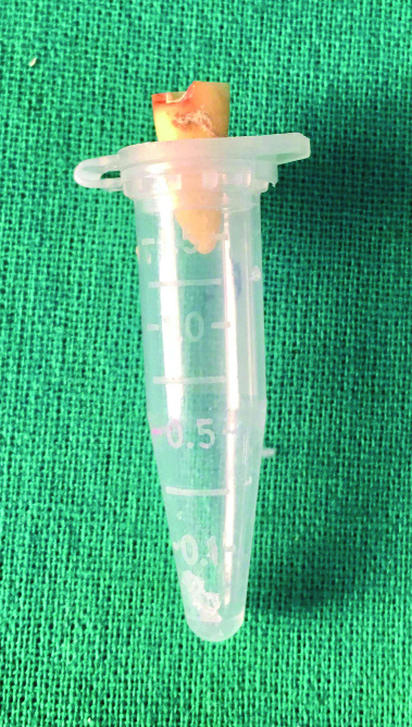
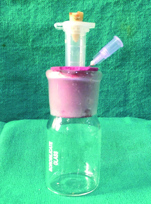
The debris collection apparatus was made according to the method described by Myers GL and Montgomery S [9]. Eppendorf tubes were taken and weighed by an electronic microbalance (Eurotech Class1 Model ETA 220I). Each individual tooth was held in a preweighed eppendorf tube which was estimated by taking three consecutive weights and computing the mean weight. The tube was then fixed inside a glass vial through a rubber plug and was seen that there was no contact between the tube and the glass vial. The tube was vented with a 25 gauge needle to equalize the pressure inside and outside [Table/Fig-1].
Decoronated sample mounted in a preweighed eppendorf tube.
The teeth were then mounted inside the eppendorf tubes and randomly divided into two groups (n=15) [Table/Fig-2].
Eppendorf tube fixed inside a glass vial through a rubber plug. A 25 gauge needle vented to equalise the pressure.
Group A-PTR group: The root fillings in this group were first softened with 0.1 mL of RC Solve (Prime Dental Products, India) and removed using PTR files: D1, D2 and D3 as per the manufacturer’s instructions with Endomate TC2 endomotor (NSK, Japan). The files were used in a brushing motion, resting against the canal walls in a crown-down direction until it reached the working length. D1 was used till the cervical third, D2 till the middle third and D3 till the entire working length. Each file was used till no filling material was seen on them.
Group B-ERE group: The root fillings in this group were similarly softened with 0.1 mL of RC Solve (Prime Dental Products, India) and removed using ERE files: 1, 2, 3 and 4 sequentially as per the manufacturer’s instructions with Endomate TC2 endomotor (NSK, Japan). The files were used in a similar brushing motion resting against the canal walls in a crown-down direction until it reached the working length. Each file was used till no filling material was seen on them, as in Group A.
Following the completion of the retreatment procedure, the periapical debris was collected by washing off the periapical area with 1 mL of distilled water into the eppendrof tube. The tube was then stored in an incubator at 70°C for five days to allow the moisture to evaporate. The tubes were then weighed thrice and the mean weight was computed [10].

Statistical Analysis
The final weight in grams of AED by two different rertreatment file systems was recorded for 15 samples in each group and analysed using independent sample t-test. The level of statistical significance was tested at p<0.05.
Results
A descriptive analysis of results with independent sample t-test is presented in [Table/Fig-3]. The independent sample t-test reveals no significant difference between the means of extruded debris of the two groups (p=0.701). The means of two distributions with different ranges may be equal. Therefore, it is pertinent to have a closer look at the distributions of two groups with descriptive statistics like minimum, maximum and quartiles [Table/Fig-4]. The medians which are the middle values of the distributions and also the interquartile range are not much different for the groups.
Independent sample t-test comparing means of extruded debris of the two groups.
| Groups | Levene’s test for equality of variances | t-test for equality of means |
|---|
| F | Sig. | t | df | Sig. (2-tailed) p-value | Mean difference | Sth. error difference | 95% Confidence interval of the difference |
|---|
| Lower | Upper |
|---|
| Apically extruded debris | Equal variances assumed | <0.001 | 0.985 | 0.388 | 28 | 0.701 | 0.000766 | 0.0019756 | -0.00328034 | 0.00481367 |
| Equal variances not assumed | 0.388 | 27.966 | 0.701 | 0.000766 | 0.0019756 | -0.00328056 | 0.00481389 |
Level of significance set as p≤0.05
Comparison of excluded debris (in grams) among groups.
| Variables | ProTaper R | Endostar RE | Total |
|---|
| No of samples | 15 | 15 | 30 |
| Mean±SD | 0.0076±0.0055 | 0.0068±0.0053 | 0.0072±0.0053 |
| p-value | 0.701 |
| Minimum | 0.0002 | 0.0004 | 0 |
| Maximum | 0.0179 | 0.0164 | 0.018 |
| 25th percentile (Q1) | 0.0027 | 0.0017 | 0.0025 |
| 50th Percentiles (Q2) (Median) | 0.0062 | 0.0050 | 0.0056 |
| 75th Percentiles (Q3) | 0.0113 | 0.0111 | 0.0112 |
Therefore, null hypothesis was rejected as there was no significant difference in the amount of debris extrusion among PTR and ERE file systems. Though statistically insignificant AED was more for PTR files as compared to ERE files while PTR files cleaned the canal walls better as compared to ERE files.
Discussion
The primary objective of the root canal retreatment depends on successful removal of the infected filling material followed by cleaning, shaping and refilling of the root canal. During retreatment procedures extrusion of infected debris into periradicular tissues may be one of the causes of postoperative pain. Microbial extrusion into periradicular tissue has potential to bring about serious systemic diseases such as endocarditis, brain abscesses and septicaemia particularly in compromised patients. Factors affecting extrusion of debris include: apical patency [11], dentin hardness [12], quantity and flow of irrigant [13], size of final apical file [14] and instrumentation techniques [15].
Mandibular first premolars were considered for this in vitro study as they are frequently extracted for orthodontic reasons. Although their canals are often straight and they are flattened mesiodistally (major variation anatomically). This study did not control apical size for retreatment, and protocol was followed according to manufacturers’ instructions. To our knowledge there are no previous studies that compared debris extrusion of PTR system (Dentsply Maillefer, Ballaigues, Switzerland) and ERE system (Endostar, Poland), which are distinctly different in their design.
Earlier study concluded that ProTaper retreatment files extruded less debris as compared to K3 files (statistically insignificant) and H-files with solvents (Significant difference) [16]. On the contrary study by Pawar AM et al., showed that PTR files used along with Waveone exhibited significantly more AED as compared to (PTR+ProTaper next) and (PTR+Self adjusting files) [14].
Statistically insignificant difference was seen in extrusion of AED for both rotary instrumentation groups as seen in previous studies [6,17]. This may be due to use of crown down technique with early flaring of the coronal part of the preparation with PTR and ERE files which improves instrument control during preparation of apical one-third of the canal. It was observed that rotary instrumentation for gutta-percha removal tend to pull the dentinal debris into the file flutes and direct it towards the coronal aspect of the canal, thus avoiding its compaction into the canal and thereby resulting in less AED. Intial use of 0.1 mL of gutta-percha solvent might have helped in better removal of the filling materials.
As per the results obtained extrusion of debris occurred independent of the type of instrument used which was more for PTR group (p>0.05) than ERE group. The PTR system have a convex triangular cross-section with three machined cutting edges which results in decreased cutting efficiency and smaller dentin chip space which inadvertently acts like a piston and forces the debris apically during instrumentation [18]. The apical D3 file with a larger taper of 7% and tip diameter 0.20 mm may attribute to more apical extrusion of debris. In ERE system file no. 1,2 have square cross-section similar to K-files with four cutting edges and good elasticity while No. 3 and No. 4 have S-cross-section with cutting taper/tip diameter of 0.06/30 and 0.04/30 respectively. Apical extrusion of debris was less for this group. This may be attributed to S-cross section design of No. 3 and No. 4 files which provides positive rake angle with two cutting edges distributed symmetrically, increased pitch length and more cross-sectional space for enhanced cutting and allowing the debris to move coronally. They also present with great cutting ability, a non cutting tip and good elasticity [19].
A study by Khedmat S et al., showed that two retreatment files with similar cross-section require significantly less time for gutta-percha removal as compared to ProTaper retreatment files [20].
Results of this study concur with earlier studies which support that differences in AED may be caused by preparation techniques and/or cross-sectional design of instruments [21-23]. Salt crystal formation occurs after dessication of NaCl solution, which interferes with experimental findings [22]. Bi-distilled (double distilled) water was used as irrigant and to rinse the periapical area after retreatment to avoid possible weight increase due to crystal formation. Caution should be taken while shifting the results to a clinical situation as back pressure created by periapical tissue resistance is not considered in this study. Though floral foam technique has been proposed as a simulation of back pressure of periapical tissues, it has many disadvantages including absorption of irrigants and debris. Hence no attempt was made in this study to simulate periapical resistance [24].
Limitation
Because of zero back pressure design, gravity may have carried the irrigant out of the canal. This is a known drawback of the in vitro designs with no periapical resistance [20]. Also, the results of this study cannot be generalised to multiple rooted teeth, curved canals, open apices and incomplete root development cases.
Further in vitro studies can be conducted using ProTaper and Endostar retreatment files with solvents to evaluate their ability in removing gutta-percha completely from root canals and also the time required for the same.
Conclusion
In present study, both PTR and ERE rotary retreatment file systems caused apical debris extrusion. Though statistically insignificant AED was less in ERE as compared to PTR systems.
Level of significance set as p≤0.05
[1]. Siqueira JF Jr, Aetiology of root canal treatment failure: why well-treated teeth can failInt Endod J 2001 34(1):01-10.10.1046/j.1365-2591.2001.00396.x11307374 [Google Scholar] [CrossRef] [PubMed]
[2]. Friedman S, Stabholz A, Tamse A, Endodontic retreatment-case selection and technique. Part 3. Retreatment techniquesJ Endod 1990 16(11):543-49.10.1016/S0099-2399(07)80219-6 [Google Scholar] [CrossRef]
[3]. Stabholz A, Friedman S, Endodontic retreatment-case selection and technique. Part 2: Treatment planning for retreatmentJ Endod 1988 14(12):607-14.10.1016/S0099-2399(88)80058-X [Google Scholar] [CrossRef]
[4]. Seltzer S, Naidorf IJ, Flare-ups in endodontics: I. Etiological factorsJ Endod 1985 11(11):472-78.10.1016/S0099-2399(85)80220-X [Google Scholar] [CrossRef]
[5]. Naidorf IJ, Endodontic flare ups– bacteriological and immunological mechanismJ Endod 1985 11(11):462-64.10.1016/S0099-2399(85)80218-1 [Google Scholar] [CrossRef]
[6]. Surakanti JR, Venkata RC, Vemisetty HK, Dandolu RK, Jaya NK, Thota S, Comparative evaluation of apically extruded debris during root canal preparation using ProTaper™, Hyflex™ and Waveone™ rotary systemsJ Conserv Dent 2014 17(2):129-32.10.4103/0972-0707.12804524778507 [Google Scholar] [CrossRef] [PubMed]
[7]. Siqueira JF Jr, Microbial causes of endodontic flare-upsInt Endod J 2003 36(7):453-63.10.1046/j.1365-2591.2003.00671.x12823700 [Google Scholar] [CrossRef] [PubMed]
[8]. Ruiz-Hubard EE, Gutmann JL, Wagner MJ, A quantitative assessment of canal debris forced periapically during root canal instrumentation using two different techniquesJ Endod 1987 13(12):554-58.10.1016/S0099-2399(87)80004-3 [Google Scholar] [CrossRef]
[9]. Myers GL, Montgomery S, A comparison of weights of debris extruded apically by conventional filing and canal master techniquesJ Endod 1991 17:275-79.10.1016/S0099-2399(06)81866-2 [Google Scholar] [CrossRef]
[10]. Burklein S, Benten S, Schafer E, Quantitative evaluation of apically extruded debris with different single-file systems: Reciproc, F360 and OneShape versus MtwoInt Endod J 2014 47(5):405-09.10.1111/iej.1216123889673 [Google Scholar] [CrossRef] [PubMed]
[11]. Deonizio MD, Sydney GB, Batista A, Pontarolo R, Guimaraes PR, Gavini G, Ifluence of apical patency and cleaning of the apicalforamen on periapical extrusion in retreatmentBraz Dent J 2013 24(5):482-86.10.1590/0103-644020130201624474289 [Google Scholar] [CrossRef] [PubMed]
[12]. Tanalp J, Kaptan F, Sert S, Kayahan B, Bayirl G, Quantitative evaluation of the amount of apically extruded debris using 3 different rotary instrumentationsystemsOral Surg Oral Med Oral Pathol Oral Radiol Endod 2006 101(2):250-57.10.1016/j.tripleo.2005.03.00216448929 [Google Scholar] [CrossRef] [PubMed]
[13]. Sheetal BG, Girish CK, Manoj GC, Akarte NR, Apical extrusion of debris and irrigant using hand and rotary systems: A comparative studyJ Conserv Dent 2011 14(2):187-90.10.4103/0972-0707.8262221814364 [Google Scholar] [CrossRef] [PubMed]
[14]. Pawar AM, Pawar M, Metzger Z, Thakur B, Apical extrusion of debris by supplementary filesused for retreatment: An ex vivo comparative studyJ Conserv Dent 2016 19(2):125-29.10.4103/0972-0707.17868627099416 [Google Scholar] [CrossRef] [PubMed]
[15]. Silva EJ, Sa L, Bellaonna FG, Neves AA, Accorsi-Mendonca T, Vieria VT, Reciprocating versus rotary systems for root filling removal: Assessment of the apically extruded materialJ Endod 2014 40(12):2077-80.10.1016/j.joen.2014.09.00925442728 [Google Scholar] [CrossRef] [PubMed]
[16]. Arora C, Bahri R, Mittal N, Comparative evaluation of debris extruded apically by using, ProTaper retreatment file, K3 file and H-file with solvent in endodontic retreatmentSaudi Endod J 2012 2(3):136-41.10.4103/1658-5984.112706 [Google Scholar] [CrossRef]
[17]. Kustarci A, Altunbas D, Akpinar KE, Comparative study of apically extruded debris using one manual and two rotary instrumentation techniques for endodontic retreatmentJ Dent Sci 2012 7:01-06.10.1016/j.jds.2011.09.011 [Google Scholar] [CrossRef]
[18]. Burklein S, Hinschitza K, Dammaschke T, Schafer E, Shaping ability and cleaning effectiveness of two single-file systems in severely curved root canals of extracted teeth: Reciproc and WaveOne versus Mtwo and ProTaperInt Endod J 2012 45(5):449-61.10.1111/j.1365-2591.2011.01996.x22188401 [Google Scholar] [CrossRef] [PubMed]
[19]. Downloads-PDF Instructions | Endostar [Internet]. Endostar.eu. 2018 [cited 29 January 2018]. Available from: http://www.endostar.eu/en/downloads [Google Scholar]
[20]. Khedmat S, Azari A, Shamshiri AZ, Fadae M, Fakhar HB, Efficacy of ProTaper and Mtwo retreatment files in removal of gutta-percha and guttaflow from root canalsIran Endod J 2016 11(3):184-87. [Google Scholar]
[21]. Joseph M, Ahlawat J, Malhotra A, Rao M, Sharma A, Talwar S, In vitro evaluation of efficacy of different rotary instrument systems for guttapercha removal during root canal retreatmentJ Clin Exp Dent 2016 8(4):e355-60.10.4317/jced.5248827703601 [Google Scholar] [CrossRef] [PubMed]
[22]. Western JS, Dicksit DD, Apical extrusion of debris in four different endodontic instrumentation systems: A meta-analysisJ Conserv Dent 2017 20(1):30-36.10.4103/0972-0707.20906628761250 [Google Scholar] [CrossRef] [PubMed]
[23]. Elmsallati EA, Wadachi R, Suda H, Extrusion of debris after use of rotarynickel-titanium files with different pitch: a pilot studyAust Endod J 2009 35(2):65-69.10.1111/j.1747-4477.2008.00128.x19703077 [Google Scholar] [CrossRef] [PubMed]
[24]. Altundasar E, Nagas E, Uyanik O, Serper A, Debris and irrigant extrusion potential of 2 rotary systems and irrigation needlesOral Surg Oral Med Oral Pathol Oral Radiol Endod 2011 112(4):e31-35.10.1016/j.tripleo.2011.03.04421778084 [Google Scholar] [CrossRef] [PubMed]