JCDR - Register at Journal of Clinical and Diagnostic Research
Journal of Clinical and Diagnostic Research, ISSN - 0973 - 709X
Dermatology Section DOI : 10.7860/JCDR/2018/35575.11766
Year : 2018 | Month : Jul | Volume : 12 | Issue : 7 Full Version Page : WH01 - WH02

Eyelid Discoid Lupus Erythematosus Misdiagnosed as Leishmaniasis

Amir Feily1, Farhang Houshmand2, Marigdalia K Ramirez-Fort3

1 Dermatologist, Department of Dermatology, Skin and Stem Cell Research Center, Tehran University of Medical Sciences, Tehran, Iran.
2 Dermatopathologist, Department of Pathology, Jahrom University of Medical Sciences, Jahrom, Iran.
3 Senior Resident, Department of Radiation Oncology, Medical University of South Carolina, South Carolina, USA.


NAME, ADDRESS, E-MAIL ID OF THE CORRESPONDING AUTHOR: Dr. Amir Feily, Dr. Feily Clinic of Dermatology, Skin and Stem Cell Research Center, Tehran University of Medical Sciences, Tehran, Iran; Moalem BLVD, Jahrom, Fars, Iran.
E-mail: dr.feily@yahoo.com
Abstract

Keywords

<p>A 42-year-old man presented to the clinic of Dermatology and Hair Transplantation, with a two-year history of right eyelid scarring and associated eyelash alopecia. Upon initial presentation of the patient’s symptomatology, he had a positive smear test for leishmania. Therefore, he was diagnosed and treated for eyelid leishmaniasis with several rounds of cryotherapy and glucantim for two years; his lesions were refractory to this initial therapy.</p><p>
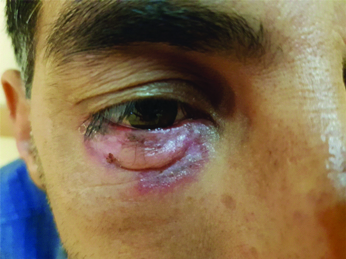
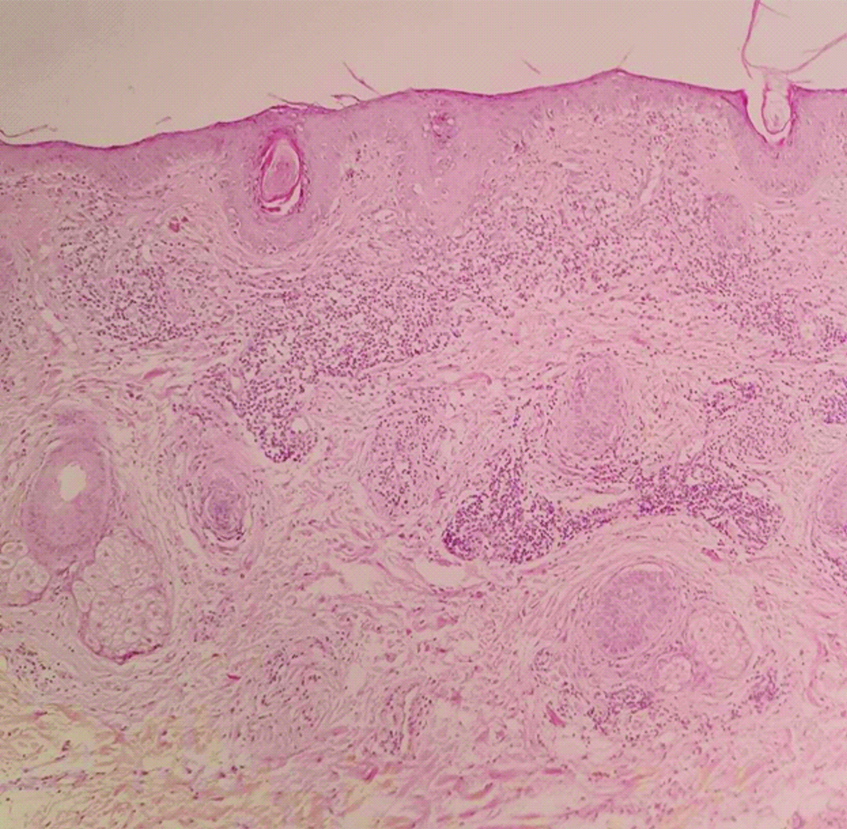
General physical examination was unremarkable. Right eye examination revealed, periocular erythematous, oedematous plaques with active borders, central scarring and lower lid madarosis [<a ref-type="fig" href="#F1">Table/Fig-1</a>]. There was no other skin lesion on the body. The lesion was clinically suspicious for cutaneous lupus and a biopsy was performed. Histopathological analysis confirmed Discoid Lupus Erythematosus (DLE) [<a ref-type="fig" href="#F2">Table/Fig-2</a>]. Specifically, the sections showed hyperkeratosis with follicular plugging. Thinning and flattening of the stratum Malpighi with focal hydropic degeneration of the basal layer was identified. A brisk perivascular and periadnexal mononuclear lymphocytic infiltrate with some admixed melanophages, around hair follicles was seen. Vasodilation of upper dermal vessels was also present.</p><fig id="F1"><label>[Table/Fig-1]:</label><div class='subhead'><p>Right eyelid examination; periocular erythematous, oedematous plaques with active borders, central scarring and lower lid madarosis.</p></div><img alt='' style="max-height:500px;max-width:600px" src="/articles/images/11766//jcdr-12-WH01-g001.jpg" /></fig><fig id="F2"><label>[Table/Fig-2]:</label><div class='subhead'><p>The skin lesion biopsy (H&E staining; Low power).</p></div><img alt='' style="max-height:500px;max-width:600px" src="/articles/images/11766//jcdr-12-WH01-g002.jpg" /></fig><p>
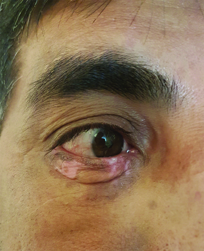
The patient was successfully treated with hydroxychloroquine 200 mg BID and prednisolone 15 mg daily, resulting in lesional resolution in six months [<a ref-type="fig" href="#F3">Table/Fig-3</a>].</p><fig id="F3"><label>[Table/Fig-3]:</label><div class='subhead'><p>Successful treatment of the patient with hydroxychloroquine and prednisolone and lesional resolution in six months.</p></div><img alt='' style="max-height:500px;max-width:600px" src="/articles/images/11766//jcdr-12-WH01-g003.jpg" /></fig><p>DLE is a chronic, autoimmune disorder that is limited to the skin; morphologically, DLE presents with characteristic acute erythema and discoid lesions [<a href="#b1">1</a>]. Rarely does DLE involve the eyelid and periocular region [<a href="#b2">2</a>]. Early diagnosis of DLE is important towards initiating the correct treatment and prevention of permanent scarring and discolouration [<a href="#b1">1</a>,<a href="#b2">2</a>]. The differential diagnosis of periocular DLE includes: psoriasis, rosacea, lupus vulgaris, sarcoidosis, Bowen’s disease, polymorphous light eruptions, lichen planopilaris, dermatomyositis, granuloma annulare, granuloma faciale and leishmaniasis [<a href="#b2">2</a>]. There are many misdiagnosed cases in the literature based on the clinical similarities of the aforementioned conditions [<a href="#b3">3</a>-<a href="#b6">6</a>]. Similarly, the present patient’s treatment was delayed due to an incorrect laboratory-based diagnosis, without noticing the clinical signs. A higher index of suspicion for DLE could have accelerated the biopsy and correct management; the presented clinical scenario, emphasises the importance of physical examination and histopathological examination in dermatological diagnoses.</p><p>Patient’s consent was obtained before publishing the clinical images.</p></p> </p> </div> <div class="subHead"> <span>References</span> </div> <div class="content" style="font-weight:normal;"> <p> <b>[1]</b>. <nlm-citation citation-type="journal"><person-group person-group-type="author"><name><surname>Uva</surname> <given-names>L</given-names>, </name><name><surname>Miguel</surname> <given-names>D</given-names>, </name><name><surname>Pinheiro</surname> <given-names>C</given-names>, </name><name><surname>Freitas</surname> <given-names>JP</given-names>, </name><name><surname>Gomes</surname> <given-names>MM</given-names>, </name><name><surname>Filipe</surname> <given-names>P</given-names>, </name></person-group><article-title>Cutaneous manifestations of systemic lupus erythematosus</article-title> <i>Autoimmune Dis</i> 2012 2012:834291<pub-id pub-id-type="doi">10.1155/2012/834291</pub-id><pub-id pub-id-type="pmid">22888407</pub-id></nlm-citation>  [<a href='https://scholar.google.co.in/scholar?hl=en&q=Uva+L%2C+Miguel+D%2C+Pinheiro+C%2C+Freitas+JP%2C+Gomes+MM%2C+Filipe+P%2C+Cutaneous+manifestations+of+systemic+lupus+erythematosus+Autoimmune+Dis+2012' target='_blank'>Google Scholar</a>]  [<a href='https://doi.org/10.1155/2012/834291' target='_blank'>CrossRef</a>]  [<a href='https://www.ncbi.nlm.nih.gov/pubmed/22888407' target='_blank'>PubMed</a>]<br/><br/><b>[2]</b>. <nlm-citation citation-type="journal"><person-group person-group-type="author"><name><surname>Yaghoobi</surname> <given-names>R</given-names>, </name><name><surname>Feily</surname> <given-names>A</given-names>, </name><name><surname>Behrooz</surname> <given-names>B</given-names>, </name><name><surname>Yaghoobi</surname> <given-names>E</given-names>, </name><name><surname>Mokhtarzadeh</surname> <given-names>S</given-names>, </name></person-group><article-title>Palpebral involvement as a presenting and sole manifestation of discoid lupus erythematosus</article-title> <i>Scientific World Journal</i> 2010 10:2130-31.<pub-id pub-id-type="doi">10.1100/tsw.2010.209</pub-id><pub-id pub-id-type="pmid">21057726</pub-id></nlm-citation>  [<a href='https://scholar.google.co.in/scholar?hl=en&q=Yaghoobi+R%2C+Feily+A%2C+Behrooz+B%2C+Yaghoobi+E%2C+Mokhtarzadeh+S%2C+Palpebral+involvement+as+a+presenting+and+sole+manifestation+of+discoid+lupus+erythematosus+Scientific+World+Journal+2010' target='_blank'>Google Scholar</a>]  [<a href='https://doi.org/10.1100/tsw.2010.209' target='_blank'>CrossRef</a>]  [<a href='https://www.ncbi.nlm.nih.gov/pubmed/21057726' target='_blank'>PubMed</a>]<br/><br/><b>[3]</b>. <nlm-citation citation-type="journal"><person-group person-group-type="author"><name><surname>Sisti</surname> <given-names>A</given-names>, </name><name><surname>Fallaha</surname> <given-names>A</given-names>, </name><name><surname>Tassinari</surname> <given-names>J</given-names>, </name><name><surname>Nisi</surname> <given-names>G</given-names>, </name><name><surname>Grimaldi</surname> <given-names>L</given-names>, </name><name><surname>Eisendle</surname> <given-names>K</given-names>, </name></person-group><article-title>Melanoma in situ mimicking a Lichen planus-like keratosis</article-title> <i>Acta Biomed</i> 2018 88:496-98.</nlm-citation>  [<a href='https://scholar.google.co.in/scholar?hl=en&q=Sisti+A%2C+Fallaha+A%2C+Tassinari+J%2C+Nisi+G%2C+Grimaldi+L%2C+Eisendle+K%2C+Melanoma+in+situ+mimicking+a+Lichen+planus-like+keratosis+Acta+Biomed+2018' target='_blank'>Google Scholar</a>]<br/><br/><b>[4]</b>. <nlm-citation citation-type="journal"><person-group person-group-type="author"><name><surname>Yaghoobi</surname> <given-names>R</given-names>, </name><name><surname>Feily</surname> <given-names>A</given-names>, </name><name><surname>Ranjbari</surname> <given-names>N</given-names>, </name><name><surname>Lotfi</surname> <given-names>J</given-names>, </name><name><surname>Yaghoobi</surname> <given-names>E</given-names>, </name></person-group><article-title>Lepromatous leprosy: a commonly misdiagnosed disease</article-title> <i>Scientific World Journal</i> 2010 10:2348-49.<pub-id pub-id-type="doi">10.1100/tsw.2010.206</pub-id></nlm-citation>  [<a href='https://scholar.google.co.in/scholar?hl=en&q=Yaghoobi+R%2C+Feily+A%2C+Ranjbari+N%2C+Lotfi+J%2C+Yaghoobi+E%2C+Lepromatous+leprosy:+a+commonly+misdiagnosed+disease+Scientific+World+Journal+2010' target='_blank'>Google Scholar</a>]  [<a href='https://doi.org/10.1100/tsw.2010.206' target='_blank'>CrossRef</a>]<br/><br/><b>[5]</b>. <nlm-citation citation-type="journal"><person-group person-group-type="author"><name><surname>Maor</surname> <given-names>D</given-names>, </name><name><surname>Ondhia</surname> <given-names>C</given-names>, </name><name><surname>Yu</surname> <given-names>LL</given-names>, </name><name><surname>Chan</surname> <given-names>JJ</given-names>, </name></person-group><article-title>Lichenoid keratosis is frequently misdiagnosed as basal cell carcinoma</article-title> <i>Clin Exp Dermatol</i> 2017 42:663-66.<pub-id pub-id-type="doi">10.1111/ced.13178</pub-id><pub-id pub-id-type="pmid">28636260</pub-id></nlm-citation>  [<a href='https://scholar.google.co.in/scholar?hl=en&q=Maor+D%2C+Ondhia+C%2C+Yu+LL%2C+Chan+JJ%2C+Lichenoid+keratosis+is+frequently+misdiagnosed+as+basal+cell+carcinoma+Clin+Exp+Dermatol+2017' target='_blank'>Google Scholar</a>]  [<a href='https://doi.org/10.1111/ced.13178' target='_blank'>CrossRef</a>]  [<a href='https://www.ncbi.nlm.nih.gov/pubmed/28636260' target='_blank'>PubMed</a>]<br/><br/><b>[6]</b>. <nlm-citation citation-type="journal"><person-group person-group-type="author"><name><surname>Feily</surname> <given-names>A</given-names>, </name><name><surname>Namazi</surname> <given-names>MR</given-names>, </name><name><surname>Seifmanesh</surname> <given-names>H</given-names>, </name></person-group><article-title>Generalized pustular psoriasis-like dermatophytosis due to Trichophyton rubrum</article-title> <i>Acta Dermatovenerol Croat</i> 2011 19:209-11.</nlm-citation>  [<a href='https://scholar.google.co.in/scholar?hl=en&q=Feily+A%2C+Namazi+MR%2C+Seifmanesh+H%2C+Generalized+pustular+psoriasis-like+dermatophytosis+due+to+Trichophyton+rubrum+Acta+Dermatovenerol+Croat+2011' target='_blank'>Google Scholar</a>]<br/><br/> </p> </div> <div id="ladiv" class="content imgdiv"> </div> </div> </div> </form> </body> </html>