Variant Insertion of Deltoid Muscle
Lydia S Quadros1, Antony Sylvan D’Souza2
1 Lecturer, Department of Anatomy, Kasturba Medical College, Manipal Academy of Higher Education, Manipal, Karnataka, India.
2 Professor and Head, Department of Anatomy, Kasturba Medical College, Manipal Academy of Higher Education, Manipal, Karnataka, India.
NAME, ADDRESS, E-MAIL ID OF THE CORRESPONDING AUTHOR: Dr. Lydia S Quadros, Lecturer, Department of Anatomy, Centre for Basic Sciences, Kasturba Medical College, Manipal Academy of Higher Education, Madhav Nagar, Manipal-576104, Karnataka, India.
E-mail: lidibudy@gmail.com
During the routine dissection for the undergraduate medical students a unilateral variation was seen in the insertion of deltoid muscle in the left arm. The muscle took origin as usual. The most anterior clavicular fibers inserted in the form of a long tendon into the medial epicondyle of the humerus. The tendon crossed the neurovascular structures of the arm. This type of variation may compress upon the neurovascular structures leading to lack of blood supply and neurological compromise of the distal parts of the upper limb.
Median nerve, Medial epicondyle, Neurovascular, Tendon
Case Report
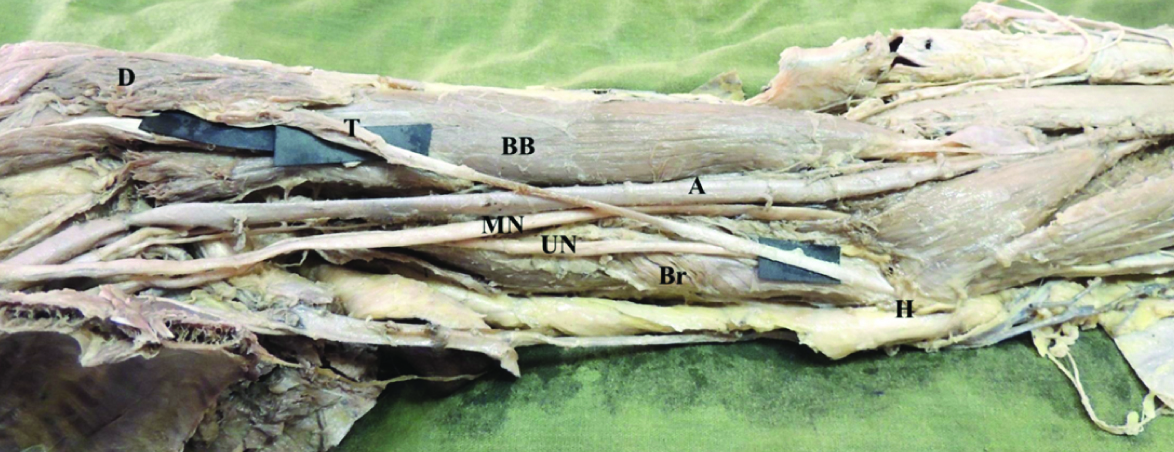
During the routine dissection of the left upper limb of a 65-year-old male cadaver for the undergraduate students, in the Department of Anatomy, a variation was observed in the pattern of insertion of the clavicular fibers of the deltoid muscle. The fibers formed a thick tendon, passing superficial to the biceps brachii muscle, brachial artery, median nerve, ulnar nerve and the brachialis muscle and finally inserted into the medial epicondyle of the humerus. The tendon measured 14.3 cm long with a uniform thickness of approximately 0.7 cm [Table/Fig-1]. No such variation was observed in the contralateral upper limb.
Dissected specimen of left upper limb arm region showing clavicular fibers of deltoid (D) muscle inserting as long tendon (T) on the medial epicondyle of humerus (H). Note the tendon passing superficial to biceps brachii (BB), brachial artery (A), median nerve (MN), brachialis (Br) and Ulnar nerve (UN).
Discussion
Variations of the deltoid muscle are quite uncommon. Deltoid muscle usually takes origin from the clavicle, the acromion process and the spine of the scapula. The fibers inserted into the deltoid tuberosity of the humerus in an inverted V-shaped manner [1]. A single fascial sheath usually encloses the whole of the deltoid muscle. Previous studies have reported the splitting of the deltoid muscle fibers enclosed within separate fascial sheath. The independent fibers arising from the acromion process of the scapula was reported as early as 1875 by Macalister A [2]. In the present case, it was observed that the clavicular fibers of deltoid muscle inserted onto the medial epicondyle of humerus.
Embryologically, the deltoid muscle originates from the dorsal muscle mass of the limb bud which is formed by the somatic mesoderm during the fifth week of intrauterine life [3]. The myogenic cells unite into two muscle masses during the fifth week of intrauterine life. One is the precursor of the flexor muscles and the other is the precursor of the extensor muscles. These muscle masses then splits to give rise to the definitive muscles of the upper limb [4]. Incorrect splitting of the dorsal muscle mass may give rise to an accessory deltoid muscle.
Separation of the deltoid muscle into numerous segments has been reported in several lower animals’ species [5]. Similar separate numerous segments have been reported in humans [6]. No such segments were observed in the present case. Posterior fibers of deltoid muscle enclosed within separate fascial sheath have been reported by previous studies [7,8]. In the present case the whole muscle was enclosed within a common fascial sheath.
The fibers of the deltoid muscle may continue into the trapezius. The fibers of deltoid may also fuse with the pectoralis major. Additional slips of deltoid may extend from the vertebral border of the scapula, infraspinous fascia and the axillary border of the scapula. These are the possible variations of the deltoid muscle reported so far [1].
The unique variation seen in the present case may be of great clinical importance. The tendon of the anterior fibers of deltoid may compresses the brachial artery causing lack of major arterial supply to the distal parts of the upper limb. The long tendon crossing the median nerve might cause neurological compression symptoms such as wasting of the muscles, numbness or paresthesia, which needs detailed evaluation for identifying and treating the causative factor. The struthers ligament seen in this region may also cause neurovascular compression. Such variation should be kept in mind while managing such cases.
Conclusion
Knowledge about this unusual insertion of the deltoid muscle may possibly serve as helpful information to the neurosurgeons and vascular surgeons. Thorough knowledge about this variation may help to prevent the diagnostic errors. It also act as a guide during the interventional procedures and avoid complication during the surgery in case of any trauma.
[1]. Standring S, Johnson D, Ellis H, Collins P, Gray’s Anatomy 2005 39th edLondonChurchill Livingstone:836 [Google Scholar]
[2]. Macalister A, Additional observations on muscular anomalies in human anatomy (third series), with a catalogue of the principal muscular variations hitherto publishedTrans Roy Irish Acad Sci 1875 25:01-134. [Google Scholar]
[3]. Larsen WJ, Human embryology 2001 3rd edEdinburghChurchill Livingstone:324 [Google Scholar]
[4]. Carlson BM, Human embryology and developmental biology 2004 3rd edMosby:224 [Google Scholar]
[5]. Brown JM, Wickham JB, McAndrew DJ, Huang XF, Testut L. Les Anomalies Musculaires chezl’homme 1884 ParisMasson:332-42. [Google Scholar]
[6]. Audenaert E, Barbaix E, Separate segments within the deltoid muscle: Anatomical variants or wishful thinking?Int J Shoulder Surg 2008 2(3):69-70.10.4103/0973-6042.4258320300320 [Google Scholar] [CrossRef] [PubMed]
[7]. Kayikcioglu A, Celik HH, Yilmaz E, An anatomic variation of the deltoid muscle (case report)Bull Assoc Anat (Nancy) 1993 77(238):15-16. [Google Scholar]
[8]. Kamburglu HO, Boran OF, Sargon MF, Kecik A, An unusual variation of deltoid muscleInt J Shoulder Surg 2008 2(3):62-63.10.4103/0973-6042.4220120300317 [Google Scholar] [CrossRef] [PubMed]