Jategaonkar’s Triple-Jerk Technique: A Safety Augmenting Method for Laparoscopic Suprapubic Trocar Insertion
Priyadarshan Anand Jategaonkar1, Smita Priyadarshan Jategaonkar2, Sudeep Pradeep Yadav3
1 Professor, Department of General Surgery, Mahatma Gandhi Institute of Medical Sciences, Sewagram, Maharashtra, India.
2 Assistant Professor, Department of Pediatrics, Mahatma Gandhi Institute of Medical Sciences, Sewagram, Maharashtra, India.
3 Senior Resident, Department of Plastic and Reconstructive Surgery, Grant’s Government Medical College & Sir J.J. Group of Hospitals, Mumbai, Maharashtra, India.
NAME, ADDRESS, E-MAIL ID OF THE CORRESPONDING AUTHOR: Dr. Priyadarshan Anand Jategaonkar, E-4, Senior Staff Quarters, Mgims, Medical Square, Sewagram, Maharashtra, India.
E-mail: jategaonkarpa@gmail.com
Suprapubic port, Surgery, Access port, Laparoscopy, Trocar
Sir,
Laparoscopic Suprapubic Port (SPT) remains a dedicated, versatile key peritoneal access-point vastly used across minimally invasive surgical specialties [1,2]. By-and-large, its insertion is considered and practiced as “routine”, just as any other abdominal port. However, loose hypogastric parietal peritoneum laden heavily with adipose tissue often renders it rather tricky, even after achieving adequate pneumoperitoneum [3]. In this context, we present a step-by-step, surgeon-friendly approach for securing SPT under direct laparoscopic vision. To our knowledge, such a technique is yet to be described in the literature.
Once pneumoperitoneum is created and primary umbilical trocar inserted, a suprapubic skin incision befitting 10 mm trocar is deepened through the subcutaneous tissue up to the anterior rectus sheath. Then the sharp tip of the trocar-cannula assembly is introduced and rested at the deepest point into the incision. At right-angle, the first controlled jerk is exerted to the trocar in order to pierce the midline in one go. Further advancement of the trocar-tip is ceased once slight peritoneal “coning effect” is appreciated under laparoscopic vision. It is then gently deviated ventrally by about 45o and stationed at a desired ergonomic peritoneal location. At this stage, the second jerk is applied in a guided manner to ensure the exit of just the trocar-tip into the peritoneal cavity. While maintaining the trocar-cannula assembly firmly in identical position, the third co-axial jerk is employed producing its immediate ingress. Finally, the trocar is completely removed and the sleeve gradually retrieved back keeping merely 2 cm length in situ [Table/Fig-1,2].
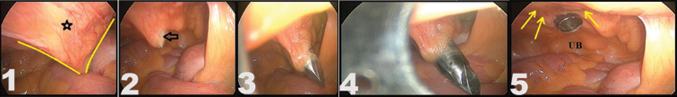
(1) Common difficulty encountered during suprapubic trocar insertion in presence of abundant pre-peritoneal adipose tissue (star). Repeated, random attempts of exiting the trocar facilitate inadvertent elevation of the lax peritoneum resulting in formation of ever increasing large peritoneal “cone” (yellow lines) that accommodates most of the trocar-length. Note its tip reaches close vicinity of the bowel, risking it for serious injury; (2) The first jerk of our technique has minimal peritoneal coning effect. This allows better in-vitro space for the trocar’s directional precision. Note that the trocar-tip is clearly visible through peritoneum (arrow); (3) The second jerk is represented by controlled propulsion of the trocar perpendicular to the peritoneum that extrudes only the trocar-tip; (4) The third jerk moves the trocar-cannula assembly further and brings out the cannula into the peritoneal cavity. Note the minimal peritoneal “stripping” and the resultant porting accuracy; (5) On partial withdrawal of the cannula, the peritoneum regains normalcy (yellow arrows). UB, Urinary Bladder.
(a) The typical problem of loosely adherent hypogastric parietal peritoneum at insertion of suprapubic trocar. Note the stretched-out peritoneal “cone” (yellow lines) without desired peritoneal access; (b) The first jerk succeeds in safe access till the peritoneum; (c) The second jerk carried in the same line (arrow), pierces only the beveled end of the trocar. Note the inadequacy of the peritoneal puncture for accommodating the sleeve; (d) The third jerk jets the sleeve out with ease. Note that the unwithdrawn trocar aids in this process; (e) The cannula is retracted back partially to get the peritoneum back to its primary position (Star, right pubic bone).

With alarming 6% morbidity, every laparoscopic trocar insertion necessitates utmost professionalism [3,4]. In this regard, our technique has following improvisations and advantages. First, the traditionally recommended gradual cork-screwing movement for trocar insertion tends to strip the peritoneum off its underlying musculature, inadvertently “coning” it excessively, thereby failing to offer adequate counter-pressure for effective trocar-puncture. Consequently, the operator often “wanders” aimlessly into the pre-peritoneal space trying multiple futile attempts of exit, rendering pre-peritoneal vessels vulnerable to trauma. But our technique’s first jerk largely “flattens” this potentially hazardous peritoneal tenting and sets a safe stage for the next stage. Second, random thrusting movements while attempting to get through such peritoneal cone by the SPT could result into a sudden give-way injuring closely underlying abdominal viscera, the most precarious being iliac vessels. However, second jerk, the key step of our technique, allows peritoneal penetration in a controlled and temperate fashion, minimising the chances of underlying organ injury. It also offers precision in tailoring the exit to an ergonomic location. Third, for reducing the trocar-related injuries, it is generally advised to keep the sharp trocar-tip withdrawn inside the blunt cannula before channelising it into the abdominal cavity [5]. Nevertheless, we have frequently observed that the trocar-induced peritoneal puncture tends to have suboptimal size for accommodating the sleeve that follows. And, forcible manipulations of threading it, as conventionally practiced, further dislodge the peritoneum. In the process, leaked pneumoperitoneum “balloons” the pre-peritoneal space eventually collapsing the operative working space almost completely. The third jerk of our method avoids this by precisely tailoring the peritoneal hiatus to fit the cannula snugly into it. Fourth, upon withdrawal of the sleeve as the last step, the surrounding peritoneum gets re-approximated to the parietes and obviates the undesired “clapper-in-the-bell” effect. Fifth, it may be efficiently practiced for obese individuals and patients having infra-umbilical scars. Sixth, it does not use any specialised instruments, hence likely to be cost-effective. Seventh, it can easily be mastered even by a novice.
Overall, it enhances the safety and is of significant clinical value in such a frequent procedure. Moreover, we have not observed any noteworthy limitations of the modification described here. With the learning curve of 5 cases and mean time for completion being 15 seconds, we have used it in over 2500 cases without any complications. In this context, a prospective skill-based study is underway at our institute. This technique is turning out to be a stepping stone for shortening the overall laparoscopic learning curve and enhancing surgical confidence of our postgraduates, hence is recommended for regular use.
[1]. Pollard RR, The suprapubic port: the key element in an enlarged fibroid uterusJ Minim Invasive Gynecol 2015 22(6S):S14510.1016/j.jmig.2015.08.51027678808 [Google Scholar] [CrossRef] [PubMed]
[2]. Gueye NA, Goodman LR, Falcone T, Versatility of the suprapubic port in robotic assisted laparoscopic myomectomyFertil Steril 2017 108(3):e110.1016/j.fertnstert.2017.06.03028865555 [Google Scholar] [CrossRef] [PubMed]
[3]. Alkatout I, Complications of laparoscopy in connection with entry techniquesJ Gynecol Surg 2017 33(3):81-91.10.1089/gyn.2016.011128663686 [Google Scholar] [CrossRef] [PubMed]
[4]. Jategaonkar PA, Yadav SP, A quick and simple method of laparoscopic port closureHellenic J Surg 2014 86:11410.1007/s13126-014-0110-4 [Google Scholar] [CrossRef]
[5]. Thepsuwan J, Huang K-G, Wilamarta M, Adlan A-S, Manvelyan V, Lee C-L, Principles of safe abdominal entry in laparoscopic gynecologic surgeryGynecology and Minimally Invasive Therapy 2013 2:105-09.10.1016/j.gmit.2013.07.003 [Google Scholar] [CrossRef]