Hypoplastic and Accessory Radial Arteries: A Case Report
Heidar Toolee1, Yousef Mohamadi2
1 PhD Student, Department of Anatomy, School of Medicine, Tehran University of Medical Sciences, Tehran, Iran.
2 PhD Student, Department of Anatomy, School of Medicine, Tehran University of Medical Sciences, Tehran, Iran.
NAME, ADDRESS, E-MAIL ID OF THE CORRESPONDING AUTHOR: Dr. Yousef Mohamadi, PhD Student, Department of Anatomy, School of Medicine, Tehran University of Medical Sciences, Tehran, Iran.
E-mail: yosef.1365@yahoo.com
Arterial variations in the upper extremity occur in a high prevalence. The radial artery is a terminal branch of the brachial artery in the cubital fossa. This artery is used in some treatment interventions and it’s variation is important for clinicians. Here, we report a cadaver with a rare pattern of two radial arteries. In this cadaver, a hypoplastic radial artery in the cubital fossa was seen as a terminal branch of the brachial artery and high origin accessory radial artery in the arm separated from the brachial artery was also found.
Brachioradialis muscle, Cadaver, Cubital fossa, Median nerve
Case Report
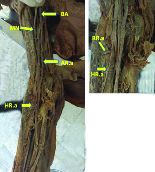
The observations were found on the upper extremity of a 57-year-old male cadaver during the routine dissection in the Department of Anatomy, Tehran University of Medical Sciences, Iran. After removal of the skin, superficial and deep fascia, a separate large unusual branch arising from the brachial artery was noticed. To determine the exact distribution of the brachial artery, the upper extremity was dissected distally. We found that the brachial artery terminated in cubital fossa by dividing into normal ulnar artery and a rudimentary hypoplastic radial artery. The rudimentary radial artery gave rise to the radial recurrent artery and then terminated deep to the brachioradialis muscle as a muscular branch. The above mentioned separate large unusual branch was a larger accessory radial artery that originated from the medial side of the brachial artery in the proximal portion of the middle third of the arm. From its origin, the accessory radial artery descended medial to the median nerve, but in the cubital fossa, the accessory radial artery related medial and anterior to the median nerve. Then, it passed superficial to the ulnar artery and arrived to the lateral side of the forearm as a radial artery [Table/Fig-1a,b]. Finally, it contributed in the formation of deep palmar arch. Other branches of the brachial artery were normal.
Branching of brachial and radial arteries.
MN: Median nerve; BA: Brachial artery; AR.a: Accessory radial artery; HR.a: Hypoplastic radial artery; RR.a: Recurrent radial artery.
Discussion
According to the main reference book of anatomy, the brachial artery is typically a continuation of the third part of the axillary artery at the lower border of teres major muscle. After giving rise to deep brachial artery, superior and inferior ulnar collateral arteries and muscular branches, the brachial artery ends in the cubital fossa at the level of the neck of radius by dividing into the radial and ulnar arteries. Radial artery is the smaller branch that descends at the lateral border of the forearm while the ulnar artery has a deeper route at the medial border of the forearm. In the upper part of the arm the median nerve runs laterally to the brachial artery, but it crosses anterior to the artery at the midpoint of the arm to arrive at the medial side of the artery [1]. Although the anatomy of each structure has been identified, but variation in anatomy is a common incidence [2]. Therefore, there is always a chance for physicians to face an unusual anatomy during the clinical interventions. This suggests a high clinical importance for anatomical variations. The arteries of upper extremity are used in different medical interventions such as angiography and doppler ultrasound [3]. Absence of the knowledge of variant dividing patterns of brachial artery jeopardizes the patients during surgical interventions and cardiac cannulation through the radial artery [4]. The present case report was an abnormal case of branching of the brachial artery that gives rise to two radial arteries.
Arterial variation in upper extremity is common. The prevalence of the variation in the branching pattern of brachial artery into ulnar and radial arteries is approximately 16.8%. Most of the variations belong to the radial artery [5]. However, Deepa TK and John MK found only two unusual distribution patterns (bilaterally in one cadaver) during the dissection of 102 upper limbs [6]. There are different patterns branching pattern of brachial artery. Some of these patterns are trifurcation of brachial artery in the arm [6], high bifurcation of the brachial artery in the arm [7], high origin of the radial artery [8], low origin of the radial artery [9], high origin of the ulnar artery [10], trifurcation of brachial artery in the cubital fossa [11] and even the absence of the brachial artery and a collateral connection between the axillary artery and radial and ulnar arteries [5]. In the present case reported here, the radial artery can retain as a small rudimentary branch [12]. Sometimes a branch originates high from the axillary artery or basically from the brachial artery and moves downward as accessory brachial artery. This artery continues as different routes distally. In some cases, it unites with the main brachial artery again, continues as a superficial ulnar artery in the forearm or divides into the radial and ulnar arteries when the main brachial artery terminates in the arm. The pattern of accessory radial artery in the present cadaver is included in a category, with a prevalence of 6% on average, in which it substitutes the radial artery in the forearm [13,14]. However, here, we also dissected the proper radial artery as a hypoplastic residual artery. From this point of view, we think it can be considered as rare pattern of branching.
The development of upper extremity, vessels is a complex process. Chemical and haemodynamic factors, genetic conditions, the fetal position in the uterus and developmental changes can induce different arterial variations [15]. In the embryo, the supplying artery for the upper extremity called axis artery arises from the seventh cervical intersegmental artery. This artery continues along the limb as the brachial and interosseous arteries upto the palm. Next, the interosseous artery gives rise to the median artery. The new branch connects to the palmar arterial plexus while the interosseous artery is disconnected. The radial and ulnar arteries develop late in the process. The ulnar artery originates from the interosseous artery in the cubital fossa, but the proper radial artery forms in two phases. Firstly, it originates from the brachial artery high in the arm and later, the final origin of the artery develops in the cubital fossa at the level of the ulnar artery [16]. This dual origin of the radial artery during the embryonic development of the vessels can be a reason for its high prevalence variation compared to other arteries and justify a high origin of radial artery in adults. Defect in the late phases of the development results in complete absence of the radial artery and a small ulnar artery but a dominant interosseous artery instead [17].
The absence of knowledge about the brachial artery variations frequently leads to disruption of treatment interventions in the patients. The radial artery is a suitable choice for creating an arterio-venous fistula for dialysis and also access to the heart when the femoral artery is not available [18]. The accessory brachial artery may be confused with basilic vein during small intravenous cannulation because of its superficial route in the lower part of the arm [19]. An accidental injection into this artery may lead to thrombosis or gangrene and a subsequent amputation [20].
Conclusion
Dissecting a rare patterned brachial artery, in the current case report, with residual hypoplastic and accessory radial arteries notifies the clinicians to look for unexpected anatomical variations during therapeutic and/or diagnostic endeavors. We think that considering possible unknown anatomical variations reduces the risk of medical interventions and also improves the efficiency
[1]. Standring S, Anand N, Jawaheer G, Tubbs RS, Birch R, Crossman AR, Gray’s Anatomy, The Anatomical basis of clinical practice 2015 41th EditionEdinburghElsevier Churchill Livingstone:827-30. [Google Scholar]
[2]. Mohamadi Y, Toolee H, Hassanzadeh G, Bilateral variation of internal iliac arteryAnat Sci J 2016 13(3):197-99. [Google Scholar]
[3]. Rao TR, Shetty P, Suresh R, Abnormal branching pattern of the axillary artery and its clinical significanceInt J Morphol 2008 26(2):389-92.10.4067/S0717-95022008000200022 [Google Scholar] [CrossRef]
[4]. Cavolli R, Eryilmaz S, Kaya B, Ozyurda U, Report of the anatomic variation of the brachial artery in a patient undergoing transradial cardiac catheterizationJ Vasc Nurs 2007 25(1):19-20.10.1016/j.jvn.2006.12.00117324765 [Google Scholar] [CrossRef] [PubMed]
[5]. Ciervo A, Kahn M, Pangilinan AJ, Dardik H, Absence of the brachial artery: report of a rare human variation and review of upper extremity arterial anomaliesJ Vasc Surg 2001 33(1):191-94.10.1067/mva.2001.11221211137944 [Google Scholar] [CrossRef] [PubMed]
[6]. Deepa TK, John MK, An anatomical study of variations in termination of brachial artery, with its embryological basis and clinical significanceInt J Med Res Health Sci 2016 5(3):85-89. [Google Scholar]
[7]. Rohilla A, Parmar P, Singh K, Rohilla J, Unilateral high division of brachial artery and its clinical significanceJ Res Med Sci 2016 4(12):5513-15.10.18203/2320-6012.ijrms20164244 [Google Scholar] [CrossRef]
[8]. Özgür M, Gölpınar M, Arifoğlu Y, Çavdar S, High origin of radial artery from the axillary artery: Case reportArtery Research 2017 17:39-42.10.1016/j.artres.2017.01.003 [Google Scholar] [CrossRef]
[9]. Wysiadecki G, Polguj M, Haładaj R, Topol M, Low origin of the radial artery: A case study including a review of literature and proposal of an embryological explanationAnat Sci Int 2016 92(2):293-98.10.1007/s12565-016-0371-927631096 [Google Scholar] [CrossRef] [PubMed]
[10]. Aharinejad S, Nourani F, Hollensteiner H, Rare case of high origin of the ulnar artery from the brachial arteryClin Anat 1997 10(4):253-58.10.1002/(SICI)1098-2353(1997)10:4<253::AID-CA7>3.0.CO;2-T [Google Scholar] [CrossRef]
[11]. Vollala VR, Nagabhooshana S, Bhat SM, Trifurcation of brachial artery with variant course of radial artery: Rare observationAnat Sci Int 2008 83(4):307-09.10.1111/j.1447-073X.2008.00235.x19159366 [Google Scholar] [CrossRef] [PubMed]
[12]. Thomson A, Notes on two instances of abnormality in the course and distribution of the radial arteryJ Anat Physiol 1884 18(Pt 3):265-68. [Google Scholar]
[13]. Chakravarthi KK, Siddaraju K, Venumadhav N, Sharma A, Kumar N, Anatomical variations of brachial artery- Its morphology, embryogenesis and clinical implicationsJ Clin Diagn Res 2014 8(12):AC17-AC20.10.7860/JCDR/2014/10418.530825653931 [Google Scholar] [CrossRef] [PubMed]
[14]. Yang HJ, Gil YC, Jung WS, Lee HY, Variations of the superficial brachial artery in Korean cadaversJ Korean Med Sci 2008 23(5):884-87.10.3346/jkms.2008.23.5.88418955798 [Google Scholar] [CrossRef] [PubMed]
[15]. Rodríguez-Niedenführ M, Burton G, Deu J, Sañudo J, Development of the arterial pattern in the upper limb of staged human embryos: normal development and anatomic variationsJ Anat 2001 199(4):407-17.10.1046/j.1469-7580.2001.19940407.x11693301 [Google Scholar] [CrossRef] [PubMed]
[16]. Singer E, Embryological pattern persisting in the arteries of the armAnat Rec 1933 55(4):403-09.10.1002/ar.1090550407 [Google Scholar] [CrossRef]
[17]. Zheng Y, Shao L, Mao JY, Bilaterally symmetrical congenital absence of radial artery: a case reportBMC Surgery 2014 14(1):1510.1186/1471-2482-14-1524650137 [Google Scholar] [CrossRef] [PubMed]
[18]. Rossi Junior WC, Esteves A, Simões JS, Fernandes GJM, Bilateral high division of the brachial artery in one human male cadaver: Case reportBraz J Morphol Sci 2011 28(3):204-07. [Google Scholar]
[19]. Hazlett J, Superficial ulnar artery with reference to accidental intra-arterial injectionCan Med Assoc J 1949 61(3):289-93. [Google Scholar]
[20]. Karamürsel S, Bagdatl D, Demir Z, Tüccar E, Çelebioglu S, Use of medial arm skin as a free flapPlast Reconstr Surg 2005 115(7):2025-31.10.1097/01.PRS.0000163321.83155.5A15923851 [Google Scholar] [CrossRef] [PubMed]