Supernumerary teeth are defined as teeth that appear in addition to the normal dental formula regardless of their location and morphology. Many individuals present with supernumerary teeth however the presence of multiple supernumerary teeth in individuals with no other associated disease or syndrome is rare. Herein we report a case of 17-year-old monozygotic twins who had presented for orthodontic treatment. Radiographic and histopathological examination revealed the presence of impacted #43 along with multiple developing supernumerary teeth in the elder twin and of impacted #32, #33, #34, #35 along with 1 supplementary tooth and 1 odontome in the younger twin without any other associated syndromes. The presence of multiple supernumerary teeth in non-syndromic monozygotic twins suggests the role of genetics in their formation.
Case-Report
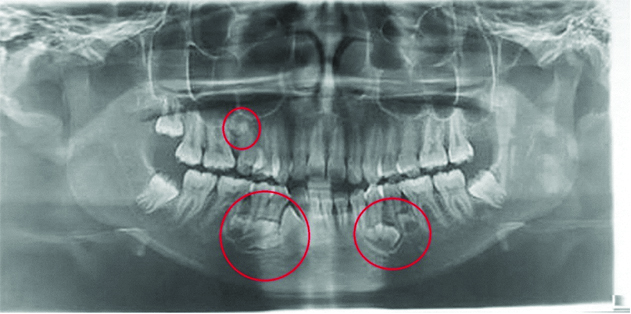
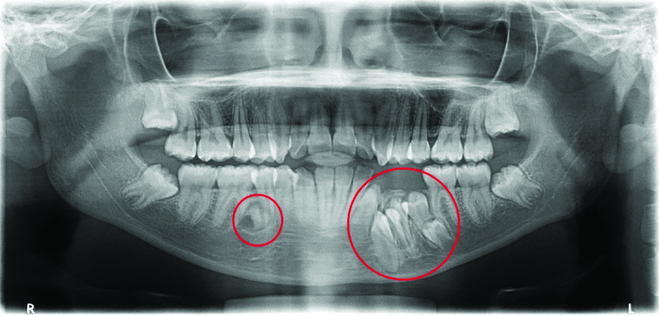
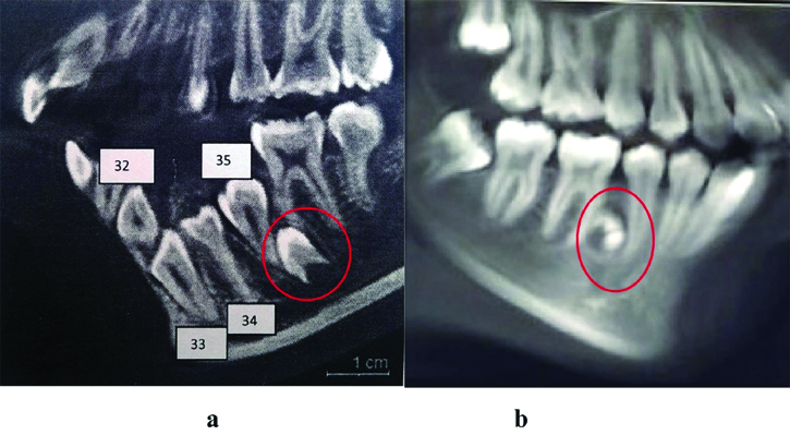
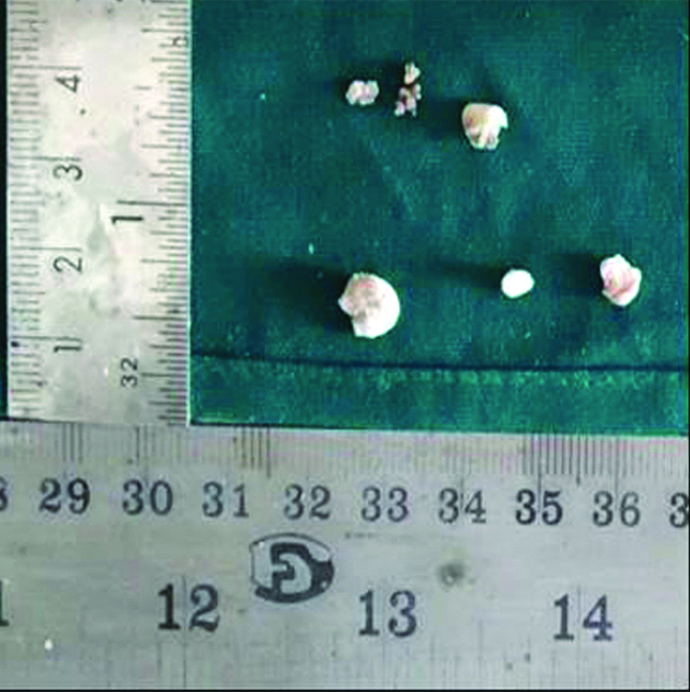
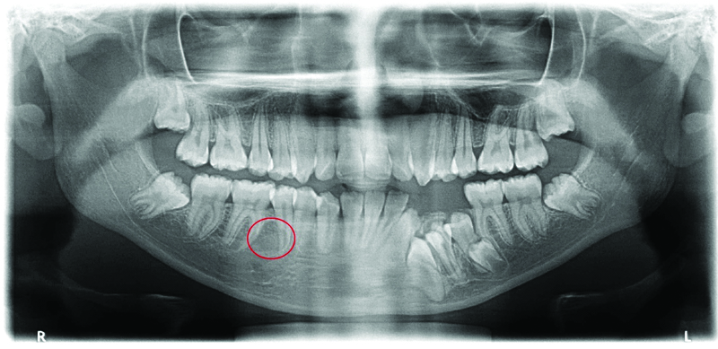
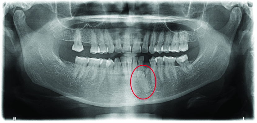
Herein we report a case of 17-year-old monozygotic twins who had presented to the Department of Oral Medicine and Radiology with a chief complaint of forwardly placed upper and lower front teeth since childhood. Medical histories of the twins were insignificant. Past dental history revealed that the younger twin underwent extraction of three retained deciduous teeth one month ago. Intraoral examination revealed that the elder twin had Angle’s Class I molar relation with anterior open bite whereas the younger twin had Angle’s Class I molar relation with partially edentulous arch in relation to #32, #33, #34, #35 and dental midline shift of 2 mm to the left. In the elder twin digital panoramic imaging revealed the presence of impacted #43, presence of a well-defined radiopacity with internal radiolucency with an open apex enclosed within a radiolucent capsule suggestive of a developing supernumerary teeth were seen in the interdental region of #35 and #36, periapical region of #34, #35 and presence of three fused developing supernumerary teeth in the #44, #45, #46 region [Table/Fig-1]. Presence of a well-defined radiopacity surrounded by a radiolucency was seen in #15, #16 region suggestive of developing supernumerary tooth or odontoma [Table/Fig-1]. Digital panoramic imaging in the younger twin revealed impacted #32, #33, #34, #35 [Table/Fig-2] and presence of a well-defined radiopacity with internal radiolucency with an open apex enclosed within a radiolucent capsule resembling a premolar suggestive of a supplementary premolar apical to the mesial root of #36 [Table/Fig-2]. Presence of two well-defined radiopacities enclosed within a radiolucent capsule suggestive of complex composite odontome was seen in the inter-radicular region of #45 and #46 [Table/Fig-2]. Radiographic differential diagnosis considered were periapical cemento osseous dysplasia, cemento ossifying fibroma, cementoblastoma. A full mouth Cone Beam Computed Tomography (CBCT) was taken in both the twins using Sirona Dental Systems Galileos Comfort CBCT Cephalometric with a Field Of View of 8×8 cm and an estimated effective dosage of 84 μSv. Images were reconstructed into axial, coronal, sagittal planes using Galileos software. In the elder twin presence of multiple developing supernumerary teeth were seen in relation to #15, #16, #34, #35, #36, #44, #45, #46 regions [Table/Fig 3a-c]. An impacted #43 was also appreciated in the elder twin [Table/Fig-3b]. In the younger twin impacted #32, #33, #34, #35 were seen and a supernumerary tooth was seen in #36 region [Table/Fig-4a]. An odontoma was also appreciated in #45, #46 region in the younger twin [Table/Fig-4b]. Patients were advised surgical extraction of the supernumerary teeth followed by orthodontic treatment of the malocclusion. The well-defined radiopacity present in the younger twin in relation to #45, #46 was extracted under local anaesthesia [Table/Fig-5] and sent for histopathological examination. Histopathological examination showed disorganised mass of tubular dentin with one area showing enamel space, dentin and pulp space in a tooth like arrangement and soft tissue section showed enamel organ like area [Table/Fig-6]. A digital panoramic image was taken post extraction in the younger twin [Table/Fig-7]. Extraction of the other radiopacities was advised under general anaesthesia in both the twins but the patients were not willing for it. Hence the elder twin did not undergo any treatment. Based on the radiographic and histological features a diagnosis of complex composite odontome was given in relation to #45, #46 region in the younger twin. Patients parents were subjected to digital panoramic imaging. Father’s radiograph revealed no impacted or supernumerary teeth [Table/Fig-8] while the mother’s radiograph revealed an impacted #33 [Table/Fig-9]. Patient’s mother gave a history of extraction of a retained deciduous tooth three years ago due to decay.
Digital panoramic image of the elder twin revealing an impacted #43 and multiple supernumerary tooth.
Digital panoramic image of the younger twin revealing impacted #32, #33, #34, #35, 1 supplementary tooth in relation to #36 and an odontome in relation to #45, #46 region.
Sagittal sections in a CBCT of the elder twin showing developing supernumerary teeth in relation to: a) #15, #16 region; b) #15, #16, #44, #45, #46 region; b,c) an impacted #43 and #34, #35, #36 regions.

Sagittal sections in a CBCT of the younger twin showing: a) impacted #32, #33, #34, #35 and a developing supernumerary tooth in #36 region; b) odontoma in #45, #46 region.
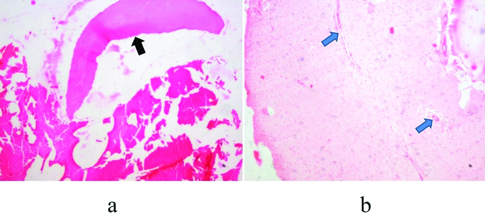
Biopsy specimen consisting of three hard tissue bits and four soft tissue bits.
Histopathological examination showing: a) enamel space, dentin (black arrow) (H&E, 4X); and b) the proliferating capillaries (blue arrow) in the pulp space arranged in a tooth like arrangement (H&E, 4X).
Digital panoramic image taken after extraction in #44, #45 region in the younger twin.
Digital panoramic image of the father.

Digital panoramic image of the mother showing an impacted #33.
Discussion
Supernumerary teeth are defined as teeth that appear in addition to the normal dental formula regardless of their location and morphology [1]. Multiple hyperdontia not associated with syndromes is rare and its prevalence is around 1.2% in South Indian paediatric population [2].
The aetiology of supernumerary teeth is unknown however, several theories such as the phylogenetic theory given by Smith et al., dichotomy theory given by Liu et al., hyperactive dental lamina given by Primosh et al., and Liu et al., and a combination of genetic and environmental factors-unified aetiologic explanation by Brook et al., in 1984 have been proposed regarding its aetiology [3]. Supernumerary teeth are of various types which include supplemental which are of normal shape and size, the conical form, tuberculate which are barrel shaped and the odontomes [4].
Adequate knowledge is available regarding tooth morphogenesis and differentiation however little is known about the molecular mechanisms underlying supernumerary tooth formation [5].
A number of genes that act at specific stages of tooth development and regulate its differentiation process have been identified via animal and human studies. The conserved signaling pathways of Bone Morphogenic Protein (BMP), Fibroblast Growth Factor (FGF), Sonic Hedgehog (SHH) and Wingless-type Mouse Mammary Tumour Virus (MMTV) integration site Wnt ligands and their receptors constitute the key pathways that are used during tooth development and mediate the epithelial-mesenchymal interactions [5].
Severe tooth development disturbances such as anadontia, supernumerary tooth occur due to disruption of genes that are part of these signaling pathways and the inhibitors of these signaling pathways. Studies have proved that tooth development arrests at lamina stage if FGF8 gene is inactivated in the dental epithelium whereas arrest of tooth development at the bud stage is seen due to over expression of BMPR1a in transgenic mice, or functional inactivation of FGFR2b or SHH [5].
Any disturbance in the inhibitors or mediators of these signaling pathways lead to formation of teeth with abnormal shape, ameloblast or odontoblast differentiation defects and reduced matrix deposition [5].
Various studies have proved that supernumerary tooth formation occurs due to loss of Ectodin causing inhibition of BMP signaling, overexpression of Wnt signaling mediators Ectodysplasin (Eda), Adenomatous polyposis coli (Apc) and β-catenin. Similarly mutation of mediators and/or inhibitors of the SHH and FGF signaling leads to formation of supernumerary tooth [5].
The prevalence of supernumerary tooth is found to be higher in the relatives of affected individuals than in the general population however, the incidence of familial occurrence of supernumerary and impacted teeth is a poorly discussed issue [6,7]. Twins provide the easiest tool to obtain heritability estimates of diseases and malformations and even help in understanding the interaction between “nature” and “nurture” [7].
Monozygotic twins originate from one fertilised egg that divides later on. An interesting aspect of concordance between monozygotic twins is the occurrence of “mirror imaging” that is the appearance of an asymmetrical feature on the right side of one twin and on the left side of the other and “duplicates ” that is when asymmetrical feature occur on the same side of the body in both the twins [7].
Sharma A reported a case of concomitant hypodontia and hyperdontia in 7-year-old monozygotic twin boys where one of the twins had two tuberculate mesiodens in relation to #51, #21 respectively however, the maxillary left second premolar was missing whereas the other twin had 1 tuberculate mesioden in the midline and a missing mandibular left second premolar [8].
Khambete N et al., reported a case of multiple supernumerary teeth in three generations where the grandfather had a history of extra teeth but he had passed away and no clinical records were available, the father had five supernumerary teeth, elder son had 8 supernumerary teeth with impacted maxillary left canine and younger son had two inverted mesiodens [1].
Liu JF et al., reported a case of seven year and seven-month-old monozygotic twin boys where the elder brother had four supernumerary teeth two in the premaxilla and two mandibular supernumerary premolars whereas the younger brother had three impacted supernumerary teeth one in the premaxilla and two mandibular supernumerary premolars [9].
Gurgel CV et al., reported a case nine-year-old monozygotic twins where both the twins presented with impacted permanent maxillary central incisors and the presence of bilateral mesiodens [10].
Reddy GSM et al., reported a case of 14-year-old monozygotic twin boys where the first twin showed double conical mesiodens and second twin showed single conical mesioden [11].
Genetic and environmental influences may be the reason for the differences observed between twins both in duplicates and mirror imaging [7].
Common problems associated with supernumerary tooth include nasal eruption, cystic degeneration of the tooth, loss of vitality, diastema formation, displacement, impaction, crowding of the adjacent teeth and compromise on implant site [12].
The type and position of the supernumerary teeth as well as its effect on adjacent teeth determine its treatment. Removal of the supernumerary tooth is indicated if it is associated with any of the above listed complications [12]. However, much debate is concerned with the merit of prophylactic removal. A compromised approach to the timing of surgical intervention based on tooth type and stage of eruption has been recommended [12]. The conical form should be observed for eruption whereas tuberculate and inverted conical forms should be removed immediately since they frequently remain unerupted and create complications. For supernumerary teeth that are located beyond the apex either an early or delayed intervention can be done [12].
Conclusion
Several authors have shown that any disturbance of genes in the networks of activators and inhibitors of signaling pathways of BMP, FGF, SHH and Wnt ligands and their receptors which control tooth formation leads to abnormalities in either number or patterning of teeth. The presence of multiple impacted, supernumerary teeth in both the twins and an impacted tooth in the mother in our patients strongly support the role of genes in tooth development disturbances.
[1]. Khambete N, Kumar R, Genetics and presence of non-syndromic supernumerary teeth: a mystery case report and review of literatureContemp Clin Dent 2012 3:499-502.10.4103/0976-237X.10745523633820 [Google Scholar] [CrossRef] [PubMed]
[2]. Anegundi RT, Tegginmani VS, Battepati P, Tavargeri A, Patil S, Trasad V, Prevalence and characteristics of supernumerary teeth in a non-syndromic South Indian pediatric populationJ Indian Soc Pedod Prev Dent 2014 32:09-12.10.4103/0970-4388.12704124531595 [Google Scholar] [CrossRef] [PubMed]
[3]. Fazliah SN, Supernumerary tooth: report of a caseArchives of Orofacial Sciences 2007 2:54-58. [Google Scholar]
[4]. Garvey MT, Barry HJ, Blake M, Supernumerary teeth-an overview of classification, diagnosis and managementJ Can Dent Assoc 1999 65:612-16. [Google Scholar]
[5]. Bei M, Molecular genetics of tooth developmentCurr Opin Genet Dev 2009 19(5):504-10.10.1016/j.gde.2009.09.00219875280 [Google Scholar] [CrossRef] [PubMed]
[6]. Subasioglu A, Savas S, Kucukyilmaz E, Kesim S, Yagci A, Munis Dundar M, Genetic background of supernumerary teethEur J Dent 2015 9(1):153-58.10.4103/1305-7456.14967025713500 [Google Scholar] [CrossRef] [PubMed]
[7]. Rodrigues MTV, Cardoso CL, Munhoz Ede A, Yaedu RYF, Sant’Ana E, Ferreira Junior O, Different impacted teeth in monozygotic twins: discussion of an unusual caseRev Clín Pesq Odonto l 2008 4(2):95-100. [Google Scholar]
[8]. Sharma A, A rare case of concomitant hypo-hyperdontia in identical twinsJ Indian Soc Pedod Prev Dent 2008 26(Suppl S2):79-81. [Google Scholar]
[9]. Liu JF, Cheng HL, Multiple maxillary and mandibular supernumerary teeth in twins: 5-year follow-upJ Dent Sci 2014 9:195-98.10.1016/j.jds.2012.05.012 [Google Scholar] [CrossRef]
[10]. Gurgel CV, Cota ALS, Kobayashi TY, Silva SMB, Machado MAAM, Rioas D, Bilateral mesiodens in monozygotic twins: 3d diagnostic and managementCase Reports in Dentistry 2013 2013:19361410.1155/2013/19361423533824 [Google Scholar] [CrossRef] [PubMed]
[11]. Reddy GSM, Mahajan B, Desai RS, Mesiodens in twins: a case reportIndian Journal of Dental Sciences 2013 5(1):88-89. [Google Scholar]
[12]. Primosch RE, Anterior supernumerary teeth-assessment and surgical intervention in childrenPediat Dent 1981 3(2):204-14. [Google Scholar]