Complete Paraplegia Due to Anterior Spinal Artery Syndrome Following Total Knee Arthroplasty under Epidural Anaesthesia: A Case Report
Nikolaos Anastasopoulos1, Trifon Totlis2, Nikolaos Lazaridis3, Konstantinos Natsis4
1 Assistant Professor, Department of Anatomy and Surgical Anatomy, School of Medicine, Faculty of Health Sciences, Aristotle University of Thessaloniki, Central Macedonia, Greece.
2 Lecturer, Department of Anatomy and Surgical Anatomy, School of Medicine, Faculty of Health Sciences, Aristotle University of Thessaloniki, Central Macedonia, Greece.
3 Lecturer, Department of Anatomy and Surgical Anatomy, School of Medicine, Faculty of Health Sciences, Aristotle University of Thessaloniki, Central Macedonia, Greece.
4 Professor, Department of Anatomy and Surgical Anatomy, School of Medicine, Faculty of Health Sciences, Aristotle University of Thessaloniki, Central Macedonia, Greece.
NAME, ADDRESS, E-MAIL ID OF THE CORRESPONDING AUTHOR: Dr. Trifon Totlis, Lecturer, Department of Anatomy and Surgical Anatomy, School of Medicine, Faculty of Health Sciences, A.U.T.H., PO Box 300, Postal Code 54 124, Thesaloniki, Central Macedonia, Greece.
E-mail: totlis@med.auth.gr
Anterior Spinal Artery Syndrome (ASAS) is an uncommon devastating neurological condition caused by ischemia in the spinal cord. We present a rare case of complete paraplegia due to ASAS following a total knee arthroplasty under lumbar epidural anaesthesia. The patient was a 78-year-old woman with mild hypertension. Epidural anaesthesia was performed at the L3-L4 interspace after a single uneventful attempt. Twenty hours postoperatively, the patient developed a flaccid paralysis of both legs, with complete loss of motor function, rectal tone and tendon reflexes, but no Babinski reflexes. Pinprick and temperature sensation were both absent bilaterally from T11 down. Proprioception and vibration sensation were intact in the ankle joint only. An immediate MRI of the spine and a second one, 48 hours postoperatively, demonstrated only a central disc herniation at T10-T11 with concomitant spinal canal stenosis. Based on neurological and MRI findings, the diagnosis of ASAS was made. The epidural catheter was removed immediately. Methylprednisolone 30 mg/kg iv bolus over 15 minutes, followed by a 5.4 mg/kg/h iv infusion for the next 23 hours was given. The patient was transferred to a rehabilitation center but no improvement was noticed and one year later the deficit was considered permanent and 2 years later the patient died. The present case alerts the orthopaedic surgeons and anaesthesiologists for the risk of ASAS in patients having predisposing factors for blood flow restriction to the spinal cord, such as elderly patients with degenerative spine disorders. The guidelines of ASAS management are analysed on a case-based approach.
Knee arthroplasty complications, Regional anaesthesia complications, Spinal cord infraction
Case Report
A 78-year-old woman suffering from end-stage degenerative osteoarthritis underwent a total knee arthroplasty with a cruciate retaining prosthesis (PFC Sigma, DePuy Orthopaedics, Warsaw, IN, USA). The patient was diagnosed with mild hypertension 16 years ago, that was effectively treated with an angiotensin II receptor antagonist- eprosartan mesylate. The lying Blood Pressure (BP) was 140 mmHg systolic and 85 mm Hg diastolic, while the standing BP was 130 mmHg systolic and 80 mm Hg diastolic.
The preoperative clinical and laboratory evaluation were unremarkable. Epidural anaesthesia was performed at the L3-L4 interspace after a single uneventful attempt. An epidural catheter was inserted, where 12 mL Ropivacaine 1% (120 mg) and 3 mL Morphine (3 mg) were administered. The blood pressure throughout the operation ranged between 95-130 mm Hg systolic and 50-70 mm Hg diastolic. The epidural catheter was connected to a continuous-infusion pump, where 100 mL Ropivacaine 0.2% and 3 mg Morphine, titrated at 8 ml/hrs was used for postoperative analgesia. The patient was started on Tinzaparin, 12.000 IU subcutaneously, 8 hours post-surgery. The patient had good analgesia seven hours postoperatively and minimal mobilization was begun, since she was able to move both lower extremities. Ten hours postoperatively, she had a brief episode of hypotension (60/40 mm Hg) that lasted approximately 30 minutes and responded well to intravenous fluid administration. This was accomplished by inserting two large-caliber (16 and 18-gauge) peripheral intravenous catheters, administrating 1000 ml of Ringer’s Lactated solution, within 15 minutes. The BP was then raised to 110/70 mm Hg.
Approximately twenty hours postoperatively, the patient complained of lower extremity discomfort and an inability to move her legs. The epidural catheter was removed immediately. The neurologic examination revealed a flaccid paralysis of both legs, with complete loss of motor function, rectal tone and tendon reflexes, while Babinski reflexes were not exerted. Pinprick and temperature sensation were both absent bilaterally from T11 down. Proprioception and vibration sensation were intact in the ankle joint only.
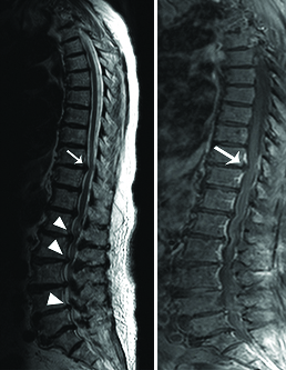
An immediate MRI of the spine demonstrated degenerative intervertebral disc protrusions at lumbar spine with spinal canal stenosis [Table/Fig-1]. At the thoracic region the MRI displayed a central disc herniation at T10-T11 with concomitant spinal canal stenosis [Table/Fig-1]. Another MRI, repeated 48 hours postoperatively demonstrated the same findings. Based on neurological and MRI findings, the diagnosis of anterior spinal artery syndrome was made.
Sagittal T2-weighted: (a) and post-contrast sagittal T1-weighted; (b) MR images on the first postoperative day showed degenerative intervertebral disc protrusions at LI-L2, L2-L3 and L4-L5 (arrow heads) with stenosis of the spinal canal. At the thoracic region the MRI displayed a central disc herniation at T10-T11 (arrow) with concomitant stenosis of the spinal canal with no spinal cord signal changes due to ischemia.
Methylprednisolone 30 mg/kg iv bolus over 15 min, followed by a 5.4 mg/kg/h iv infusion for the next 23 hours was given. The neurologic deficits were evaluated daily, the patient was transferred to a rehabilitation center but no improvement was noticed and one year later the deficit was considered permanent. The patient died 6 months before the preparation of the manuscript.
Discussion
Total Knee Arthroplasty (TKA) is an effective procedure for end stage osteoarthritis of the knee with a high success rate. Lumbar epidural anaesthesia is frequently used in both knee and hip arthroplasties. Complications related to epidural anaesthesia are very rare, but may be devastating and seriously compromise the outcome of the operation.
The blood supply to the spinal cord normally derives from a single anterior spinal artery and two posterior spinal arteries. ASAS is an uncommon devastating neurological condition caused by occlusion of the anterior spinal artery and subsequent ischemia in the spinal cord. It is characterized by a sudden, painless onset of flaccid paraplegia, loss of sensation to pain and temperature below the lesion level, bowel and urinary incontinence, while proprioception, light touch and vibration senses remain intact [1-4]. Diagnosis of the ASAS is based on the neurologic clinical picture and exclusion of other conditions, since there is no specific laboratory test to confirm the diagnosis and the MRI may not demonstrate pathognomonic for ASAS spinal cord lesions in the acute stage [5,6]. In our case, the clinical findings were typical of ASAS from T11 down. Furthermore, two factors that might have contributed to an ASAS were present; the spinal stenosis in the thoracolumbar region found in the MRI and the postoperative hypotension episodes. According to Dell’Isola D et al., spinal venous pressure may increase due to spinal stenosis and this may also affect susceptibility of the spinal cord to ischemia [7].
Both MRIs performed in our patient did not show any signal changes due to ischemia. According to the literature, in the acute setting of ASAS, MRI is used to exclude surgically treatable causes of direct cord compression. Repeat MRI, at least 4 hour after clinical onset may reveal spinal cord signal changes due to ischemia, such as hyperintensities on T2-weighted images [1,2]. However, these changes are non-specific for ischemia and according to Masson C et al., the MRI may remain normal up to 21 days after clinical onset [5].
In the literature, there are few reports of ASAS following epidural anaesthesia for both major orthopaedic operations [1,8] and non-orthopaedic cases such as cardiovascular surgery [2], oesophagectomy [9], laparoscopic surgery [10] and even partum [11] or caesarean delivery [12] included. Although there are no certain guidelines for the treatment of anterior spinal artery syndrome, a high dose of methylprednisolone may be administrated because it has been shown to improve neurological outcome in other forms of acute spinal cord injury [13]. However, no improvement was noticed in the neurological deficits of our patient following iv steroid infusion. The neurological status remained unchanged in other ASAS cases in the literature as well [1,8,9], whereas in some cases eventual partial improvement in motor function and bowel and bladder control may occur following rehabilitation [2,14,15].
The patient died six months before the preparation of this manuscript and her husband had died before her. They had no children and it is impossible to reach any relatives. Unfortunately, there are no longer any relatives to contact right now. Hence, we could not provide consent for publication of this case report. Even without informed consent of the patient, we report this case because the predisposing factors of ASAS may be preoperatively recognized and preventive measures can be taken, in order to decrease its incidence.
Conclusion
The present case alerts the anaesthesiologists and orthopaedic surgeons for the risk of ASAS in patients who undergo epidural anaesthesia for a major orthopaedic operation while having predisposing factors for blood flow restriction to the spinal cord, such as elderly patients with degenerative spine disorders. The guidelines of ASAS management were analysed on a case-based approach.
[1]. Hong DK, Lawrence HM, Anterior spinal artery syndrome following total hip arthroplasty under epidural anaesthesiaAnaesth Intensive Care 2001 29:62-66.10.1177/0310057X010290011311261915 [Google Scholar] [CrossRef] [PubMed]
[2]. Yoshida S, Nitta Y, Oda K, Anterior spinal artery syndrome after minimally invasive direct coronary artery bypass grafting under general combined epidural anaesthesiaJpn J Thorac Cardiovasc Surg 2005 53(4):230-33.10.1007/s11748-005-0114-715875563 [Google Scholar] [CrossRef] [PubMed]
[3]. Singh U, Silver JR, Welply NC, Hypotensive infarction of the spinal cordJ Paraplegia 1994 32:314-22.10.1038/sc.1994.548058348 [Google Scholar] [CrossRef] [PubMed]
[4]. Davies A, Solomon B, Levene A, Paraplegia following epidural anaesthesiaBr Med J 1958 2(5097):654-57.10.1136/bmj.2.5097.65413572858 [Google Scholar] [CrossRef] [PubMed]
[5]. Masson C, Pruvo JP, Meder JF, Cordonnier C, Touzé E, De La Sayette V, Spinal cord infarction: clinical and magnetic resonance imaging findings and short term outcomeJ Neurol Neurosurg Psychiatry 2004 75:1431-35.10.1136/jnnp.2003.03172415377691 [Google Scholar] [CrossRef] [PubMed]
[6]. Lee YS, Teh HS, Anterior spinal artery infraction: MR imaging and clinical findingsEuropean Journal of Radiology Extra 2006 60:49-50.10.1016/j.ejrex.2006.08.006 [Google Scholar] [CrossRef]
[7]. Dell’Isola B, Vidailhet M, Gatfosse M, Wechsler B, Dormont D, Le Thi Hvong DU, Recovery of anterior spinal artery syndrome in a patient with systemic lupus erythematosus and antiphospholipid antibodiesBr J Rheumatol 1991 30:314-15.10.1093/rheumatology/30.4.3141863835 [Google Scholar] [CrossRef] [PubMed]
[8]. Mutch JA, Johansson JE, Occlusion of the artery of Adamkiewicz after hip and knee arthroplastyJ Arthroplasty 2011 26:505 e5-8.10.1016/j.arth.2010.03.03020570093 [Google Scholar] [CrossRef] [PubMed]
[9]. Djurberg H, Haddad M, Anterior spinal artery syndrome. Paraplegia following segmental ischaemic injury to the spinal cord after oesophagectomyAnaesthesia 1995 50:345-48.10.1111/j.1365-2044.1995.tb04614.x7747855 [Google Scholar] [CrossRef] [PubMed]
[10]. Houri K, Hamasaki S, Tsujimoto T, Uchida T, Iwamoto T, Shirai T, A spinal cord infarction that occurred after laparoscopic gastrectomy performed under general anaesthesia and epidural analgesiaJA Clin Rep 2018 4(1):0610.1186/s40981-017-0138-x29457116 [Google Scholar] [CrossRef] [PubMed]
[11]. Eastwood DW, Anterior spinal artery syndrome after epidural anaesthesia in pregnant diabetic patient with sclerodermaAnaesth Analg 1991 73:90-91.10.1213/00000539-199107000-000171858996 [Google Scholar] [CrossRef] [PubMed]
[12]. Gong J, Gao H, Gao Y, Yin W, Jin Y, Huang Y, Anterior spinal artery syndrome after spinal anaesthesia for caesarean delivery with normal lumbar and thoracic magnetic resonance imagingJ Obstet Gynaecol 2016 36(7):855-56.10.1080/01443615.2016.117482227184089 [Google Scholar] [CrossRef] [PubMed]
[13]. Bracken MB, Shepard MJ, Collins WF, Holford TR, Young W, Baskin DS, A randomized, controlled trial of methylprednisolone or naloxone in the treatment of acute spinal-cord injury. Results of the Second National Acute Spinal Cord Injury StudyN Engl J Med 1990 322:1405-11.10.1056/NEJM1990051732220012278545 [Google Scholar] [CrossRef] [PubMed]
[14]. Linz SM, Charbonnet C, Mikhail MS, Vadehra N, Zelman V, Katz RL, Spinal artery syndrome masked by postoperative epidural analgesiaCan J Anaesth 1997 44(11):1178-81.10.1007/BF030133419398958 [Google Scholar] [CrossRef] [PubMed]
[15]. Klakeel M, Thompson J, Srinivasan R, McDonald F, Anterior spinal cord syndrome of unknown etiologyProc (Bayl Univ Med Cent) 2015 28(1):85-87.10.1080/08998280.2015.1192920125552812 [Google Scholar] [CrossRef] [PubMed]