Palmer Freckling as a New Clinical Marker: Clinico-epidemiological Study of 26 Cases of Neurofibromatosis Type-1
Rahul Ahar1, Ishad Aggarwal2, Ayan Samanta3, Arghyaprasun Ghosh4, Gobinda Chatterjee5, Neha Sharma6
1 Assistant Professor, Department of Dermatology, SSS Medical College, Dehradun, Uttarakhand, India.
2 Senior Resident, Department of Dermatology, IPGMER, Kolkata, West Bengal, India.
3 Senior Resident, Department of Dermatology, NRS Medical College, Kolkata, West Bengal, India.
4 Professor, Department of Dermatology, BS Medical College, KOLKATA, West Bengal, India.
5 Professor and Head, Department of Dermatology, IPGMER, Kolkata, West Bengal, India.
6 Junior Resident, Department of Dermatology, IPGMER, Kolkata, West Bengal, India.
NAME, ADDRESS, E-MAIL ID OF THE CORRESPONDING AUTHOR: Dr. Arghyaprasun Ghosh, CF-230, Sector-1, Salt Lake City, Kolkata-700064, West Bengal, India.
E-mail: arghyap_ghosh@yahoo.com
Introduction
Neurofibromatosis Type 1 (NF-1) is an autosomal dominant genodermatosis characterised by Café Au lait Macules (CALMs), multiple neurofibromas, iris Lisch nodules, axillary and inguinal freckling and bony abnormalities. Axillary and inguinal freckling has been considered under the major criteria for diagnosis, however we found unusually high occurrence of hitherto under-reported patterns of freckling in our patients.
Aim
To study the patterns of freckling in patients with NF-1.
Materials and Methods
A total of 26 consecutive patients presenting with features of NF-1 to the dermatology OPD of a tertiary care centre of eastern India were examined for various features of NF-1 including the presence of freckling in various parts of the body.
Results
Out of 26 patients in this cross-sectional observational study, 76.92% patients had axillary freckling or inguinal freckling or both; whereas 88.46% patients had palmer freckling.
Conclusion
The unconventional patterns of freckling like palmer and facial freckling may be occurring at a higher frequency than reported and in some scenarios, may be more than the established criteria like axillary freckling.
Axillary freckling, Genodermatosis, Melanosis, Pigmentation, Von recklinghausen disease
Introduction
Neurofibromatosis is an autosomal dominant genodermatosis with an incidence of 1 in 3000 births, presenting with many cutaneous and extra cutaneous features. Spontaneous mutation in the gene located on the long arm of chromosome 17 is reported to occur in 50% of patients [1-3]. This gene encodes a 327 kDa and 2818 amino acid containing protein called neurofibromin which normally suppresses the tumour producing Ras signaling by hydrolysing active Ras-Guanosine Triphosphate (GTP) to inactive Ras-Guanosine Diphosphate (GDP) [4-6]. Neurofibromin is known to regulate learning, and cell growth and proliferation in addition to preventing the formation of neurofibroma [5]. Although Riccardi’s original classification included eight types of neurofibromatosis, von Recklinghausen disease or NF-1 is the most well documented [4].
Different patterns of pigmentation in patients with NF-1 include CALMs and freckling in axillary and inguinal region, both of which are included in the diagnostic criteria for NF-1 [4,5]. CALMs are the first lesions to appear in NF-1 anywhere on the skin surface almost since birth and keep increasing in size (from 5 to 15 mm) and number as the child grows older. In contrast, freckles are less than 5 mm in diameter and reported to occur especially over the intertriginous areas of the body [5]. However, it has been observed that palmer, plantar and facial freckling is also found in a rather large subset of patients with NF-1, however these have not been included in the diagnostic criteria. Through this cross-sectional observational study, we intended to find the relative occurrence of palmer, plantar and facial freckling in patients with NF-1 in comparison to the well recognized axillary and inguinal freckling.
Materials and Methods
This cross-sectional observational study was conducted at the dermatology OPD of a tertiary care centre of eastern India from September 2012 to August 2013. Inclusion criteria included subsequent patients presenting with features of NF-1 and satisfying the National Institute of Health Consensus Development Conference Statement, 1988 and in whom, the age of onset of symptoms of NF-1 was before 18 years. Patients unwilling to take part in the study were excluded. A detailed history, including age of onset of symptoms, involvement of family members and history of consanguinity were recorded. They were examined for various features of NF-1 as included in the criteria mentioned above. Palmer, plantar and facial freckling was sought and graded according to [Table/Fig-1]. Those with number of freckles zero (none) were graded 0 (zero), those with freckles in either palm or sole less than 20 were graded 1 (mild), those with freckles between 21 and 40 were graded 2 (moderate) and those with more than 41 were graded 3 (severe). Their relative prevalence with other features of NF-1 was compared, since this was the primary aim of the study. Due permission was sought from the Institutional Ethics Committee to conduct the study. All the patients were referred to the ophthalmology OPD for slit lamp examination to detect iris hamartomas called Lisch nodules and other possible eye changes. Histopathological examination was done wherever necessary. Using proper aseptic precautions, a 5 mm punch biopsy was taken from the site of palmar or plantar freckle. Histopathological examination showed a normal stratum corneum with increased basal melanin pigmentation in sharp contrast to an adjacent normally pigmented basal layer [Table/Fig-2].
Depicting the grading of freckling.
| Number of freckles | Grade |
|---|
| None | 0 (Zero) |
| 1 to 20 | 1 (Mild) |
| 21 to 40 | 2 (Moderate) |
| >41 | 3 (Severe) |
Histopathology from palm showing increased basal pigmentation in a freckle (H&E, 100X) (black arrow) in contrast to normal adjacent basal pigmentation (red arrow).

Results
Of the 26 consecutive patients attending the OPD in the study duration and satisfying the inclusion criteria, the mean age of onset of features of NF-1 was 11.24 years (ranging from 4 months to 17 year). There were 14 males (53.86%) and 12 females (46.14%). The youngest patient was a seven-year-old girl and the oldest a 46-year-old male with an average age of 22.07 years. Family history of NF-1 was present in 9 (34.62%) cases. CALM were present in 23 (88.46%) patients, axillary freckling was present in 18 (69.23%) of the patients, inguinal freckling in 18 (69.23%) patients, at least one of axillary or inguinal freckling in 20 (76.92%) patients, palmer freckling in 23 (88.46%) [Table/Fig-3,4], plantar freckling in 5 (19.23%) patients, facial freckling in 12 (46.15%) patients [Table/Fig-5] and at least one of palmer, plantar or facial freckling was present in 24 patients (92.30%) [Table/Fig-6]. Surprisingly, there were 5 (19.23%) cases in which both axillary and inguinal freckling were absent in the presence of palmer freckling [Table/Fig-7,8].
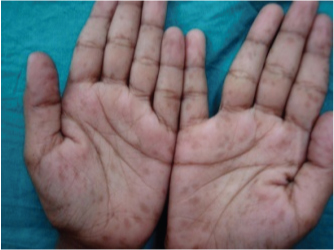
Picture showing palmer freckling in a child.
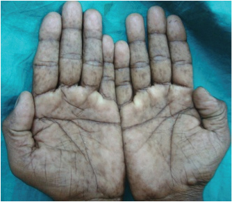
Picture showing palmer freckling in an adult.
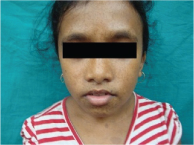
Picture showing facial freckling along with CALM over upper chest.
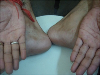
Picture showing both palmer and plantar freckling in an adult.
Depicting the occurrence of freckling according to body sites.
| Freckling over body sites | Occurrence (Percentage) |
|---|
| CALM | 23 (88.46%) |
| Axillary freckling | 18 (69.23%) |
| Inguinal freckling | 18 (69.23%) |
| Either axillary or inguinal freckling | 20 (76.92%) |
| Palmer freckling | 23 (88.46%) |
| Plantar freckling | 5 (19.23%) |
| Facial freckling | 12 (46.15%) |
| Palmer or plantar or facial freckling | 24 (92.31%) |
N = 26 (M=14, F=12)
Showing the details of each patient.
| S. No. | Initials | Religion | Sex | Age | Onset | CALM | Nerofib | Plex | Lisle | Axil | Inguin | Palm | Plan | Facial | Fam | Ass |
|---|
| NF | Nod | Fr | Fr | Fr | Fr | Fr | His | Feat |
|---|
| 1 | DD | H | F | 14Y | 9Y | P | A | A | P | 2 | 2 | 2 | 0 | 2 | A | N |
| 2 | MM | M | M | 42Y | 14Y | A | P | P | P | 2 | 2 | 2 | 0 | 0 | P | H |
| 3 | MB | M | F | 22Y | 12Y | P | P | A | A | 1 | 1 | 1 | 0 | 0 | A | N |
| 4 | JS | M | M | 20Y | 8Y | P | P | A | P | 1 | 1 | 1 | 0 | 0 | P | N |
| 5 | RB | H | F | 25Y | 17Y | P | P | A | A | 1 | 1 | 1 | 0 | 0 | A | N |
| 6 | A | M | M | 16Y | 8Y | P | P | P | A | 1 | 1 | 1 | 0 | 0 | A | H+S |
| 7 | PM | H | F | 20Y | 16Y | P | P | A | A | 0 | 0 | 1 | 0 | 0 | A | N |
| 8 | UN | M | F | 7Y | 4Y | P | A | A | P | 0 | 0 | 1 | 0 | 3 | P | N |
| 9 | MM | H | M | 20Y | 16Y | A | P | A | P | 0 | 0 | 2 | 0 | 0 | A | N |
| 10 | SJ | H | M | 13Y | 2Y | P | P | A | P | 0 | 0 | 1 | 0 | 0 | A | N |
| 11 | BB | H | M | 18Y | 7Y | P | P | P | P | 0 | 0 | 2 | 0 | 0 | P | H |
| 12 | PB | H | F | 16Y | 7Y | P | P | P | P | 2 | 1 | 3 | 1 | 3 | P | H |
| 13 | KB | H | F | 21Y | 7Y | P | P | P | P | 3 | 1 | 2 | 0 | 3 | P | H |
| 14 | SG | H | M | 35Y | 5Y | P | P | A | A | 0 | 0 | 0 | 0 | 0 | A | N |
| 15 | NP | H | M | 46Y | 16Y | P | P | A | A | 1 | 0 | 0 | 0 | 0 | A | N |
| 16 | AY | H | M | 16Y | 9Y | P | P | A | A | 2 | 2 | 2 | 0 | 2 | A | N |
| 17 | NK | M | F | 18Y | 12Y | P | P | A | P | 2 | 0 | 1 | 0 | 2 | A | H |
| 18 | TKM | H | M | 30Y | 16Y | P | P | P | P | 2 | 2 | 2 | 1 | 0 | A | H |
| 19 | AB | M | F | 30Y | 15Y | P | P | A | A | 1 | 1 | 2 | 0 | 1 | P | N |
| 20 | HR | M | M | 19Y | 9Y | P | P | A | P | 1 | 2 | 1 | 2 | 0 | P | H |
| 21 | R | H | M | 18Y | 12Y | P | P | A | A | 2 | 1 | 2 | 1 | 1 | P | H |
| 22 | MB | M | F | 32Y | 12Y | A | P | A | A | 2 | 2 | 2 | 0 | 2 | A | H |
| 23 | AA | M | M | 21Y | 14Y | P | P | A | P | 0 | 2 | 2 | 0 | 0 | A | N |
| 24 | AA | M | M | 24Y | 14Y | P | P | P | A | 0 | 1 | 2 | 1 | 0 | A | N |
| 25 | DB | H | F | 22Y | 16Y | P | P | A | A | 1 | 1 | 1 | 0 | 2 | A | H |
| 26 | RM | H | F | 9Y | 8Y | P | A | A | A | 2 | 1 | 0 | 0 | 2 | A | H |
A: Absent; P: Present; N: Nil; H: Hypertelorism; S: Scoliosis; Y: Year
Nerofib: Neurofibroma; Plex NF: Plexiform NF; Lisle Nod: Lisch nodules; Axil Fr: Axillary freckling; Inguin Fr: Inguinal freckling; Palm Fr: Palmer freckling; Plan Fr: Plantar freckling; Facial Fr: Facial freckling; Fam His: Family history; Ass Feat: Associated features
Discussion
Neurofibromatosis type 1 (von Recklinghausen’s disease) is diagnosed by following criteria laid down by the National Institute of Health Consensus Development Conference Statement, 1988 [7]. Two or more criteria need to be fulfilled for the diagnosis of NF-1:
Six or more CALMs of over 5 mm in greatest diameter in prepubertal individuals and over 15 mm in greatest diameter in post pubertal individuals.
Two or more neurofibromas of any type or one plexiform neurofibroma.
Axillary or inguinal freckling.
Optic glioma.
Two or more iris Lisch nodules.
A distinctive osseous lesion such as sphenoid dysplasia or thinning of the cortex of the long bones with or without pseudoarthrosis.
A first degree relative (parent, sibling, offspring) with NF-1 by above criteria.
Palmer, plantar and facial freckling has been mentioned to be present in patients of NF-1 but has not been included in the diagnostic criteria [4]. In the present study, it was seen that palmer freckling alone is more common than both axillary and inguinal freckling combined.
Freckles usually appear by the age of 4 to 6 years just after the appearance of CALMs but usually before the appearance of neurofibromas and are a useful diagnostic sign in a child suspected of NF-1 [8]. In contrast to the freckles that occurs in fair skinned individuals over the sun exposed sites, freckling in NF-1 is predominantly seen in the sun protected sites [5]. Although freckles are seen in the groins, neck and other areas of the body, the axillae are affected in about 80% of cases [5,9]. The patients in this study noticed the appearance of the cutaneous features at an average of almost 11.24 years of age. Out of the 23 patients with palmer freckling only 7 patients either noticed or were concerned about it. Only 2 patients out of 5 had noticed the plantar freckling. Such patients were advised sun protection and topical retinoids for the same. Freckling can occur in a dermatomal pattern along with neurofibromas and CALMs in segmental NF-1 which was previously called NF-5 [4]. Segmental NF presents with neurofibroma in a segmental distribution of a nerve like trigeminal, thoracic or others and presents with major cosmetic concerns sometimes requiring excision. Three localised CALMs in the right inguinal region along with multiple neurofibromas all over and a plexiform neurofibroma over scalp has been reported [10]. However, none of the patients in the current study had segmental NF-1. There are few studies regarding these pigmentation patterns in NF-1. Yesudian P et al., reported palmer freckling in 84% of patients with NF-1 [11]. Jeevankumar B et al., reported the frequency of CALM in 82.5%, intertriginous freckling in 77.5%, palmer freckling in 90.0% and plantar freckling in 45% [12]. The results of the present study are similar to the previous ones as far as the frequency of palmer freckling is concerned.
However, plantar freckling was seen in only 19.23% of cases in the current study. Although the freckling can extend beyond the intertriginous areas, no treatment is required [13] because it is purely a cosmetic concern to the patients. Sangolli PM reported a solitary case of NF-1 with partial bilateral radial artery stenosis along with axillary and palmer freckling [14].
Among the various criteria of NF-1, axillary freckling (Crowe’s sign) is reported to be present in about 81% of cases ultimately diagnosed as NF-1 [5]. On the other hand, palmer freckling, as reported by various studies, including the present one, ranges from 84% to 90% [8,9]. Similarly, CALM occurs in around 82.5% to 88.46% of patients. Both CALM and intertriginous freckling have been traditionally considered to be the diagnostic markers of NF-1. However, palmer freckling has been reported to occur in similar or higher frequency than these two pigmentary changes. The present study also revealed, in addition, a few cases of NF-1 which showed palmer freckling and lacked inguinal and axillary freckling.
Conclusion
In spite of considerable advances made in the understanding of the pathogenesis, there is limited data as to the causation of the pigmentary changes occurring in NF-1. Although freckling is not of any prognostic significance its presence early in childhood along with CALMs might point to a clinical diagnosis of NF-1 even in the absence of other pathognomic signs of this genodermatosis. Palmer freckling may be more common than CALM and intertriginous freckling in patients with NF-1 as was evident from the current study. It can start appearing gradually, early childhood onwards, just like the other signs. Therefore, it could be considered to be included in the diagnostic criteria since its occurrence is comparable to the other characteristic signs of NF-1. Further, palms and soles are easily accessible sites before the clinician proceeds to examine other areas. However, larger studies may be needed to further evaluate the prevalence of this finding in NF-1.
N = 26 (M=14, F=12)
A: Absent; P: Present; N: Nil; H: Hypertelorism; S: Scoliosis; Y: Year
Nerofib: Neurofibroma; Plex NF: Plexiform NF; Lisle Nod: Lisch nodules; Axil Fr: Axillary freckling; Inguin Fr: Inguinal freckling; Palm Fr: Palmer freckling; Plan Fr: Plantar freckling; Facial Fr: Facial freckling; Fam His: Family history; Ass Feat: Associated features
[1]. Samuelsson B, Axelsson R, Neurofibromatosis. A clinical and genetic study of 96 cases in Gothenberg, SwedenActa Derm Venereol Suppl (Stockh) 1981 95:67-71. [Google Scholar]
[2]. Huson SM, Compston DA, Clark P, Harper PS, A genetic study of von Recklinghausen’s neurofibromatosis in south east Wales.1. Prevalence, fitness, mutation rate, and effect of parental transmission on severityJ Med Genet 1989 26:704-11.10.1136/jmg.26.11.7042511318 [Google Scholar] [CrossRef] [PubMed]
[3]. Ledbetter DH, Rich DC, O’Connell P, Leppert M, Carey JC, Precise localization of NF-1 to 17q11.2 by balanced translocationAm J Hum Genet 1989 44:20-24. [Google Scholar]
[4]. Tsao H, Luo S, Neurofibromatosis and tuberous sclerosis. In: Bolognia JL, Jorizzo JL, Schaffer JV edsDermatology 2012 3rd edElsevier Saunders:925-41. [Google Scholar]
[5]. Listernick R, Charrow J, The neurofibromatosis. In: Goldsmith LA, Katz SI, Gilchrist BA, Paller AS, Leffell DJ, Wolff K edsFitzpatrick’s dermatology in general medicine 2008 8th edNew YorkMcGraw-Hill:1680-90. [Google Scholar]
[6]. Shen MH, Harper PS, Upadhaya M, Molecular genetics of neurofibromatosis type 1(NF 1)J Med Genet 1996 33:2-17.10.1136/jmg.33.1.28825042 [Google Scholar] [CrossRef] [PubMed]
[7]. National Institutes of Health Consensus Development Conference Statement. Neurofi bromatosisArch Neurol 1988 45:575-78.10.1001/archneur.1988.005202901150233128965 [Google Scholar] [CrossRef] [PubMed]
[8]. Korf BR, Diagnostic outcome in children with multiple café au lait spotsPediatrics 1992 90:924-27. [Google Scholar]
[9]. McGaughran JM, Harris DI, Donnai D, Teare D, MacLeod R, Westerbeek R, A clinical study of type 1 neurofibromatosis in north west EnglandJ Med Jenet 1999 36:197-203. [Google Scholar]
[10]. Ibrahim A, Asuku ME, Images in clinical medicine. NeurofibromatosisN Engl J Med 2011 365(21):202010.1056/NEJMicm110385922111720 [Google Scholar] [CrossRef] [PubMed]
[11]. Yesudian P, Premlatha S, Thambiah AS, Palmar melanotic macules. A sign of neurofibromatosisInt J Dermatol 1984 23:468-71.10.1111/ijd.1984.23.7.4686436184 [Google Scholar] [CrossRef] [PubMed]
[12]. Jeevankumar B, Thappa DM, Narasimahan R, Sahai A, Cutaneous manifestations of neurofibromatosis type 1 in south IndiaIndian J Dermatol 2001 46:218-26. [Google Scholar]
[13]. Williams VC, Lucas J, Babcock MA, Gutmann DH, Korf B, Maria BL, Neurofibromatosis Type 1 revisitedPediatrics 2009 123(1):124-33.10.1542/peds.2007-320419117870 [Google Scholar] [CrossRef] [PubMed]
[14]. Sangolli PM, Neurofibromatosis-1 with partial bilateral radial artery stenosisIndian Dermatol Online J 2012 3:51-53.10.4103/2229-5178.9350623130265 [Google Scholar] [CrossRef] [PubMed]