Root Canal Treatment of Three Rooted Maxillary First and Second Premolar using CBCT: A Case Report
Parul Mour Agarwal1, Sonali Taneja2
1 Private Practitioner, Guwahati, Assam, India.
2 Professor and Head, Department of Conservative Dentistry and Endodontics, I.T.S Dental College, Ghaziabad, U.P., India.
NAME, ADDRESS, E-MAIL ID OF THE CORRESPONDING AUTHOR: Dr. Parul Mour Agarwal, Dr. J.P Gynae Clinic, Behind Hotel Maitri Residency, Paltan Bazaar, G.S Road, Guwahati-781008, Assam, India.
E-mail: parulmour@yahoo.in
A discrete knowledge of the innate anatomy of a tooth is imperative for favourable endodontic treatment. Meticulous understanding of tooth morphology, judicious study of angled radiographs, emphasis on access cavity modifications and thorough exploration of the pulpal floor under magnification are important factors that contribute to successful treatment outcome. The present case report outlines the diagnostic approach and endodontic management of maxillary 1st and 2nd premolar with three root canals, each treated with the aid of operating microscope and Cone-Beam Computed Tomography (CBCT) image.
Radiculous molars, Endodontic treatment, Operating microscope
Case Report
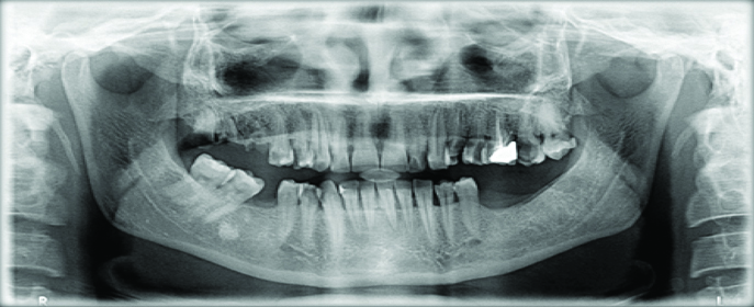
A 40-year-old female patient reported with chief complaint of moderate dull aching, intermittent pain on the right side of her upper jaw for the past one month. Patient’s medical history was non-contributory. Radiographically there was a Class-II disto-occlusal caries involving pulp of tooth no. #14 and mesio-occlusal caries involving pulp of tooth no. #15 with no signs of periapical pathology [Table/Fig-1]. Electronic pulp testing showed a delayed response but the teeth were tender on percussion. Based on these findings, a diagnosis of acute irreversible pulpitis with apical periodontitis was made for both the premolar teeth, necessitating endodontic therapy.
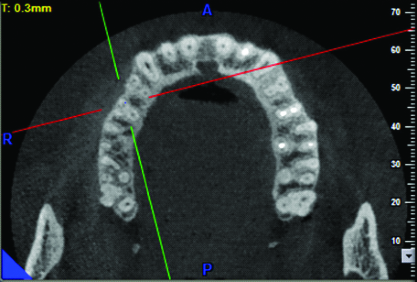
After local anaesthesia administration (Lidocaine 2% with epinephrine 1:80,000) and rubber dam isolation, all carious tissue was excavated and an adequate coronal access was made. Careful exploration of the pulpal floor was done with a DG-16 explorer (Hu-Friedy, Chicago, USA) under operating microscope. Magnification revealed three orifices which were reconfirmed using CBCT [Table/Fig-2]. Working length was determined using apex locator (Root Zx, J Morita, Japan) and confirmed radiographically [Table/Fig-3,4]. Patency was maintained in all the three canals with a 10 K-file (Maillefer, Dentsply, Switzerland).
The CBCT images of teeth #14, #15 showing three canals.
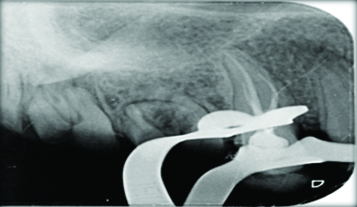
Working length radiograph of #15.
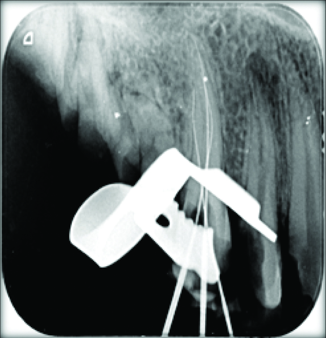
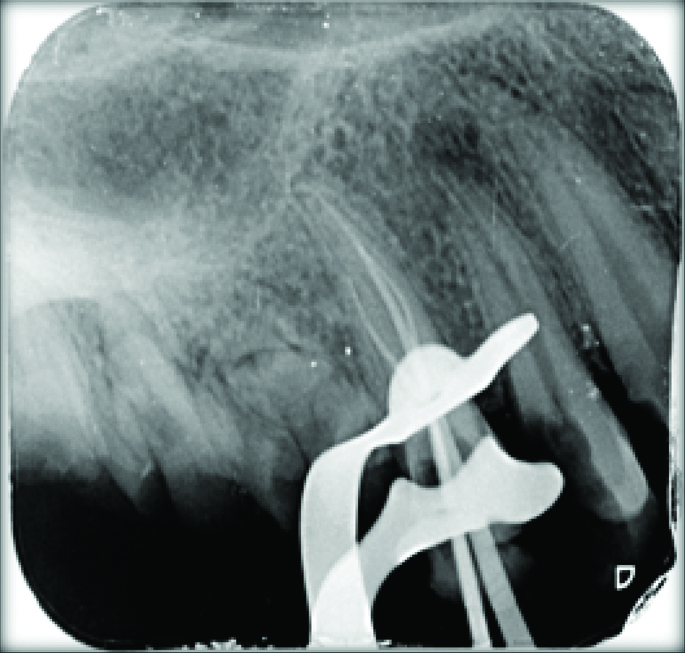
Working length radiograph of tooth #14.
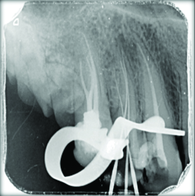
The preparation was completed with Hyflex files (Coltene, UK) using 5.25% NaOCl (Novo Dental Products Pvt., Ltd., India) as the main irrigant during instrumentation, EDTA (Glyde, Maillefer, Dentsply, Switzerland) and subsequently flushed with sterile saline. Canals were dried using paper points and root canal filling was done using gutta percha and AH-Plus sealer (Dentsply, Konstanz, Germany) by cold lateral condensation technique [Table/Fig-56,7 and 8]. The access cavity was then sealed with IRM (Dentsply Caulk, Milford, USA) and restored with composite after seven days. Two months later the patient was reviewed and the teeth were asymptomatic.
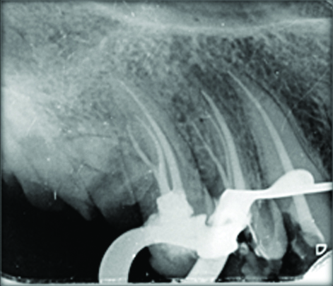
Master cone radiograph of tooth #15.
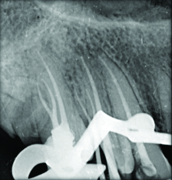
Master cone radiograph of tooth #14.
Discussion
Probable variations in the anatomy of human teeth should be known to assure favourable endodontic treatment. From the previous work to the studies till date, it has been acknowledged that a tooth with a graceful, tapering canal and a single apical foramen is an exception rather than a rule.
Three-rooted maxillary premolars resemble its adjacent maxillary molars, and are referred as “small molars” or “radiculous” The incidence of three-canalled maxillary premolars has been observed as 5-6% for first premolars and 1% for second premolars [1-3]. In a study by Vertucci FJ and Gegauff A, 5% incidence of maxillary first premolars with three canals was reported [1]. Carns and Skidmore reported that out of 100 maxillary premolar tooth, six tooth had three separate canals in three separate roots [2]. Gupta S et al., conducted a study on 250 maxillary first premolar teeth in North Indian population, out of which 0.4% had three canals [4].
The present case report emphasises the importance of correctly determining root canal morphology to enhance endodontic treatment outcomes. Missed root canals and spaces can lead to inadequate elimination of microorganisms which may contribute to failure of therapy.
An accurate preoperative radiograph as well as two radiographs taken at different angles is important for the evaluation of the canal configuration and the presence of any additional roots canals. Indicators of likely aberrant anatomy are unusual location or size of canal openings, indistinct X-ray images and modified coronal access. However, radiograph being two-dimensional image of a three dimensional structure has its inherent limitations and therefore an accurate evaluation of root canal number and morphology can be done using advanced diagnostic methodology like CBCT. The CBCT is a valuable diagnostic tool as it provides a three-dimensional imaging of the root canal system with much reduced patient radiation dose [5,6]. Sudhanva ME et al., and Matus D et al., reported the root canal treatment of three rooted maxillary second premolar using CBCT [7,8].
Till date, the incidence of three rooted maxillary premolar has been illustrated in a few clinical case reports [7,8]. Even then, a clinician should inspect the tooth and identify aberrant anatomy for predictable endodontic treatment.
Conclusion
Endodontists should always consider the possibility of anatomical aberrations before beginning the treatment. The knowledge of such aberrancy as well as thorough clinical and radiographical diagnosis is prerequisite for quality endodontic treatment. Use of advanced diagnostic tools like operating microscope and CBCT may help a clinician in diagnosing and managing teeth with aberrant anatomy.
[1]. Vertucci FJ, Gegauff A, Root canal morphology of the maxillary first premolarJ Am Dent Assoc 1979 99(2):194-98.10.14219/jada.archive.1979.0255287737 [Google Scholar] [CrossRef] [PubMed]
[2]. Carns EJ, Skidmore AE, Configurations and deviations of root canals of maxillary first premolarsOral Surg 1973 36(6):880-86.10.1016/0030-4220(73)90340-X [Google Scholar] [CrossRef]
[3]. Vertucci F, Seelig A, Gillis R, Root canal morphology of the human maxillary second premolarOral Surg 1974 38(3):456-64.10.1016/0030-4220(74)90374-0 [Google Scholar] [CrossRef]
[4]. Gupta S, Sinha DJ, Gowhar O, Tyagi SP, Singh NN, Gupta S, Root and canal morphology of maxillary first premolar teeth in north Indian population using clearing technique: An in vitro studyJ Conserv Dent 2015 18(3):232-36.10.4103/0972-0707.15726026069411 [Google Scholar] [CrossRef] [PubMed]
[5]. Chhabra N, Singbal KP, Chhabra TN, Type I canal configuration in a single rooted maxillary first molar diagnosed with an aid of cone beam computed tomographic technique: A rare case reportJ Conserv Dent 2013 16(4):385-87.10.4103/0972-0707.11434623956547 [Google Scholar] [CrossRef] [PubMed]
[6]. Ngan DC, Kharbanda OP, Geenty JP, Darendeliler MA, Comparison of radiation levels from computed tomography and conventional dental radiographsAust Orthod J 2003 19(2):67-75. [Google Scholar]
[7]. Sudhanva ME, Sisodia M, Nagaraj T, Pasha S, Root canal treatment of three rooted maxillary second premolar using cone beam computed tomography for diagnosis a case reportInternational Journal of Medical and Dental Case Reports 2015 :1-3. [Google Scholar]
[8]. Matus D, Garay I, Betancourt P, Root canal preparation of maxillary second premolar with three roots and three separate root canals: a micro-computed tomography studyBiomedical Research 2017 28(10):4657-59. [Google Scholar]