Introduction
Patients with diabetes mellitus are prone to develop angle closure resulting in glaucoma which is one of the leading causes of blindness.
Aim
The aim of the present study was to find the impact of diabetes mellitus on anterior segment parameters in relation to the duration of the disease and glycaemic control.
Materials and Methods
A total of 100 consecutive diabetic patients aged 40 years and above were included in the present observational cross sectional study. After obtaining informed consent, complete ocular and medical history was taken, followed by clinical evaluation. Relevant laboratory investigations were carried out. Anterior Chamber Depth (ACD) was measured with IOL Master. Central Corneal Thickness (CCT) and Lens Thickness (LT) were measured using A-Scan Biometry and Ultrasound Pachymetry respectively. SAS 9.2 was used for analysis. Microsoft Word and Excel were used to generate graphs, tables etc. Significance was assessed at 5% level of significance.
Results
A total of 100 patient’s eyes were studied. Group analyses in terms of duration of diabetes and glycaemic control were done. Patients with diabetes more than five years duration and poor control of the disease had lower mean ACD (p=0.056 and p<0.0001 respectively), high mean CCT (p=0.0094 and p=0.022 respectively) and high LT (p<0.0001).
Conclusion
The present study showed that diabetic patients with more than five years duration and poor glycaemic control had shallower anterior chamber, thicker central cornea and thicker lens than that of those who had diabetes less than five years duration and good glycaemic control.
Angle closure, Anterior Chamber depth, Central corneal thickness, Glycaemic control, Lens thickness
Introduction
Eye, undisputedly the most important sense organ of human being, is one of the greatest gifts from the almighty to mankind and it is the duty of every health care provider to play every possible role in preventing any threat to the vision. Diabetes Mellitus affects every part of the eye and eventually leads to blindness. It is the leading cause of irreversible blindness throughout the world [1]. Glaucoma is one of the major causes of blindness and diabetic patients have increased risk of developing glaucoma [2]. Glaucoma occurrence is more frequent in around 5% of patients with diabetes as against 2% in the general population [3]. Diabetic patients have 1.6-4.7 times higher risk than non-diabetic patients [4]. Blue Mountains and Beaver Dam Eye studies found participants with diabetes had two times higher risk to have glaucoma [5]. However, studies focused on association between diabetes in Caucasians and open angle glaucoma [5]. Whether diabetes is also a risk factor for Angle Closure Glaucoma (ACG) is less clear, and has not been thoroughly investigated. Studies have shown that diabetes can decrease the ACD [6,7], which can be a predisposing factor for the development of ACG. Probably there have been no population based data available in India. The purpose of the present study was to prove the existence of correlation between anterior segment parameters and diabetes mellitus in relation to the duration of the disease and glycaemic control. Then the, measurement of anterior chamber depth and lens thickness can be used as a screening tool for detecting angle closure glaucoma in diabetic patients.
Materials and Methods
The observational cross-sectional study was performed in accordance to the tenets of the Declaration of Helsinki and cleared by the local Ethics Committee. A total of 100 eyes of 100 Consecutive diabetic patients aged 40 years and above who attended the Department of Ophthalmology were included in this observational cross-sectional study. Around 1000 diabetic patients are referred to the Department of Ophthalmology in six months; the sample size calculated was 96 for a 95% confidence level. We included 100 consecutive patients reported during January 2016 to June 2016.
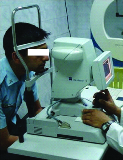
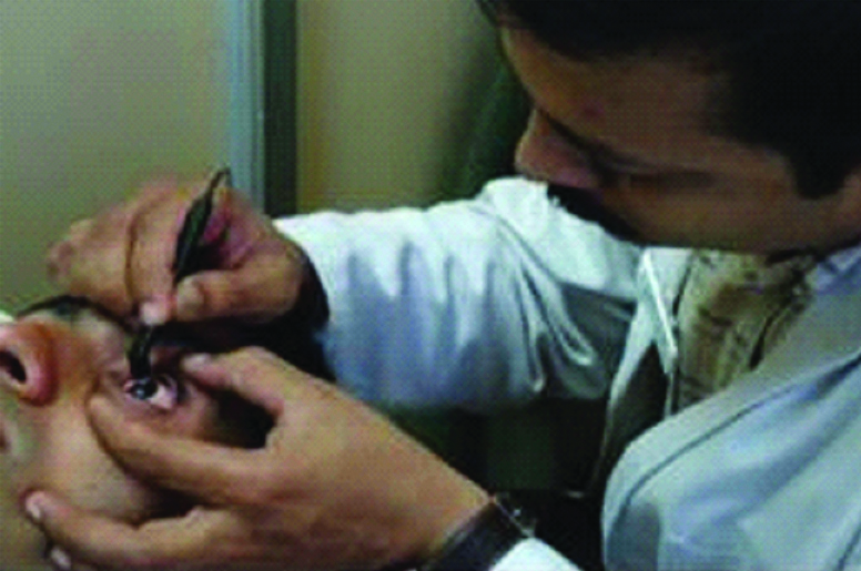
Exclusion criteria included age less than 40 years, intraocular or corneal surgery, corneal diseases, uveitis, congenital anomalies of eye, extreme degrees of refractive error and glaucoma. After a written informed consent all the eligible patients underwent complete ophthalmic evaluation including visual acuity, refraction, external eye examination, and anterior segment evaluation by slit lamp, intraocular pressure measurement by Goldmann applanation tonometer, gonioscopy and posterior segment examination. The ACD was measured using partial coherence laser interferometry (IOL Master) [Table/Fig-1]. The CCT was measured using ultrasound pachymetry. The LT was measured using A-scan biometry [Table/Fig-2]. All measurements were performed by single trained personnel. Average of three readings was taken for analysis. For the sake of uniformity, each eye was measured after the administration of 1.0% cyclopentolate and 5% phenylephrine eye drops to obtain maximal pupillary dilation and paralysis of accommodation.
Measurement of anterior chamber depth.
Measurement of central corneal thickness and lens thickness.
Patients were grouped into different categories based on the following criteria:
Duration of Diabetes:
Less than five years.
More than five years.
Glycaemic control:
Controlled: HbA1c <7.0%.
Poorly controlled: HbA1c >7.0%
Statistical Analysis
The statistical spreadsheet software program namely SAS 9.2 was used for the analysis of the data and Microsoft Word and Excel were used to generate graphs, tables etc. Descriptive statistical analysis has been carried out in the present study. Results on continuous measurements are presented on Mean±SD. Student’s t-test was applied to find the statistical significance of study parameters between group differences.
Significance is assessed at 5% level of significance.
Significant figures:
Suggestive significance: p-value 0.05<p<0.10
Moderately significant: p-value 0.01<p
Strongly significant: p-value p<0.01
Results
A total of 100 right eyes of 100 patients were studied. Patients were predominantly males and most of them had diabetes less than five years duration [Table/Fig-3]. Group analyses in terms of duration of testdiabetes and glycaemic control were done. The mean age of the patients was 49.81±8.34.
Distribution of patients.
| Male | Female | According to Duration of Diabetes | According to Control of Diabetes |
|---|
| | | More than 5 years | Less than 5 years | HbA1c >7.0% | HbA1c <7.0% |
|---|
| No. of patients | 78 | 22 | 24 | 76 | 56 | 44 |
The mean ACD of patients with diabetes more than five years was less when compared to the mean ACD of patients with less than five years duration which was statistically significant (p=0.056). Mean CCT of patients with diabetes more than five years duration was 550±24.65 μm as against 537±29.41 μm of patients with less than five years duration which was statistically significant (p=0.0094). Mean LT of patients with diabetes more than five years duration was more when compared to the mean LT of patients with less than five years duration which was statistically significant (p<0.0001) [Table/Fig-4].
Impact of diabetes on anterior segment parameters.
| Diabetes Duration | n | ACD | p-value | CCT | p-value | LT | p-value |
|---|
| Mean (mm) | SD | Mean (μm) | SD | Mean (mm) | SD |
|---|
| Less Than 5 Yrs | 76 | 2.74 | 0.33 | 0.0561 | 537 | 29.41 | 0.0094 | 4.05 | 0.38 | <0.0001 |
| More Than 5 Yrs | 24 | 2.59 | 0.42 | 550 | 24.65 | 4.76 | 0.50 |
ACD: Anterior chamber depth; CCT: Central corneal thickness; LT: Lens thickness; SD: Standard deviation; p-value: Probablility value
Test used: Student t-testdiabetes
In patients with controlled diabetes the mean ACD was more than that of patients with poorly controlled diabetes which was statistically significant (p<0.0001). Poorly controlled diabetic patients had thicker central corneas (543±27.69 μm) than well controlled patients (537±30.01 μm) which was statistically significant (p=0.022). Poorly controlled diabetic patients had thicker lens (4.27±0.60 mm) than well controlled patients (4.15±0.35 mm) which was statistically significant (p<0.0001) [Table/Fig-5]. The present study showed that diabetic patients with more than five years duration and poor glycaemic control had shallower anterior chamber, thicker central cornea and thicker lens than that of those who had diabetes less than five years duration and good glycaemic control.
Anterior segment parameters according to control of diabetes.
| Diabetes Duration | n | ACD | p-value | CCT | p-value | LT | p-value |
|---|
| Mean (mm) | SD | Mean (μm) | SD | Mean (mm) | SD |
|---|
| Controlled (HbA1c<7.0%) | 44 | 2.96 | 0.21 | <0.0001 | 537 | 30.01 | 0.0225 | 4.15 | 0.35 | <0.0001 |
| Poorly controlled (HbA1c>7.0%) | 56 | 2.50 | 0.33 | 543 | 27.69 | 4.27 | 0.60 |
HbA1c: Glycated haemoglobin; ACD: Anterior chamber depth; CCT: Central corneal thickness; LT: Lens thickness; SD: Standard deviation; p-value: Probablility value
Test used: Student t-test
Discussion
Diabetes mellitus is associated with increased corneal thickening and enhanced endothelial cell permeability [8]. Covalent cross-linking bonds caused by accumulation of end products of advanced Maillard reaction is thought to result in corneal biomechanical changes and thickening of cornea [8]. Diabetes Mellitus affects corneal biomechanics and various biometric parameters of the eye [9,10].
Studies have found that diabetic patients have shallower anterior chamber, thicker central cornea and lens [6,7]. Though, in diabetic patients, acute hyperglycaemic status does not cause any significant changes in anterior chamber parameters, but when compared to non diabetic patients they are found to have significant changes in ocular biometrics [11]. Saw SM et al., in the Tanjong Pagar Survey stated that Singapore Chinese population with diabetes had shallower anterior chambers and thicker lenses than those without diabetes [6]. In a study which involved 150 canine eyes, ACD was significantly reduced in eyes with diabetic cataracts compared with eyes with non cataractous eyes (p<0.05) but not in other eyes [12].
Increasing LT and decreasing ACD with increasing duration of diabetes have been confirmed in a population based twin study [13]. In Insulin dependent diabetes mellitus patients, statistically significant positive correlation between the duration of diabetes and LT and a negative correlation between the duration and ACD were found among all zygosity groups [13]. Probably, there have been no Indian studies done on the impact of diabetes with respect to duration of diabetes and glycaemic control.
Many studies have found that central corneas of diabetic patients were thicker when compared to those of non-diabetic patients [14-18]. Ozdamar Y et al., found no significance in central corneal thickness in respect to the level of glycosylated haemoglobin and disease duration [19]. However, Lee JS et al., had found that the central corneal thickness was significantly correlated with diabetic duration. Patients with diabetic duration of over 10 years had more corneal morphological abnormalities when compared with the normal subjects. The mean corneal thickness was significantly higher in diabetic patients of over 10 years duration than for those with diabetes of less than 10 years duration [20]. The present study also found strong significance in CCT in respect to disease duration (p=0.0094) and glycaemic control (p=0.0225).
Limitation
In view of the prevalence of diabetes, it would have been better if the sample size was higher. We had calculated the sample size based on the number of diabetic patients visiting the department of ophthalmology.
Conclusion
The present study has found statistically significant difference in the ACD, CCT and LT of diabetic patients in respect to duration of disease and glycaemic control. Though, the duration of diabetes cannot be reversed, the importance of glycaemic control has to be reiterated. Since, shallower anterior chamber can be a predisposing factor for developing angle closure glaucoma, measurement of anterior segment parameters such as ACD, CCT and LT can be used as screening tool for diabetic patients particularly in those who have poor glycaemic control and longer duration of disease.
ACD: Anterior chamber depth; CCT: Central corneal thickness; LT: Lens thickness; SD: Standard deviation; p-value: Probablility valueTest used: Student t-testdiabetes
HbA1c: Glycated haemoglobin; ACD: Anterior chamber depth; CCT: Central corneal thickness; LT: Lens thickness; SD: Standard deviation; p-value: Probablility valueTest used: Student t-test
[1]. Kaji Y, Prevention of diabetic keratopathyBr J Ophthalmol 2005 89:254-55.10.1136/bjo.2004.05554115722297 [Google Scholar] [CrossRef] [PubMed]
[2]. Zhao YX, Chen XW, Diabetes and risk of glaucoma: systematic review and a Metaanalysis of prospective cohort studiesInt J Ophthalmol 2017 10(9):1430-35. [Google Scholar]
[3]. Bernth-Petersen P, Bach E, Epidemiologic aspects of cataract surgery. III. Frequencies of diabetes and glaucoma in a cataract populationActa Ophthalmol 1983 61(3):406-16.10.1111/j.1755-3768.1983.tb01439.x6624407 [Google Scholar] [CrossRef] [PubMed]
[4]. Reynolds DC, Relative risk factors in chronic open-angle glaucoma: an epidemiological studyAm J Optom Physiol Opt 1977 54(2):116-20.10.1097/00006324-197702000-00010326056 [Google Scholar] [CrossRef] [PubMed]
[5]. Mitchell P, Smith W, Chey T, Healey PR, Open-angle glaucoma and diabetes: The Blue Mountains Eye Study, AustraliaOphthalmology 1997 104(4):712-18.10.1016/S0161-6420(97)30247-4 [Google Scholar] [CrossRef]
[6]. Saw SM, Wong TT, Ting S, Foong AW, Foster PJ, The relationship between anterior chamber depth and the presence of diabetes in the Tanjong Pagar surveyAm J Ophthalmol 2007 144(1):325-26.10.1016/j.ajo.2007.03.03817659975 [Google Scholar] [CrossRef] [PubMed]
[7]. Kocatürk T, Zengin MÖ, Cakmak H, Evliçoglu GE, Dündar SO, Omürlü IK, The ocular biometric differences of diabetic patientsEur J Ophthalmol 2014 24(5):786-89.10.5301/ejo.500044624557759 [Google Scholar] [CrossRef] [PubMed]
[8]. Sady C, Khosrof S, Nagaraj R, Advanced Maillard reaction and cross linking of corneal collagen in diabetesBiochem Biophys Res Commun 1995 214(3):793-97.10.1006/bbrc.1995.23567575546 [Google Scholar] [CrossRef] [PubMed]
[9]. Bao F, Deng M, Zheng X, Li L, Zhao Y, Cao S, Effects of diabetes mellitus on biomechanical properties of the rabbit corneaExp Eye Res 2017 161:82-88.10.1016/j.exer.2017.05.01528603017 [Google Scholar] [CrossRef] [PubMed]
[10]. Sahin A, Bayer A, Ozge G, Mumcuoğlu T, Corneal biomechanical changes in diabetes mellitus and their influence on intraocular pressure measurementInvest Ophthalmol Vis Sci 2009 50(10):4597-604.10.1167/iovs.08-276319443722 [Google Scholar] [CrossRef] [PubMed]
[11]. Tai MC, Lin SY, Chen JT, Liang CM, Chou PI, Lu DW, Sweet hyperopia: refractive changes in acute hyperglycemiaEur J Ophthalmol 2006 16(5):663-66.10.1177/11206721060160050117061215 [Google Scholar] [CrossRef] [PubMed]
[12]. Williams DL, Lens morphometry determined by B-mode ultrasonography of the normal and cataractous canine lensVeterinary Ophthalmol 2004 7(2):91-95.10.1111/j.1463-5224.2004.04005.x14982588 [Google Scholar] [CrossRef] [PubMed]
[13]. Logstrup N, Sjolie AK, Kyvik KO, Green A, Long-term influence of insulin dependent diabetes mellitus on refraction and its components: a population based twin studyBr J Ophthalmol 1997 8(5):343-49.10.1136/bjo.81.5.3439227196 [Google Scholar] [CrossRef] [PubMed]
[14]. Herse PR, Corneal hydration control in normal and alloxan induced diabetic rabbitsInvest Ophthalmol Vis Sci 1990 31:2205-13. [Google Scholar]
[15]. Meyer LA, Ubels JL, Edelhauser HF, Corneal endothelial morphology in the ratInvest Ophthalmol Vis Sci 1988 29:940-48. [Google Scholar]
[16]. Yee RW, Matsuda M, Kern TS, Engerman RL, Edelhauser HF, Corneal endothelial changes in diabetic dogsCurr Eye Res 1985 4(7):759-66.10.3109/027136885090200314028800 [Google Scholar] [CrossRef] [PubMed]
[17]. Sanchis-Gimeno JA, Alonso L, Rahhal M, Bastir M, Perez-Bermejo M, Belda-Salmeron L, Corneal thickness differences between type 2 diabetes and nondiabetes subjects during preoperative laser surgery examinationJ Diabetes Complications 2017 31(1):209-12.10.1016/j.jdiacomp.2016.08.02427623389 [Google Scholar] [CrossRef] [PubMed]
[18]. El-Agamy A, Alsubaie S, Corneal endothelium and central corneal thickness changes in type 2 diabetes mellitusClin Ophthalmol. 2017 11:481-66.10.2147/OPTH.S12621728280298 [Google Scholar] [CrossRef] [PubMed]
[19]. Ozdamar Y, Cankaya B, Ozalp S, Acaroglu G, Karakaya J, Ozkan SS, Is there a correlation between diabetes mellitus and central corneal thickness?J Glaucoma 2010 19(9):613-16.10.1097/IJG.0b013e3181ca7c6220051882 [Google Scholar] [CrossRef] [PubMed]
[20]. Lee JS, Oum BS, Choi HY, Lee JE, Cho BM, Differences in corneal thickness and corneal endothelium related to duration in DiabetesEye 2006 20(3):315-18.10.1038/sj.eye.670186815832184 [Google Scholar] [CrossRef] [PubMed]