Introduction
Multislice Computed Tomography (MCT) is the main radiographic examination to evaluate mastoid air cell system. Hounsfield Unit density (HU), determined by MCT is useful to evaluate mastoid pneumatization, but HU values in different genders, right/left mastoid sides, as well as their association with age have not been studied yet.
Aim
To evaluate the difference in Hounsfield density values between genders as well as between right and left sides, and also to correlate HU with age.
Materials and Methods
A total of 102 skull MCT examinations that included mastoid process of temporal bone were evaluated (47 from male and 55 from female patients). The HU was measured at mastoid cavity applying exclusively axial slices in a standardized region of interest. All statistical analyses were performed at a level of significance of 5%, using IBM SPSS Statistics 17, SPSS®, Inc, Chicago, IL. Linear regression was used to determine the relationship between age and HU; Non-parametric tests were performed to evaluate differences between right and left sides, as well as between genders.
Results
No statistical significant differences was observed between left and right sides HU values (p-value=0.676) nor between male and female (p-value=0.155), according to Mann-Whitney test. Age was not correlated with HU values (p-value=0.06).
Conclusion
Within the limitations of the present investigation, we concluded that the Mastoid Air Cells System (MACS) HU values do not vary among male and female individuals and left-right sides. Age is advocated to be associated with the volume of mastoid pneumatization, but it may not be related to differences in HU values.
Introduction
The mastoid process is a posterior bone projection of temporal bone, which contains a large number of tiny cavities named as “mastoid cells” that are markedly diverse in number and volume [1]. The grouped mastoid cells constitute the MACS, which include the posterior extension of the Middle Ear (ME), continuously to the air-phase with the tympanum. MACS represents one of the most important structures to the functional balance of the ME [2].
The functional role of the MACS is not well established [2,3]. It is accepted that this highly cellular temporal bone area is an air reservoir for the ME [2] and probably protects the sensitive inner ear structures from external temperature changes, as well as regulates pressure [3] by the impact of the considerable surface area in accordance with gas exchange [4-6].
Mastoid pneumatization is a combination of two different processes [1]: the process of the epithelial growth from the Eustachian tube into the middle ear with the disappearance of the space-keeping mesenchyme [7]; and the process of a preformation of the bone spaces around the mastoid antrum [8]. Moreover, MACS volume and pneumatization is inversely related to the frequency of ME chronic pathological conditions, such as otitis media [9] and cholesteatoma [10].
Complete MACS development is at nearly 10-15 years of age [11] and its pneumatization is faster in earlier second decade and slower until the third decade of life [2]. A gradual reduction in the mastoid pneumatization degree proceeds until the seventh decade [2].
Imaging methods can provide great assess to mastoid process [12] and computed tomography is the main radiographic examination to evaluate mastoid pneumatization [13]. The HU density is an essential feature of MCT imaging technique [14]. HU is the numeric translation result of the linear attenuation coefficient and it is usually applied to biological tissues [14]. Each biological tissue has its particular HU [14], with particular HU ranges. Various advances have notably simplified the clinical application of HU, and due to this fact, it is applied to the study of anatomical areas with difficult approach.
There are many papers regarding the radiological measurement of the mastoid pneumatization volume [11]. Nevertheless, although there was developed a particular algorithm for the measurement of volume and surface MACS area, using -200HU as a threshold [15], no previous study investigated HU in MACS, neither considered HU differences between gender or its association with age. Thus, the aim of the present study was to evaluate the difference in Hounsfield density values between genders as well as between right and left sides and also to correlate HU with age.
Materials and Methods
This retrospective study was conducted in Department of Stomatology, School of Dentistry, University of São Paulo (Brazil) and has initiated in October 2014 and ended in July 2017. The Ethics Committee approval was previously obtained (number: 835.694). Sample size was determined according to the number of skull MCT examinations available in the research time period (convenience sample).
A total of 102 skull MCT examinations that included mastoid process of temporal bone were analysed (47 from males and 55 from females patients). Patient data (age, gender) were recorded. Patients with history of chronic pathological conditions affecting ME or MACS were excluded.
Non contrast enhanced, high resolution MCT with 16 slices (Toshiba Activion, Medical Systems Corporation, Japan) was used for MCT imaging of temporal bone. Acquisition imaging parameters was: 0.5 mm slice thickness; 1.0 mm spacing between slices; 250 mm field of view; 120 kV peak and 250 mA.
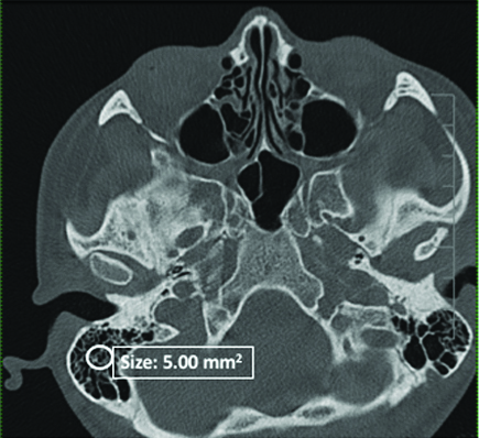
All the images were assessed on Osirix 64. The HU was measured at mastoid cavity applying exclusively axial slices. Region of Interest (ROI) size was standardized to 5.00 mm2. The ROI circle was placed at mastoid antrum as centrally as possible. Mean HU values for each determined ROI were recorded and statistical analysis were applied. [Table/Fig-1] shows an example of the data evaluation according to the specific ROI.
Example of mastoid area evaluation according to specific ROI. Axial slice.
Statistical Analysis
All statistical analyses were performed at a level of significance of 5%, using IBM SPSS Statistics 17, SPSS®, Inc, Chicago, IL. Mean HU values normality was assessed using Shapiro-Wilk test. Linear regression was used to determine the relationship between age and HU values.
Results
Two hundred and four mastoid process images were assessed from 102 skull CT examinations. Minimum age of the patients was 9 years; maximum age was 83. The number of MCT skulls examinations according to the gender of the patients, age and median HUs from left and right sides are available in [Table/Fig-2].
Number of skull multislice computed tomography examinations according to gender (male and female), Age (median, maximum, minimum), Median HU from left and right sides.
| Sex | Number of patients | Agea | Median HU left side | Median HU right side |
|---|
| Median | Maximum | Minimum |
|---|
| Male | 47 | 59.5 (IR26.25) | 83 | 9 | 397.785 (IR282.940) | 510.520 (IR308.018) |
| Female | 55 | 51.0 (IR16.25) | 73 | 21 | 414.350 (IR247.540) | 377.635 (IR655.385) |
a: Age in years, Abbreviations: IR: Interquartile Range; HU: Hounsfield Unit
Mann-Whitney test showed no statistical significant difference between the left and right sides HU values for the total sample, which included male and female MCT examinations (p-value=0.676). Additionally, no statistical significant difference was detected between male and female HU values, according to the same non-parametric test for independent groups (p-value=0.155). The linear regression analysis did not show statistical significance between age and HU values (p-value=0.06) [Table/Fig-3].
Statistical assessment results.
| Comparsion group | p-value |
|---|
| Left versus right side HU differencea | 0.676 |
| Male versus female HU differencea | 0.155 |
| Correlation between age and HU valuesb | p=0.06 |
aAccoding to Mann-Whitney test, significant if p-value <0.05
bLinear regression analysis, significant if p-value <0.05
Abbreviations: HU: Housfield Unit
Discussion
In this study, we described the median HU values of MACS of left and right side, considering male and female MCT examinations. Moreover, we observed that HU values did not differ among males and females, nor between the left and right sides. Finally, we noticed that HU values are not associated with the age of the individuals also.
Changes in bone density and hard tissues that may indicate the presence of any pathological alteration, identified in radiographic examinations like MCT, should be detected as early as possible. The diagnosis of density chances in two-dimensional radiographic techniques is based on darkness and brightness of images and its gray scale [16]. In MCT, the gray scale is translated by the Hounsfield scale, which is proportional to X-ray attenuation degree and it is assigned to each pixel in the image to express the particular tissue density [16]. Hounsfield scale provides HU values that can be accurately measured in MCT examinations [17].
Many investigators tried to measure the extent of mastoid pneumatization by many methods like water-weight, acoustic, pressurized transducer method [4] with an aim to find differences between individuals. Although the mastoid cells are all interconnected, the assessment of the MACS pneumatization degree is a complex task to be performed directly. Not withstanding, a number of radiological tools are able to measure mastoid pneumatization [11,18,19]. HU values can be easily analysed when an MCT is performed. To our knowledge, this is the first study focused exclusively on differences among HU values to the aforementioned criteria. As we have not found any other investigation that considered HU deeply in the results, we compared our results with studies using different methodologies but explained analogous points.
Considering the MACS left-right, our results found no HU differences between the sides, which may show us the symmetry among MACS sides. This result is in concordance with investigations that used other methodologies, but aimed to verify the mastoid process geometric symmetry [4,20-22]. Also, HU differences between genders were not statistically significant in our study which is in concordance with other studies which have demonstrated the absence of differences between male and female individuals [4,22,23].
Surprisingly, we have not established the relationship between age and HU values. A preceding research considering three-dimensional reconstruction of CT images has demonstrated that MACS pneumatization changes across lifespan, which is not in agreement with our results [4]. Nevertheless, our results are in concordance with another study when volume was measured in MCT. Kim J et al., observed no changes in the volume of MACS with ageing; no statistical differences between left and right MACS as well as no differences between genders [22].
These findings resemble the inheritance theory that states that the mastoid pneumatization degree is genetically regulated, leading to higher levels of sides symmetry and supporting a genetic component to these measures and also to the HU values [21].
Limitation
The limitations of the present study were the small sample size and its retrospective design. Further prospective and larger sample investigations may help to clarify our findings.
Conclusion
Within the limitations of the present investigation, we concluded that MACS HU values do not vary among male and female individuals and left-right sides. Age is advocated to be associated with the volume of mastoid pneumatization, but it may not be related to differences in HU values.
a: Age in years, Abbreviations: IR: Interquartile Range; HU: Hounsfield Unit
aAccoding to Mann-Whitney test, significant if p-value <0.05
bLinear regression analysis, significant if p-value <0.05Abbreviations: HU: Housfield Unit
[1]. Kozerska M, Skrzat J, Szczepanek A, Application of the temporal bone for sex determination from the skeletal remainsFolia Med Cracov 2015 55(2):33-39. [Google Scholar]
[2]. Yegin Y, Çelik M, Šimşek BM, Olgun B, Karahasanoğlu A, Çolak C, Impact of the Degree of the Mastoid Pneumatization on Cartilage Type 1 Tympanoplasty SuccessJ Craniofac Surg 2016 27(7):e695-e98.10.1097/SCS.000000000000302227564066 [Google Scholar] [CrossRef] [PubMed]
[3]. Doyle WJ, The mastoid as a functional rate-limiter of middle ear pressure changeInt J Pediatr Otorhinolaryngol 2007 71(3):393-402.10.1016/j.ijporl.2006.11.00417174408 [Google Scholar] [CrossRef] [PubMed]
[4]. Lee DH, Jun BC, Kim DG, Jung MK, Yeo SW, Volume variation of mastoid pneumatization in different age groups: a study by three-dimensional reconstruction based on computed tomography imagesSurg Radiol Anat 2005 27(1):37-42.10.1007/s00276-004-0274-715349696 [Google Scholar] [CrossRef] [PubMed]
[5]. Sadé J, Hyperectasis: the hyperinflated tympanic membrane: the middle ear as an actively controlled systemOtol Neurotol 2001 22(2):133-39.10.1097/00129492-200103000-0000311300258 [Google Scholar] [CrossRef] [PubMed]
[6]. Gaihede M, Dirckx JJ, Jacobsen H, Aernouts J, Søvsø M, Tveterås K, Middle ear pressure regulation--complementary active actions of the mastoid and the Eustachian tubeOtol Neurotol 2010 31(4):603-11.10.1097/MAO.0b013e3181dd13e220393372 [Google Scholar] [CrossRef] [PubMed]
[7]. Clark JA, Kelly WM, Common artifacts encountered in magnetic resonance imagingRadiol Clin North Am 1988 26(5):893-920. [Google Scholar]
[8]. Piza J, Northrop C, Eavey RD, Embryonic middle ear mesenchyme disappears by redistributionLaryngoscope 1998 108(9):1378-81.10.1097/00005537-199809000-000239738761 [Google Scholar] [CrossRef] [PubMed]
[9]. Hill CA, Ontogenetic change in temporal bone pneumatization in humansAnat Rec (Hoboken) 2011 294(7):1103-15.10.1002/ar.2140421618436 [Google Scholar] [CrossRef] [PubMed]
[10]. Valtonen HJ, Dietz A, Qvarnberg YH, Nuutinen J, Development of mastoid air cell system in children treated with ventilation tubes for early-onset otitis media: a prospective radiographic 5-year follow-up studyLaryngoscope 2005 115(2):268-73.10.1097/01.mlg.0000154731.08410.b815689748 [Google Scholar] [CrossRef] [PubMed]
[11]. Koç A, Ekinci G, Bilgili AM, Akpinar IN, Yakut H, Han T, Evaluation of the mastoid air cell system by high resolution computed tomography: three-dimensional multiplanar volume rendering techniqueJ Laryngol Otol 2003 117(8):595-98.10.1258/00222150376819990612956911 [Google Scholar] [CrossRef] [PubMed]
[12]. Lima MA, Farage L, Cury MC, Bahmad Júnior F, Mastoid surface area-to-volume ratios in adult Brazilian individualsBraz J Otorhinolaryngol 2013 79(4):446-53.10.5935/1808-8694.2013008023929144 [Google Scholar] [CrossRef] [PubMed]
[13]. Park MS, Yoo SH, Lee DH, Measurement of surface area in human mastoid air cell systemJ Laryngol Otol 2000 114(2):93-96.10.1258/002221500190496910748822 [Google Scholar] [CrossRef] [PubMed]
[14]. Brooks RA, A quantitative theory of the Hounsfield unit and its application to dual energy scanningJ Comput Assist Tomogr 1977 1(4):487-93.10.1097/00004728-197710000-00016615229 [Google Scholar] [CrossRef] [PubMed]
[15]. Byun SW, Lee SS, Park JY, Yoo JH, Normal mastoid air cell system geometry: has surface area been overestimated?Clin Exp Otorhinolaryngo 2016 9(1):27-32.10.21053/ceo.2016.9.1.2726976023 [Google Scholar] [CrossRef] [PubMed]
[16]. Razi T, Niknami M, Alavi Ghazani F, Relationship between Hounsfield Unit in CT Scan and Gray Scale in CBCTJ Dent Res Dent Clin Dent Prospects 2014 8(2):107-10. [Google Scholar]
[17]. Patrick S, Birur NP, Gurushanth K, Raghavan AS, Gurudath S, Comparison of gray values of cone-beam computed tomography with hounsfield units of multislice computed tomography: An in vitro studyIndian J Dent Res 2017 28(1):66-70.10.4103/ijdr.IJDR_415_1628393820 [Google Scholar] [CrossRef] [PubMed]
[18]. Luntz M, Malatskey S, Tan M, Bar-Meir E, Ruimi D, Volume of mastoid pneumatization: three-dimensional reconstruction with ultrahigh-resolution computed tomographyAnn Otol Rhinol Laryngol 2001 110(5 Pt 1):486-90.10.1177/00034894011100051611372935 [Google Scholar] [CrossRef] [PubMed]
[19]. Vrabec JT, Champion SW, Gomez JD, Johnson RF, Chaljub G, 3D CT imaging method for measuring temporal bone aerationActa Otolaryngol 2002 122(8):831-35.10.1080/003655402100002808512542201 [Google Scholar] [CrossRef] [PubMed]
[20]. Swarts JD, Doyle BM, Doyle WJ, Relationship between surface area and volume of the mastoid air cell system in adult humansJ Laryngol Otol 2011 125(6):580-84.10.1017/S002221511000281121208489 [Google Scholar] [CrossRef] [PubMed]
[21]. Swarts JD, Foley S, Alper CM, Doyle WJ, Mastoid geometry in a cross-section of humans from infancy through early adulthood with a confirmed history of otitis mediaInt J Pediatr Otorhinolaryngol 2012 76(1):137-41.10.1016/j.ijporl.2011.10.02122119147 [Google Scholar] [CrossRef] [PubMed]
[22]. Kim J, Song SW, Cho JH, Chang KH, Jun BC, Comparative study of the pneumatization of the mastoid air cells and paranasal sinuses using threedimensional reconstruction of computed tomography scansSurg Radiol Anat 2010 32(6):593-99.10.1007/s00276-009-0618-420047049 [Google Scholar] [CrossRef] [PubMed]
[23]. Kaymakçı M, Yanık B, Erel F, Bayar Muluk N, Cingi C, Association between atopy, mastoid pneumatization and tympanometric findingsEur Arch Otorhinolaryngol 2015 272(1):15-21.10.1007/s00405-014-3006-624647495 [Google Scholar] [CrossRef] [PubMed]