In the last few decades, rapid industrialisation and the fast pace of life have brought both comforts and catastrophe like road traffic accidents and crippling many lives [1]. The incidence of distal femoral fractures is approximately 37 per 1,00,000 person per year [2]. Distal femur fracture accounts to 6-7% of the fractures of femur [2]. In India, distal femur fractures are quite common and leads to loss of manpower in rapid growing nation and due to paucity in indian literature in comparing nailing versus plating for distal femur fractures. The present study will help to decide the better surgical option available. Most of the surgeons agree that distal femur fractures need to be treated operatively to achieve optimal patient outcomes [3]. Accounting for more than 85% of distal femur fractures, the incidence is expected to raise with the increasing age of mortality [4]. Fixation of intra-articular fractures is a challenge even to most experienced surgeons. Earlier, the treatment options for these fractures were ranging from above knee amputation to closed reduction with cast application for all reducible fractures [5,6]. Operative fixation was avoided due to increased chances of implant failure, non-union and mal-union [6]. However, with evolution of implants and fixation, the modes of treatment have changed to Locked plating, Intramedullary nailing or Total knee replacement with angled blade plates [7,8].
Fixation of these fractures are very essential for proper functioning of the knee. The quality of life can be severely affected following the surgery. A retrograde intramedullary nail aligns the femoral shaft with condyles, reducing the tendency to place varus movement at the fracture site and has the advantage of preservation of fracture haematoma, decreased blood loss, minimal soft tissue dissection, less operative time and reduced rate of infection [9]. The locked compression plate is a single beam construct where the strength of its fixation is equal to the sum of all screw-bone interfaces rather than a single screws axial stiffness or pullout resistance as seen in unlocked plates [10].
There has been a paucity in literature about comparison between the better fixation for distal femur fracture leading to confusion. In the present study, we compared the results of locked compression plating and retrograde nailing which we evaluated in terms of range of movements, early mobilisation, fracture union and full weight bearing.
Materials and Methods
The present prospective study was conducted in KLES Dr. Prabhakar Kore Hospital and Medical Research Centre, Belagavi, Karnataka, India, (July 2015 to June 2017). Institutional Ethical Clearance for the study was obtained.
Inclusion Criteria
We compared prospectively 40 patients who underwent Retrograde nailing (20) or Locked compression plating (20) for intra-articular and extra-articular distal femur fractures with age group 18 years and above. Patients with Grade 1 and 2 fractures (Gustillo-Anderson classification), who were medically fit, both males and females were included in the study.
Exclusion Criteria
Exclusion Criteria included Grade 3 fractures (Gustillo-Anderson classification), children <18 years of age, patients having pathological fractures, who were not fit for surgery, was not willing for surgery, lost in follow up and distal femoral fractures with neurovascular compromise.
A total of 40 patients were randomly allocated in two groups of Retrograde nailing (20) and Locked compression plating (20). All patients were followed up at 6th week, 3 months, 6 months and 1 year postoperatively and thereafter till fracture union was noted. Each subject filled a questionnaire, providing details regarding demographic details, medical history and nutritional status, at the baseline evaluation during each follow up.
All the patients were assessed using Modified Neer’s criteria which assigns points for pain, function, working ability, joint movements, gross and radiological appearance [11].
Supracondylar nail system made up of 316L stainless steel was used for retrograde nailing. The nails were available with outer diameter of 10, 11 and 12 mm and length of 200, 250, 300 mm. The distal end was expanded to outer diameter of 13 mm. The implant used in plating was manufactured from 316L stainless alloy with gun drilling technique. The locking compression plates were available from 5-14 hole with 4.5 mm thickness plate for lower end of femur and anatomically precontoured plate head with soft edges.
Statistical Analysis
Statistical tests like chi-square test and fisher’s exact test were used to find the p-value. Significant is p-value <0.05.
Results
There were 31 male and 9 female patients whose age range from 18 to 65 years with a mean age of 38 years. A total of 26 fractures involved the right side and14 involved the left side. The cause was motor vehicle accident in 36 patients and domestic fall in remaining four. All of them had acute fresh fractures [Table/Fig-1].
Demographics in both the groups.
| Parameters | Retrograde Nailing (n=20) | Locked Compression Plating (n=20) | p-value |
|---|
| Gender | | | |
| Male | 15 | 16 | 0.999 |
| Female | 5 | 4 |
| Mean Age | 39.6 | 37.4 | 0.999 |
| Side | | | |
| Right | 13 | 13 | 1 |
| Left | 7 | 7 |
| Road Traffic Accident (RTA) | 17 | 19 | 0.6 |
| Nature of fracture | | | |
| Closed | 13 | 11 | 0.5404 |
| Open | 7 | 9 |
| Associated injuries | 4 | 8 | 0.1 |
| Medical Co-morbidities | 6 | 7 | 0.7 |
| Average Injury Surgery Interval (days) | 2.3 | 3 | 0.7 |
| Average Duration of Surgery (min) | 105 | 110 | 0.1 |
According to AO Muller’s classification of distal femur, two were Muller’s type A1; four were type A2; 10 were type A3; three were type C1; 14 were type C2; and seven were type C3 [12].
There were 38 patients who were operated within seven days of injury. Of the two patients, for whom surgery was delayed more than seven days, one from nailing group, had head injury who was operated after 14 days of injury and second patient from plating group, had open wound which was managed initially with AO external fixator and then after 15 days open reduction internal fixation with locked compression plating was performed. The operative time ranged from 90-140 minutes with an average of 110 minutes for locked compression plating and 80-135 minutes with an average of 105 minutes for retrograde nailing which was not statistically significant. Two patients had primary bone grafting at the time of surgery. Both underwent Locked compression plating.
Postoperatively, in the wards, limb elevation was done with injectable analgesics and antibiotics for five days and oral antibiotics for another five days. Patients were started on static quadriceps on 1st postoperative day and/or active assisted bedside knee mobilisation was started from second postoperative day. Suture removal was done on 10th postoperative day.
Patients were discharged on 5th postoperative day and were advised to follow up after five days for suture removal. In the meantime, before discharge, patients were made ambulatory on walking frames without weight bearing. Toe touch walking was allowed by the sixth week. Further, weight bearing was allowed depending on the clinical and radiological picture.
The initial fracture geometry, comminution and stability of fixation were the major factors considered while advising progressive weight bearing. At each follow up, patient was assessed as regards clinico-radiological union in the form of pain at fracture site, thickening at fracture site, warmth at fractures site, radiographic alignment, evidence of callus seen, knee range of motion, extensor lag and shortening. Unprotected weight bearing was not allowed till there was good clinical and radiological evidence of progressive fracture healing. Clinically, fracture was considered to be united when there was no pain on palpation and no discomfort on weight bearing. Radiological evidence of callus and consolidation were analysed. For each fracture type, the long-term results were evaluated using Neer’s rating system which assigns points for pain, working ability and walking capacity, range of movement, gross and radiological appearance.
In the present study, average knee flexion in group A was 113.45°, and in group B it was 106.3°. Knee range of movement exercise was started on 2nd postoperative day. The final knee flexion achieved was mainly depended on the patients compliance, their education level and their application to physiotherapy. [Table/Fig-2,3 and 4].
Results in terms of average knee flexion, average full weight bearing, average radiological union time and modified Neer’s criteria score (not statistically significant).
| Parameters | Retrograde Nailing | Locked Compression Plating |
|---|
| Average knee flexion | 113.4° | 106.3° |
| Average full weight bearing | 12.4 weeks | 14.4 weeks |
| Average radiological union time | 14.6 weeks | 16.2 weeks |
| Modified Neer’s criteria score | 70% | 65% |
| Knee Flexion (Degrees) | Group A (n=20) | Group B (n=20) |
|---|
| Number | Percentage | Number | Percentage |
|---|
| <90 | 0 | 00.00 | 1 | 05.00 |
| 90 to 100 | 2 | 10.00 | 4 | 20.00 |
| 101 to 110 | 6 | 30.00 | 8 | 40.00 |
| 110 to 120 | 9 | 45.00 | 6 | 30.00 |
| >120 | 3 | 15.00 | 1 | 05.00 |
| Total | 20 | 100.00 | 20 | 100.00 |
p=0.313
Time at which full weight bearing achieved.
| Full Weight Bearing (weeks) | Group A (n=20) | Group B (n=20) |
|---|
| Number | Percentage | Number | Percentage |
|---|
| Less than 12 | 1 | 05.00 | 1 | 05.00 |
| 12 to 13.9 | 11 | 55.00 | 9 | 45.00 |
| 14 to 15.9 | 8 | 40.00 | 9 | 45.00 |
| >16 | 0 | 0.00 | 1 | 05.00 |
| Total | 20 | 100.00 | 20 | 100.00 |
p=0.525
In the present study, fracture union was noted earlier in nailing group as compared to plating group but was not statistically significant. Fracture union was noted in 70% of the patient in Group A at 13-14.9 weeks and in 25% of the patients, the fracture union was noted between 15-16.9 weeks. In Group B, 50% of the patients had fracture union with 13-14.9 weeks and in 30% of the patients, the fracture union was noted between 15-16.9 weeks. [Table/Fig-5].
| Fracture Union (weeks) | Group A (n=20) | Group B (n=20) |
|---|
| Number | Percentage | Number | Percentage |
|---|
| 13 to 14.9 | 14 | 70.00 | 10 | 50.00 |
| 15 to 16.9 | 5 | 25.00 | 6 | 30.00 |
| 17 to 18.9 | 1 | 05.00 | 3 | 15.00 |
| >19 | 0 | 0 | 1 | 05.00 |
| Total | 20 | 100.00 | 20 | 100.00 |
p=0.278
The present study based modified Neer’s score, 70% of the patients had excellent functional outcome in Group A compared to 65% in Group B. [Table/Fig-6].
| Function outcome | Group A (n=20) | Group B (n=20) |
|---|
| Number | Percentage | Number | Percentage |
|---|
| Excellent (>85) | 14 | 70.00 | 13 | 65.00 |
| Good (70 to 85) | 5 | 20.00 | 5 | 13.33 |
| Fair (55 to 69) | 1 | 0.00 | 1 | 6.67 |
| Poor (less than 55) | 0 | 6.67 | 1 | 0.00 |
| Total | 20 | 100.00 | 20 | 100.00 |
p=0.523
Patients who underwent retrograde nailing showed an extensor lag of 3 degree and patients who underwent locked compression plating showed an extensor lag of 4.5 degree.
According to modified Neer’s criteria 70% excellent results were found in retrograde nailing and 65% excellent results were found in locked compression plating. Average follow up period was 45.3 weeks. [Table/Fig-7,8].
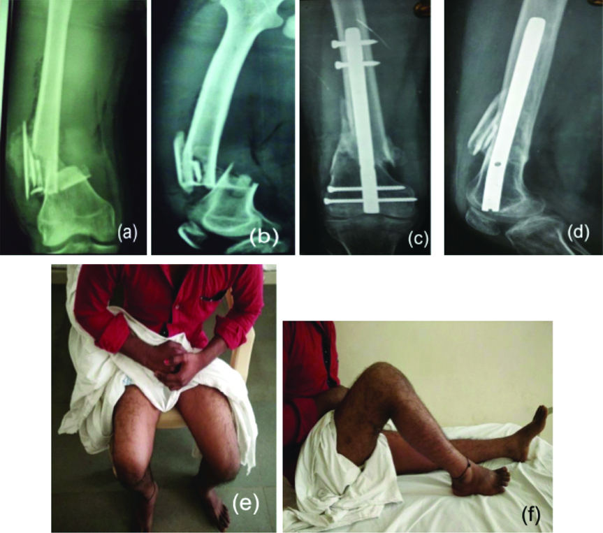
a) X-ray preoperative AP view, b) X-ray shows preoperative lateral view, c) X-ray shows 6 month follow up AP view post retrograde nailing, d) X-ray shows 6 month follow up lateral view post retrograde nailing, e) and f) shows clinical picture of range of motion at knee joint at 6 months follow up.
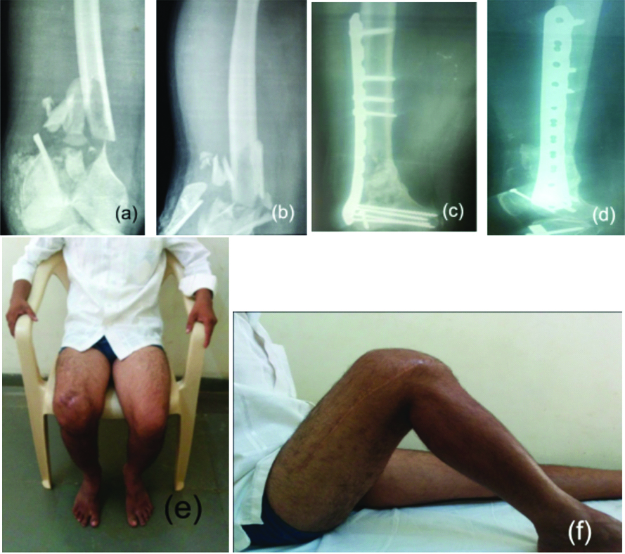
a) X-ray preoperative AP view, b) X-ray shows preoperative lateral view, c) X-ray shows 6 month follow up AP view post locked compression plating, d) X-ray shows 6 month follow up lateral view post locked compression plating, e) and f) shows clinical picture of range of motion at knee joint at 6 months follow up.
In present study, we found better results in retrograde nailing group but none of the results were statistically significant i.e., p-value was not less than 0.05.
In the present study, five patients had shortening two in nailing group and three in plating group with an average 1.5 cm and could be compensated by giving a shoe raise. There was one case of delayed union in plating group and fracture union occurred after seven months and also there were five cases of mal-union, three in nailing group and two in plating group which were given physiotherapy. There were four patients who complained pain at the site of distal screws and two patients who had snapping and which required screw removal after fracture healing.
Discussion
According to the results, retrograde nailing was found to be a better fixation for distal femur fracture in terms of knee flexion, early mobilisation, and less operative time, but the results were not statistically significant.
Mean age and sex incidence between the present study and various other studies has been compared [Table/Fig-9].
Comparison of mean age group and sex incidence among various studies.
| Study | Mean age group in years | Sex incidence in percentage |
|---|
| Male | Female |
|---|
| Watanabe Y et al., [13] | 64 | 16.6 | 83.3 |
| Yeap EJ et al., [14] | 44 | 63.6 | 36.36 |
| Gao K et al., [15] | 50 | 69.4 | 30.5 |
| Gupta SKV et al., [16] | 54 | 71.8 | 28.15 |
| Present study | 38 | 77.5 | 22.5 |
In the present series, the mean age was 39.6 in nailing group and 37.45 in plating group. And the male:female ratio was 3:1 in nailing group and 4:1 in plating group.
In the present study, road traffic accidents accounted for 36 cases out of 40 while four had domestic fall which was found similar in other studies. In the present study, 16 were compound injuries and 24 patients had closed injuries. Most of the fractures that came to present hospital were either Grade 2 modified Gustillo Anderson fractures or had intra-articular extension.
Among 40 patients in the study group, most of the patients in nailing group as well as plating group presented with fracture type C2, seven patients each. Six fractures belonged to A-3 category in nailing group and four in plating group, while two belonged to C-3 category in nailing group and five in plating group.
The final outcome in terms of knee flexion was that AO Muller type A3 fractures showed the best knee flexion range while type C3 showed the worst among different fracture patterns.
Average operative time in the present study was 105 minutes in retrograde nailing group and 110 minutes in plating group. Operative time varied with operating surgeon, implants and instruments at disposal and operation theatre standards.
Gill S et al., concluded a significant difference in terms of mean duration of surgery and intraoperative blood loss in favour of plating group [17]. They found mean duration of union to be 26.5 weeks in the locked plating and 22.6 weeks in the retrograde nail group. The difference came out to be statistically insignificant. Fractures in nailed patients united earlier but the difference was not statistically significant, and they deduced that surgical planning and expertise rather than the choice of implant are more crucial for optimal results. [Table/Fig-10].
Comparison of average union and knee flexion between various studies.
| Gao K et al., [15] | Gupta SKV et al., [16] | Present study |
|---|
| RN | LCP | RN | LCP | RN | LCP |
|---|
| Average union in weeks/rate | 84.2% | 94.1% | 29.6 | 27.2 | 14.69 | 16.25 |
| Knee flexion in degrees | 103.5 | 98.2 | 117.2 | 124.4 | 113.45 | 106.3 |
RN: Retrograde nailing; LCP: Locked compression plating
Limitation
There were limited number of patients which were not large enough to strengthen the significance of the differences. Participating surgeons had less experience with locked compression plating at their early learning stage, results probably were biased in favor of retrograde nailing fixation. Short follow up period that was not adequate for obtaining long-term outcomes. The operating surgeon was not the same for every surgery.
Conclusion
Retrograde intramedullary supracondylar nail is a good fixation system for distal third femoral fractures intra-articular as well as extra-articular with less operative-time, reduced blood loss and closed reduction without disturbing fracture haematoma and soft tissue. Even with open reduction, soft tissue trauma is less. Distal screw related local symptoms is a common problem and is related to implant and technique; and has a definite learning curve. There are less chances of non-union, minimal chances of delayed unions and angular or rotational malunion. There is no requirement for bone graft. Early surgery, closed reduction and atleast two screw in each fragment and early postoperative knee mobilisation are essential for good union and good knee range of motion. Both Locking compression plate and Retrograde intramedullary nailing are optimal tools for fractures of distal femur. They provide good fixation in the region of femur, where a widening canal, thin cortices and frequently poor bone stock make fixation difficult.
In the present study, retrograde nailing was found to be a better fixation system for both extra as well as intra-articular fractures of distal femur with better outcome in terms of range of movements, early mobilisation and less operative time and blood loss.
In future, the present study can be taken to the next level where comparison between nailing and plating will be done for osteoporotic patients only.
p=0.313
p=0.525
p=0.278
p=0.523
RN: Retrograde nailing; LCP: Locked compression plating