Cell Phones have become an important part of every day’s life. Research on cell phone and its foetal effects is relatively new. There are concerns about the safety of their use in pregnancy as they operate by creating an electromagnetic field. If an association is noticed future studies may be conducted to confirm the findings and limit the use of cell phones in pregnancy [1,2].
Being wireless devices, they operate by creating electromagnetic fields. Animal studies, report some biological effect on target tissues like oxidative damage by increasing the extent of lipid peroxidation and increasing iron levels. Wi-Fi and mobile phone induced electromagnetic radiations may cause precocious puberty and oxidative kidney and testis injury in growing rats [1]. In another study by Haghani M et al., exposure of rats to 900 MHz for 6 hours/day resulted in decreased neuronal excitability of Purkinje cells [2]. Mobile phone exposure in adolescents exposed to 2G mobile phone raised the alpha activity as detected by Electroencephalography (EEG) [3].
According to Sauter C et al., individuals were exposed to mobile phone in three different modes including sham, Global System for Mobile Communication (GSM) and Wideband Code Division Multiple Access (WCDMA). The results revealed effects on the selective attention as well as working memory [4]. Another data from Cairo, Egypt, observed the effect of mobile phone usage on FHR pattern and the researchers reported that baseline FHR increased by 5 beats/minute above the base line. However, this study had no control group [5]. Celick O and Hascalik S observed the effect of electromagnetic field of mobile phone on FHR pattern of 40 pregnant women and found no significant change in FHR pattern [6].
There is paucity of evidences about the safety of mobile phone use in pregnancy, and in fact little is known on their biological effects. Evidence based data remain lacking. The present study, serve as a pilot study from which further clinical research on identified effects could be conducted. The primary aim of the present study was to highlight the effect of mobile phone use in pregnancy on foetal cardiac activity. Future interpretations can be made from the results of present study which can serve as the basis for further research.
Materials and Methods
The research hypothesis was:
H0=The FHR does not changes with the use of mobile phone
Alternate=The FHR changes by the effect of mobile phone
The present study was conducted as an interventional study at the Department of Obstetrics and Gynaecology, Sir Ganga Ram Hospital, Lahore, Pakistan (October 2016). The present study was approved by Ethical board of Sir Ganga Ram Hospital, Lahore, Pakistan. All women who were willing to participate after informed consent were included in the present study, presenting between 28-37 weeks of gestation. Women whether, primiparous or multiparous, carrying a singleton pregnancy were included in the present study. They were instructed not to use mobile phones, 24 hours before the start of test. There were 138 patients who were willing to participate during this month in antenatal period. Considering this the sample had a confidence level of 95% with confidence interval of five. All high risk women with any medical or obstetrical disorders and women in labour were excluded from the present study. Single mobile phone with Specific Absorption Rate (SAR) rating of 0.99 W/kg was used in the present study. First, the women underwent CTG without mobile phone (controls) and then exposed to mobile phone in calling mode (served as cases) for 10 minutes in a separate room where no other mobile phone was placed. All women had a CTG performed by the same machine (BISTOS BT-300 Korea) for 20 minutes. Data was collected on self-designed proforma. The CTG tracings were analysed blindly without the information of the patient. The variables, measured for CTG were baseline FHR, beat to beat variability, accelerations and decelerations. Baseline FHR of 110-160 beats/minute is considered to be normal. Acceleration was defined as transient rise in FHR above the base line more than 15 beats/minute and lasting at least 15 seconds. Decelerations were transient slowing of FHR below the base line, more than 15 beats/minute lasting more than 15 seconds and the Baseline variability was defined as transient oscillations of FHR between 5-15 beats/minute [7].
Statistical Analysis
The data was statistically analysed with SPSS version 23.0. Microsoft Office for Windows 7 was used. Inferential statistics were used to draw conclusions from the sample tested. The Statistical Package for the Social Sciences (SPSS) was used to code and tabulate scores collected from the survey and provide summarised values where applicable including the mean, central tendency, variance, and standard deviation. Multivariate Analysis of Variance (MANOVA) was used to evaluate the research question.
Results
The hypothesis was evaluated using MANOVA analysis to determine if any significant differences in participants baseline FHR, acceleration, deceleration, and variability existed between those with a mobile phone in calling mode and those with a mobile phone in silent mode. The dependent variables were participants baseline FHR, acceleration, deceleration, and variability scores. Response parameters for participants baseline FHR scores were measured on a six-point interval scale where 1=110-120, 2=121-130, 3=131-140, 4=141-150, 5=151-160, and 6=161 and above. Response parameters for participants acceleration and deceleration scores were measured on a three-point interval scale where 1=absent, 2=1-3, and 3=more than 3. Lastly, response parameters for participants variability scores were measured on a three-point interval scale where 1=absent, 2=reduced (5-9), and 3=good (10-15). The independent variable was whether the participants had a mobile phone or not.
Data Cleaning and Tests of Parametric Assumptions
Before the data was evaluated, the data was screened for missing data, univariate and multivariate outliers. Missing data was investigated using frequency counts, and no cases existed. Thus, 138 responses from participants were received evaluated by the MANOVA model (n=138). Descriptive statistics of participants dependent variable scores by mobile phone groups are shown in [Table/Fig-1].
Descriptive statistics of the dependent variables by mobile phone groups.
| Variable | n | Minimum | Maximum | Mean | Standard Deviation | Skewness | Kurtosis |
|---|
| With mobile phone |
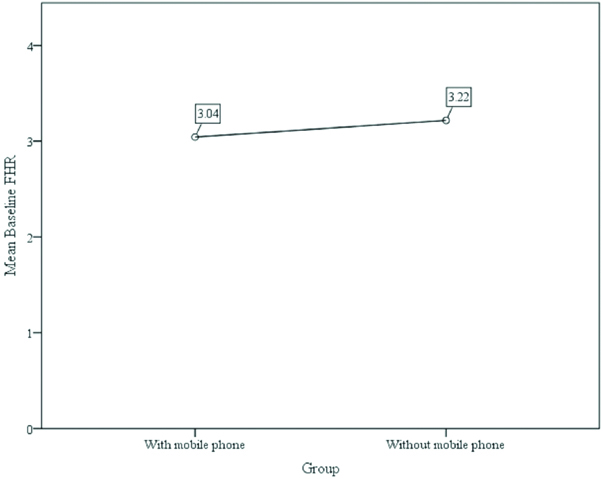
| Baseline FHR | 69 | 1.000 | 5.000 | 3.040 | 1.300 | -0.207 | -1.049 |
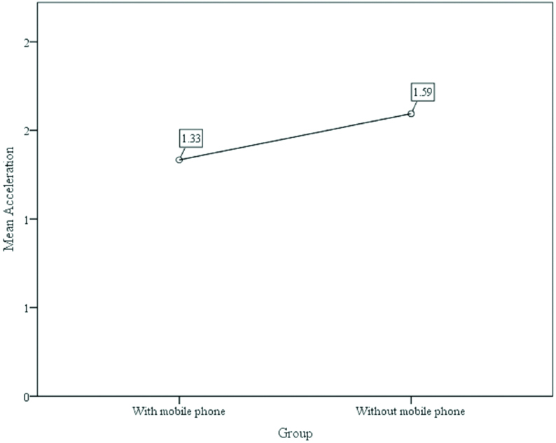
| Acceleration | 69 | 1.000 | 3.000 | 1.330 | 0.560 | 1.475 | 1.287 |
| Decelerations | 69 | 1.000 | 1.000 | 1.000 | N/A | N/A | N/A |
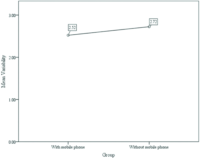
| Variability | 69 | 2.000 | 3.000 | 2.522 | 0.503 | -0.089 | -2.052 |
| Without mobile phone |
| Baseline FHR | 69 | 1.000 | 5.000 | 3.220 | 1.083 | -0.307 | -0.490 |
| Acceleration | 69 | 1.000 | 3.000 | 1.590 | 0.551 | 0.157 | -0.948 |
| Decelerations | 69 | 1.000 | 1.000 | 1.000 | N/A | N/A | N/A |
| Variability | 69 | 2.000 | 3.000 | 2.725 | 0.450 | -1.028 | -0.972 |
Note: n=138
FHR=Foetal Heart Rate
The dependent variables (baseline FHR, acceleration, deceleration, and variability) were evaluated for basic parametric assumptions including normality, homogeneity of variance, homogeneity of variance-covariance matrices, and multicollinearity. However, since all participants scored a ‘1’ (absent) on deceleration, the variable was removed from all further analyses. The distributions were examined for normality using ‘Skew’ and ‘Kurtosis’ coefficients and results indicated that two distributions (acceleration and variability) were significantly skewed and one distribution was significantly kurtotic, (variability) was significantly kurtotic. Therefore, the distributions were transformed using square root transformations. Although, the transformed data was found to be normally distributed, results from the MANOVA analysis using the transformed data was similar, compared to those found using the raw (untransformed) scores. Therefore, the raw data was used in the MANOVA model. Skewness and kurtosis statistics of the dependent variables (raw scores) by mobile phone groups are displayed in [Table/Fig-2].
Skewness and Kurtosis Statistics of the dependent variables by mobile phone groups.
| Variable | n | Skewness | Skew Std. Error | z-skew | Kurtosis | Kurtosis Std. Error | z-kurtosis |
|---|
| With mobile phone |
| Baseline FHR | 69 | -0.207 | 0.289 | -0.716 | -1.049 | 0.570 | -1.840 |
| Acceleration | 69 | 1.475 | 0.289 | 5.104* | 1.287 | 0.570 | 2.258 |
| Variability | 69 | -0.089 | 0.289 | -0.308 | -2.052 | 0.570 | -3.600* |
| Without mobile phone |
| Baseline FHR | 69 | -0.307 | 0.289 | -1.062 | -0.490 | 0.570 | -0.860 |
| Acceleration | 69 | 0.157 | 0.289 | 0.543 | -0.948 | 0.570 | -1.663 |
| Variability | 69 | -1.028 | 0.289 | -3.557* | -0.972 | 0.570 | -1.705 |
Note: *Variable is significantly skewed/kurtotic. n=138
FHR=Foetal Heart Rate
Results of MANOVA Analysis
The null and alternate hypothesis were:
H0=The FHR does not changes with the use of mobile phone
Alternate=The FHR changes by the effect of mobile phone
Results indicated that there was a significant multivariate differences between groups (with mobile phone and without mobile) on a model containing three dependent variables (baseline FHR, accelerations, and variability), Wilks’ Lambda=0.918, F=(3, 134)=3.997, p.=0.009, η2=0.082. Displayed in [Table/Fig-3].
Summary of the MANOVA multivariate test conducted for hypothesis.
| Effect | Wilks’ Lambda | F | Hypothesis df | Error df | Sig. (p) | Partial Eta Squared (η2) |
|---|
| Intercept | 0.025 | 1758.415 | 3 | 134 | <0.001 | 0.975 |
| Mobile Phone Group | 0.918 | 3.977 | 3 | 134 | 0.009 | 0.082 |
Note. Dependent variables=baseline FHR, acceleration, and variability. n=138
Results from the tests of between-subject effects indicated there were individual significant differences in two of the three dependent variables (acceleration p=0.007 and variability p=0.014) between mobile phone groups. That is, participants with mobile phones had significantly lower acceleration scores (M=1.330, SD=0.560) as compared to those without a mobile phone (M=1.590, SD=0.551). Additionally, participants with mobile phones had significantly lower variability scores (M=2.522, SD=0.503) as compared to those without a mobile phone (M=2.725, SD=0.450). Regarding the variability variable, results from the non-parametric Kruskal-Wallis test affirmed the results from the between-subject effects analysis (p=0.014). Lastly, there was no significant difference in participants baseline FHR scores (p=0.395) among those with a mobile phone (M=3.040, SD=1.300) and those without a mobile phone (M=3.220, SD=1.083). Thus, since there were significant differences in two of the three dependent variables, the null hypothesis was partially rejected in favor of the alternative hypothesis. A model summary of the individual tests of between-subject effects has displayed in [Table/Fig-4].
Model summary of the MANOVA tests of between-subjects effects for hypothesis 1.
| Source | Type III Sum of Squares | df | Mean Square | F | Sig. (p) | Partial Eta Squared (η2) |
|---|
| Corrected Model | 1.043 | 1 | 1.043 | 0.729 | 0.395 | 0.005 |
| Baseline FHR | 2.348 | 1 | 2.348 | 7.608 | 0.007 | 0.053 |
| Acceleration Variability | 1.420 | 1 | 1.420 | 6.234 | 0.014 | 0.044 |
| Intercept |
| Baseline FHR | 1352.348 | 1 | 1352.348 | 945.072 | <0.001 | 0.874 |
| Acceleration | 295.681 | 1 | 295.681 | 958.105 | <0.001 | 0.876 |
| Variability | 949.594 | 1 | 949.594 | 4167.910 | <0.001 | 0.968 |
| Certification Type |
| Baseline FHR | 1.043 | 1 | 1.043 | 0.729 | 0.395 | 0.005 |
| Acceleration | 2.348 | 1 | 2.348 | 7.608 | 0.007 | 0.053 |
| Variability | 1.420 | 1 | 1.420 | 6.234 | 0.014 | 0.044 |
| Error |
| Baseline FHR | 194.609 | 136 | 1.431 | | | |
| Acceleration | 41.971 | 136 | 0.309 | | | |
| Variability | 30.986 | 136 | 0.228 | | | |
| Total |
| Baseline FHR | 1548.000 | 138 | | | | |
| Acceleration | 340.000 | 138 | | | | |
| Variability | 982.000 | 138 | | | | |
| Corrected Total |
| Baseline FHR | 195.652 | 137 | | | | |
| Acceleration | 44.319 | 137 | | | | |
| Variability | 32.406 | 137 | | | | |
Note: n=138
FHR=Foetal Heart Rate
Means plots of the dependent variables by mobile phone groups are displayed in [Table/Fig-5,6 and 7].
Mean plots of participants base line fetal heart rate score between mobile phone and non mobile phone groups.
Mean plots of participants fetal heart rate accelerations score between mobile phone and non mobile phone groups.
Mean plots of participants fetal heart rate variability scores between mobile phone and non mobile phone groups.
Discussion
The FHR tracing depicts foetal response. Four main features are noted from the trace including the base line FHR, variability, presence or absence of decelerations and accelerations. When reviewing the CTG trace, four features are noted baseline heart rate, variability and presence of accelerations and decelerations. Normal trace constitutes a baseline heart rate of 100-160 beats/minute, beat to beat variability of 5-15 beats/minute and no decelerations. Absence of accelerations has no significance in another wise normal trace [7]. The presence of FHR accelerations is generally a sign that the baby is healthy.
In the present study, among participants there were significant differences in two of the three dependent variables. Women with mobile phones had significantly lower acceleration scores as compared to those without mobile phones. Additionally, participants with mobile phones had significantly lower variability scores as compared to those without a mobile phone. Lastly, there was no significant difference in participants baseline FHR scores. We noticed no change in baseline FHR. However, in another study by Rezk AY, et al., reported an increased FHR in fetuses who were exposed to cell phone in calling mode [8].
Physiologically accelerations reflect foetal alertness and Central Nervous System (CNS) activity and depicts healthy fetus with intact Autonomic Nervous System (ANS). They are commonly seen in association with foetal movements and external stimuli. Their absence is the indication of CNS depression, seen with certain drugs and foetal sleep [9]. A typical acceleration has two components, first a gradual rise to the peak which is under sympathetic control and the second a rapid return to base line which is mediated through parasympathetic system. Van Geijn HP, states that absence of accelerations above 45 minutes is suspicious of fetal distress [9]. Lower acceleration score in the present study raises concerns; therefore about the health of foetal CNS. Results in the present study were in contrast to Celick O and Hasclik S, who noticed no change in acceleration pattern among 40 pregnant women who had cell phone in dialing mode for five minutes [6]. There is a difference in the timing of exposure between present study and Celick O and Hasclik S study. In the present study, timing of exposure was 10 minutes in contrast to Celick O and Hasclik S study [6].
Additionally, participants with mobile phones had significantly lower variability scores as compared to those who were without a mobile phone. Base line variability also has two components short and long term, short term variability is indicative of vagal and the long term is under the sympathetic control. Therefore, heart rate variability is an assessment tool for monitoring cardiac conditions in adults with ANS abnormalities [10]. There is evidence to suggest that FHR variability is depressed by factors that depress foetal brain functioning or myocardial activity [10]. Therefore, in the present study, lower variability score in women using mobile phone may be an indirect indicator of CNS and ANS, depression under the effect of mobile phone radiations. Heart rate variability is a sensitive indicator of ANS modulation. A change in its patterns indicates foetal health [11]. It is affected by many physiological conditions like posture, age, sex, breathing and other daily activities [12]. Therefore, it can be assumed that mobile phone use can affect it as seen in the present study.
There are concerns about the long term health consequences of mobile phone used by pregnant women and appearance of behavioral disturbances in children [13-15]. Foetal safety is the primary objective of obstetric care. Hence, the results of foetal heart pattern are linked to CTG changes. Further research is required to interpret the results and if proven recommendations can be made for safe use of mobile phones in pregnancy periods.
Limitations
The present study had strength of having a good sample size with confidence level of 95% with confidence interval of 5. However, the study had the limitation of using one type of mobile phone only and the world is full of cellular phones with different SAR.
Conclusion
Mobile phone affects the FHR pattern. We observed a significant multivariate differences between groups (with mobile phone and without mobile phone). Participants with mobile phones had significantly lower acceleration scores as well as lower variability scores.