Radiomorphometeric Evaluation of Clivus in Indian Paediatric Population Visiting a Tertiary Dental Hospital- A Cone Beam Computed Tomography Study
Akhilanand Chaurasia1, Ranjitkumar Patil2, Gaurav Katheriya3
1 Assistant Professor, Department of Oral Medicine and Radiology, King George Medical University, Lucknow, Uttar Pradesh, India.
2 Professor, Department of Oral Medicine and Radiology, King George Medical University, Lucknow, Uttar Pradesh, India.
3 Resident, Department of Oral Medicine and Radiology, King George Medical University, Lucknow, Uttar Pradesh, India.
NAME, ADDRESS, E-MAIL ID OF THE CORRESPONDING AUTHOR: Dr. Akhilanand Chaurasia, Flat No-701, New Faculty Residence, T.G. Hostel Campus, Khadra, Lucknow-226003, Uttar Pradesh, India.
E-mail: chaurasiaakhilanand49@gmail.com
Introduction
The clivus is the part of the skull base situated between the foramen magnum and the dorsum sellae. It results from the fusion of the synchondrosis between the basioccipital and exoccipital bones which grows and ossify from three to 25 years of age to form the basisphenoid and the basiocciput. The clivus is a dense part of skull, so mostly it is recovered intact from a damaged or incinerated skull. So, clivus can be used for morphometeric evaluation of humans.
Aim
To determine the age and sex related changes in clivus length and clivus width. The clivus width and clivus length was also evaluated for age prediction of individuals.
Material and Methods
The CBCT images of 150 study subjects obtained from Carestream 9000cc (USA) CBCT machine at 90 Kvp, 4 mA for 11.3 seconds at FOV (17″×13.5″), voxel size of 300 in the age group of six to 17 years (76 male, 74 female) were chosen prospectively. The clivus length and clivus width were measured using Trophy Dicom Ink software programme on axial and saggital images (DICOM images). Categorical variables was presented in number and percentage (%) and continuous variables was presented as mean and SD. Unpaired t-test and ANOVA test was used for comparison between groups. Pearson correlation coefficients were used to evaluate the correlations. The data analysis was done using SPSS version 21.0.
Results
Irrespective of age and sex, the mean clivus width was 28.8±3.98 and mean clivus length was 42.7±3.98. Statistically no significant difference was observed between male and female (p-value>0.05) with clivus width. However, the clivus length was statistically significant (p<0.001) in both male and female population. The clivus width and clivus length was directly associated with age. The mathematical equations derived from linear regression analysis can be used in prediction of age of an individual if the clivus width/clivus length is known.
Conclusion
The clivus measurements on CBCT can be used to predict the age of an individuals in medicolegal disputes. As pearson correlation coefficient showed strong association between the clivus measurements, age and gender, clivus measurements can be used to differentiate between sex of humans and helps in age determination. Clivus dimensions can be used as an additional parameter in age and sex determination in inconclusive medicolegal cases.
Foramen magnum, Skull, Synchondrosis
Introduction
The identification of deceased humans from hard tissues in disasters is a difficult task which anatomists, forensic experts and anthropologist encounter in day-to-day practice [1]. So, sexual dimorphism in different biological population is considered one of the most reliable tool for identification of a deceased individual [2,3]. The enamel and bones are last to disintegrate after death so these two skeletal remains can also be used as a tool for sex determination of an individual [1]. As most of time forensic anthropologists are encountered with decomposed bodies and disintegrated skeleton. It is now need of time to look for alternative methods of sex determination [4]. In physical anthropology, skull is most studied skeletal remain for sex determination [5]. The clivus is the part of the skull base situated between the foramen magnum and the dorsum sellae. Clivus is relatively denser than other part of skull. The clivus can be recovered intact from an incinerated or damaged skull. Due to dense nature and intactness even after incineration, clivus can be alternatively used for gender determination medico legally by anthropometric measurement [4]. The formation of clivus results from the fusion of the synchondrosis between the basioccipital and exoccipital bones. It starts growing after fusion between the basioccipital and exoccipital bones. The ossification process in clivus starts from third year of age and persist till 25th years of age to form the basisphenoid and basiocciput. The anterior margin of the clivus corresponds to the sphenoidal sinus however, the posterior surface forms the anterior limit of the prepontine and premedullary cisterns. The inferior margin of clivus corresponds to posterior nasopharyngeal surface. Laterally clivus is bounded by the petro-occipital fissure which starts near the cavernous sinus and extends to the jugular foramen inferiorly [6].
Clivus is best seen in the axial and sagittal sections of CT scan. The anterior and posterior boundaries of clivus is formed by compact cortical bone however, its central portion is made of cancellous bone. In adults, the clivus is filled up with fatty narrow in its cancellous part [7]. Clinically, it is a crucial surgical site for its pathology due to its location in posterior cranial fossa and its relation with brainstem, clivus is rarely involved in invasive pathologies like giant cell tumours, meningiomas, pituitary adenoma and monostotic fibrous dysplasia [8-11]. In present study, clivus is evaluated quantitatively by Cone Beam Computed Tomography (CBCT) by measuring its width and length in axial and sagittal section respectively to determine its forensic and medicolegal significance in morhphometric analysis. Till date no study has been conducted worldwide on CBCT for evaluation of clivus in paediatric population in determination of age and sex for forensic identification in mass disaster and medicolegal cases. In light of meagre literature on paediatric clivus, this radiomorphometric study was conducted to highlight the importance of clivus in age and sex determination.
Materials and Methods
This study was an observational and prospective study which was carried out in Department of Oral medicine and Radiology. The ethical clearance was not needed as the study subjects included in the study were subjected to CBCT for other diseases referred from other departments in which Head and PNS (Paranasal Sinus images) CBCT images of 150 study subjects (76 males and 74 females) in the age group of six to 17 years were chosen prospectively. The clivus matures before 17 years of age i.e., the length and width of clivus reaches its final limitation by 17 years of age of an individual. So, the paediatric patients aged less than 17 years, no history of trauma or pathology of middle cranium were included in our study. Any CBCT with obvious pathology, trauma and facial asymmetry were excluded from this study. An informed consent was obtained from each patient or their guardians in case of minors. All the study subjects were scanned on CS9300 carestream CBCT machine. The axial and sagittal images were obtained at 90 Kvp, 4 mA for 11.3 seconds at FOV (17″×13.5″) voxel size of 300. The measurement of clivus dimensions were done directly on DICOM images using Trophy Dicom Ink software programme.
The measurements were done as follows:
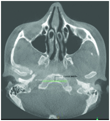
The width of clivus was defined as the longest distance from left to right side near the anterior peripheral margin of foramen magnum inferiorly and it was measured on axial sections in CBCT [Table/Fig-1].
Axial CBCT image showing the clivus width.
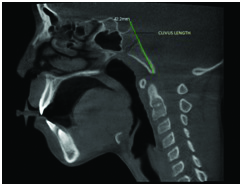
The length of clivus was defined as the longest distance superio-inferiorly from the upper point of dorsum sellae to the lowest point on anterior margin of foramen magnum and it was measured on sagittal section in CBCT [Table/Fig-2].
Sagittal CBCT image showing the clivus length.
Statistical Analysis
Categorical variables was presented in number and percentage (%) and continuous variables was presented as mean and SD. Quantitative variables was compared using Unpaired t-test between two groups and ANOVA test between three groups. Pearson correlation coefficients were used to determine the relationship between two scale parameters while correlation was defined as a measure of the strength of a linear relationship between two variables. A p-value of <0.05 was considered statistically significant. The data was analysed by using SPSS version 21.0.
Results
The study sample consists of 150 study subjects aged between six and 17 years with a mean age of 12.15±3.29 years. Majority of the study subjects were between 10 to 12 years of age (34.7%) followed by subjects 15 to 17 years (28.7%), subjects six to nine years (21.3%) and 13 to 14 year (15.3%) [Table/Fig-3]. The sex ratio in our study population showed that male subjects (50.7%) outnumber the female subjects (49.3%). Irrespective of age and sex, the mean clivus width was 28.8±3.82 and mean clivus length was 42.7±3.98 [Table/Fig-4]. The unpaired t-test for significance was used to find the correlation between sex and clivus width and clivus length. Statistically no significant difference was observed between male and female (p-value>0.05) with clivus width. However, the clivus length was statistically significant (p<0.001) in both male and female population [Table/Fig-5]. The two tailed t-test was used to know the association between clivus length, clivus width and age. It was found that the clivus width and clivus length was directly associated with age [Table/Fig-6]. The Pearson correlation between age and clivus length, clivus width demonstrates a significant positive relation (i.e. r=0.465, p<.001 and r=0.285, p=0.001 respectively) [Table/Fig-7]. The Pearson correlation coefficient shows that the clivus length and clivus width was directly associated with age.
Showing distribution of study subjects in age groups.
| Age intervals (years) | Frequency | Percentage (%) |
|---|
| 6 to 9 | 32 | 21.3 |
| 10 to 12 | 52 | 34.7 |
| 13 to 14 | 23 | 15.3 |
| 15 to 17 | 43 | 28.7 |
| Total | 150 | 100.0 |
Showing mean age, mean clivus width and mean clivus length in study population.
| n | Minimum | Maximum | mean±SD |
|---|
| Age (years) | 150 | 6 | 17 | 12.15±3.293 |
| Clivus width | 150 | 2.80 | 38.40 | 28.87±3.827 |
| Clivus length | 150 | 29.20 | 52.00 | 42.71±3.980 |
Showing the association between sex and clivus width, clivus length.
| Gender | n | mean±SD | p-value |
|---|
| Clivus width | Male | 76 | 29.2171±4.27824 | 0.264 |
| Female | 74 | 28.5162±3.29262 |
| Clivus length | Male | 76 | 43.9908±3.81450 | <0.001* |
| Female | 74 | 41.3851±3.72863 |
Applied unpaired t-test for significance. *Significant
Showing the association between age groups and and clivus width, clivus length.
| Study parameters | Age (years) | n | mean±SD | p-value |
|---|
| Clivus width | 6 to 9 | 32 | 28.1906±3.85448 | 0.006* |
| 10 to 12 | 52 | 27.8462±4.44648 |
| 13 to 14 | 23 | 29.2435±3.49608 |
| 15 to 17 | 43 | 30.4186±2.53163 |
| Total | 150 | 28.8713±3.82730 |
| Clivus length | 6 to 9 | 32 | 38.9281±4.37458 | <0.001* |
| 10 to 12 | 52 | 42.9827±2.86601 |
| 13 to 14 | 23 | 44.6522±3.64453 |
| 15 to 17 | 43 | 44.1395±3.16297 |
| Total | 150 | 42.7053±3.98044 |
Applied one way ANOVA for significance. *Significant
Showing the strong positive relation and mathematical equation used for age prediction.
| Study parameters | Pearson correlation coefficients (r) | p-value | Mathmatical equations (Linear regression analysis) |
|---|
| Clivus width | 0.285 | <0.001 | Age=5.080+0.245*(Clivus width) |
| Clivus length | 0.465 | <0.001 | Age=(-4.290)+0.385*(Clivus length) |
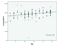
On the basis of strong positive correlation between clivus width, clivus length and age it was concluded that age plays an important role in determination of clivus length and clivus width. The linear regression analysis has been done for clivus width in relation to age and mathematical equation derived was age=5.080+0.245* (clivus width) [Table/Fig-8]. Hence, it was concluded that if the clivus width of an individual is known, the age can be predicted with help of mathematical equation derived from linear regression analysis.
Showing the distribution of study subjects in relation to clivus width and strong positive relation with age.
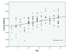
Linear regression analysis was done for clivus length in relation to age and mathematical equation derived was age=(-4.290)+0.385* (clivus length) [Table/Fig-9]. Therefore, it was concluded that if the clivus length of an individual is known, the age can be predicted with help of mathematical equation derived from linear regression analysis.
Showing the distribution of study subjects in relation to clivus length and strong positive relation with age.
Discussion
O’Higgins P et al., and Richtsmeier JT et al., conducted many morphometric studies to investigate sexual dimorphism in different primate species however, the morphometric studies involving human skull is sparse and based on relatively small sample sizes [12-14].
In both sexes, the clivus matures by 11th years of age i.e., final length of the clivus was reached by this age. Afterwards it remains constant throughout life [4]. Eleventh year of postnatal life is very crucial as final adult width of the clivus is first reached at this age. Male have higher mean clivus length and width than female however, this difference was statistically significant (p<0.0001) [4].
Krogman WM et al., concluded that the shape of the cranial base was affected by clefting from sellar angle however, the anterior cranial base length and clivus length varies significantly in both sexes [15]. So, dimensional analysis of the size of clivus and its sexual dimorphism was investigated in present study.
However, many studies were conducted to evaluate clivus length and correlated clinically with the soft tissue pathology. Dufton JA et al., studied clivus length in chiari malformation type-I patients [16]. They concluded that clivus length was shorter in chiari malformation type-I subjects (4.02±0.45 cm) than controls (4.23±0.42 cm) and there was no correlation between tonsillar herniation and clivus length (r=-0.30, p<0.001). They also concluded that a shorter clivus length is associated with greater degree of cerebellar tonsillar herniation. Heiss JD et al., also conducted a study on chiari malformation type-I subjects and concluded that clivus and basiocciput lengths were significantly shorter than control group [17]. The result was similar in both studies i.e., Dufton JA et al., and Heiss JD et al., [16,17]. The chiari malformation type-I group and similar groups were excluded from our study. In most of studies, morphometric evaluation of clivus was done by CT scan however, this is the first study till date which has been conducted on CBCT which emphasise on gender and age determination on the basis of morphometric evaluation of clivus.
In our study, irrespective of age and sex, the mean clivus width was 28.8±3.98 and mean clivus length was 42.7±3.98. Statistically no significant difference was observed between male and female (p-value>0.05) with clivus width. However, the clivus length was statistically significant (p<0.001) in both male and female population. The clivus length was higher in male population. The Pearson correlation between age and clivus length, clivus width demonstrates a significant positive relation (i.e., r=0.285, p<0.001 and r=0.465, p=0.001 respectively). The finding in present study correspond with study conducted by Chaurasia A et al., [18]. On the basis of strong positive correlation between clivus width, clivus length and age it was concluded that age plays an important role in determination of clivus length and clivus width. The mathematical equations derived by linear regression analysis for clivus width and clivus length we can predict the age of the subject if the clivus width and clivus length is known. This is very revolutionary step in the field of forensic medical sciences.
Limitation
The clivus is not routinely evaluated anatomic structure for morphometric analysis i.e., age and sex determination. The operator should have good knowledge of anatomical structure of skull and should be able to identify it in CBCT. Our study had small sample size. It should be conducted on larger sample size to eliminate inter-observer and intra-observer bias in data collection.
Conclusion
The CBCT measurement of the clivus dimensions can be used to predict the age of individuals. The clivus dimensions are directly related to age and gender. It may also be helpful to the radiologist in radiological diagnosis of clivus pathology and may act as a guide to the surgeons for the surgery of clivus and related region.
Applied unpaired t-test for significance. *Significant
Applied one way ANOVA for significance. *Significant
[1]. Deshmukh AG, Devershi DB, Comparison of cranial sex determination by univariate & multivariate analysisJ Anat Soc India 2006 55(2):48-51. [Google Scholar]
[2]. Hanken J, Hall BK, Mechanisms of skull diversity and evolution. In: Hanken J, Hall BK, editorsThe skull 1993 ChicagoUniversity of Chicago Press:01-36. [Google Scholar]
[3]. Plavcan JM, Comparison of four simple methods for estimating sexual dimorphism in fossilsAm J Phys Anthropol 1994 94:465-76.10.1002/ajpa.13309404037977674 [Google Scholar] [CrossRef] [PubMed]
[4]. Jehan M, Kumar RK, Sexual dimorphism of clivus dimension by computed tomography scanIndian J App Res 2014 4(9):379-81. [Google Scholar]
[5]. Krogman WM, Iscan MY, The human skeleton in forensic medicine 1986 2nd edCharles C. Thomas, Springfield. Illinois [Google Scholar]
[6]. Hofmann E, Prescher A, The clivus: anatomy, normal variants and imaging pathologyClin Neuroradiol 2012 22(2):123-39.10.1007/s00062-011-0083-421710384 [Google Scholar] [CrossRef] [PubMed]
[7]. Bravo AP, de Caceres A, Ibanez L, de Villoria JA, Ossaba S, Crespo J, Tumors and tumor-like conditions of the clivus. A comprehensive reviewEuropean society of radiology 2012 :C-1581 [Google Scholar]
[8]. Mahale A, Dhananjaya KVN, Pai M, Poornima V, Sahu KK, MRI sequence and characteristic features in ‘Giant Cell Tumor’ of clivusJ Clin Diagn Res 2013 7(6):1197-1200. [Google Scholar]
[9]. Sallabanda K, Bustos JC, Gutiérrez-Díaz JA, Beltrán C, Peraza C, Delgado JM, Long-term follow-up of 32 meningiomas of the clivus and foramen magnum subjected to stereotactic radiosurgery. McDermott MW (ed): Radiosurgery 2010 7Basel, Karger:202-11.10.1159/000288731 [Google Scholar] [CrossRef]
[10]. Chen X, Dai J, Ai L, Ru X, Wang J, Li S, Clivus invasion on multi-detector CT in 390 pituitary macroadenomas: correlation with sex, subtype and rates of operative complication and recurrenceAJNR 2011 32(4):785-89.10.3174/ajnr.A236421436342 [Google Scholar] [CrossRef] [PubMed]
[11]. Atalar M, Ozum U, Monostotic fibrous dysplasia of the clivus: imaging findingsTurkish Neurosurgery 2010 20(1):77-81. [Google Scholar]
[12]. O’Higgins P, Dryden IL, Sexual dimorphism in hominoids: further studies of craniofacial shape differences in Pan, Gorilla and PongoJ Hum Evol 1993 24:183-205.10.1006/jhev.1993.1014 [Google Scholar] [CrossRef]
[13]. Richtsmeier JT, Cheverud JM, Danahey SE, Corner BD, Lele S, Sexual dimorphism of ontogeny in the crab eating macaque (Macaca fasciularis)J Hum Evol 1993 25(1):01-30.10.1006/jhev.1993.1035 [Google Scholar] [CrossRef]
[14]. Wood CG, Lynch JM, Sexual dimorphism in the craniofacial skeleton of modern humans. In: Marcus LF, Corti M, Loy A, Naylor GJP, Slice DE (eds)Advances in Morphometrics. NATO ASI Series (Series A: Life Sciences) 1996 284Boston, MASpringer:407-14.10.1007/978-1-4757-9083-2_34 [Google Scholar] [CrossRef]
[15]. Krogman WM, Jain RB, Long RE, Sex differences in craniofacial growth from one month to ten years of cleft lip and palateCleft Palate J 1982 19(1):62-71. [Google Scholar]
[16]. Dufton JA, Habeeb SY, Heran MK, Mikulis DJ, Islam O, Posterior fossa measurements in patients with and without chiari I malformationCan J Neurol Sci 2011 38(3):452-55.10.1017/S031716710001186021515505 [Google Scholar] [CrossRef] [PubMed]
[17]. Heiss JD, Suffredini G, Bakhtian KD, Sarntinoranont M, Oldfield EH, Normalization of hindbrain morphology after decompression of chiari malformation type IJ Neurosurg 2012 117(5):942-46.10.3171/2012.8.JNS11147622978540 [Google Scholar] [CrossRef] [PubMed]
[18]. Chaurasia A, Katheriya G, Patil R, Radio-morphometric evaluation of clivus in Indian ethinicity-a cone beam computed tomography studyJ Oral Med, Oral Sur, Oral Pathol and Oral Radiol 2017 3(1):35-41. [Google Scholar]