When non-surgical root canal treatment proved to be unsuccessful to achieve the apical seal, surgical endodontic approach followed by retrograde restoration is recommended [1-3]. A wide variety of root end filling materials have been tried, eg: Gold foil, Silver amalgam, Gallium alloys, IRM, Cavit, Zinc phosphate, Zinc polycarboxylate, GIC, Super EBA and Resin modified GIC [2-4]. Though, the above materials have satisfied most of the specifications for root end fillings, the biocompatibility and sealing ability had been always in question. However, in previous published research the thickness of the cements used for the study was an uncontrolled variable [2-4]. The methodology used for the testing of material had also evolved with an evolution of biomaterial science. However, none of the materials mentioned and used in present clinical scenario have not completely met ideal properties of root sealants [1,5]. They are in use invariably with developing technology. Newer class of sealants have been marketed with the better sealing ability because of advanced production technology and understanding of material science [6,7]. Hence, the latest innovation towards bioactive and biocompatible materials like MTA, calcium phosphate cement and bone cement was thought of [8].
So, the present study was planned to assess recently introduced materials for sealing the root canals. Hence, this in vitro study was aimed to assess and compare the influence of different thicknesses of MTA, IRM and RMGIC on the sealing ability of root end fillings. The null hypothesis is considered in the present study.
Materials and Methods
The in vitro study was performed in the Department of Conservative and Endodontics in Drs Sudha and Nageswara Rao Siddhartha Institute of Dental Sciences, Gannavaram, Andhra Pradesh, India during the year December 2014 to June 2015. The study was approved by the institutional review board. Sixty three human maxillary anterior teeth with no caries, fractures and without root curvatures were collected and stored. They were sterilized and handled according to Occupational Safety and Health Administration (OSHA) and the Centre for Disease control and prevention (CDC) recommendations and guidelines.
Methodology
The complete study protocol was carried out by dividing 63 teeth randomly into three groups containing 21 teeth in each group. [Table/Fig-1]. Each group was then divided into three sub-groups with seven teeth in each.
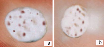
Showing randomization of samples in three groups depending on depth of cavity and materials used.
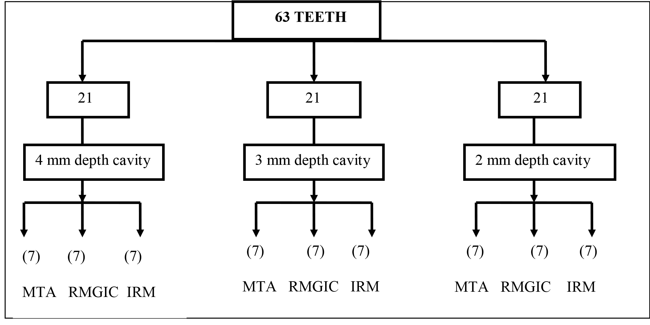
Group 1: The cavities of 4 mm in depth,
Group 2: The cavities of 3 mm in depth,
Group 3: The cavities of 2 mm in depth,
Standard access cavities were made with diamond burs (21 mm, Size 2 Dentsply Maillefer, USA) and coronal portions of canals were flared with Gates–Glidden burs (28 mm, size 1 Dentsply Maillefer, USA). The apical region was prepared up to No. 30 K-file (Dentsply Maillefer, USA) to standardize the diameter. The preparation was completed by using the step back technique in all the teeth. Two milliliters (ml) of 2% NaOCl solution was used as an irrigant between each file to eliminate debris. The cleaned and shaped canals were dried with paper points and obturated with laterally condensed gutta-percha and zinc oxide eugenol sealer. Verification of obturation was made with radiographs. Under a continuous water spray, the apical 3 mm of root was resected at 900 to the long axis of the teeth. [Table/Fig-2a]. Two coats of varnish were applied to the external surfaces of all the teeth except at the apex to prevent leakage through the tooth surface.
Showing sample cavity preparation in three groups depending on depth of cavity a) root end resection b) retrograde cavity preparation c) sectioning of root using hard tissue microtome.
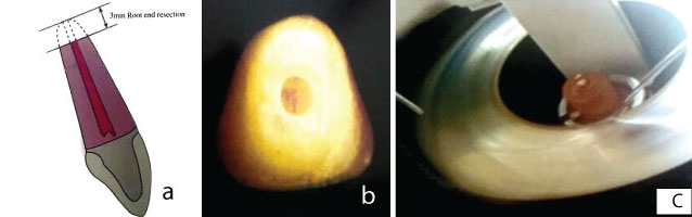
After root-end resection, retrograde cavities of different depths ‘4 mm, 3 mm, and 2 mm’ were prepared [Table/Fig-2b] and cavities were filled with MTA (MTA; Pro-root; Dentsply/Tulsa Dental, Tulsa Ok), Resin Modified Glass Ionomer Cement (RMGIC; Vitremer GC America, Alsip, IL, USA), Intermediate restorative material (IRM Dentsply, USA) according to the manufacturer’s instructions. Then teeth were placed in 50% weight silver nitrate solution for one hour and kept in the absence of light. Later, rinsed for one min in running distilled water to remove the silver ions from the surface. The photo developing solution was used for teeth immersion and exposed to light for 12 hours. Samples were then washed in distilled water and roots were transversely sectioned [Table/Fig-2c] with a hard tissue microtome (LEICA). Each tooth was sectioned per mm up to the total depth of the cavity. The sections were examined under stereo microscope (LEICA WILD M-32) at 30X magnification by two independent observers. The observers followed Cristina Braga Xavier et al., criteria for scoring; which is as follows: 0- No microleakage, 1- up to 25%, 2- 25 – 50%, 3-50 – 75%, 4-75 – 100% [9].
Statistical Analysis
The observations were tabulated using Excel and statistical analysis was done using Statistical Package for the Social Sciences (SPSS) 20.0. Statistical analysis of the result was performed using Kruskal-Wallis test (distribution free) for interexaminer variability and inter group comparison was analysed by Mann-Whitney U test. All statistical analysis were set with a significant level of p-value <0.05.
Results
The scores of different depths were compared for microleakage. The first millimeter sections showed statistically significant higher microleakage than the second, third and fourth millimeter sections. The stereo microscopic images of different depths of dye penetration are shown in [Table/Fig-3,4,5]. The MTA and RMGIC showed median score of 0 for 2 mm thickness, whereas IRM showed median score of 0 at 4 mm thickness for microleakage. For depth of 4 mm the mean and SD of microleakage score for the MTA was 0.57±0.53 at 1mm, whereas RMGIC and IRM were 1±1.15, 2±1.53 respectively. MTA showed highly significant value p<0.01 with H-value 13.5 [Table/Fig-6]. For IRM presented higher leakage of mean 1.67±1.37, 1.00±0.89, 0.50±0.84 at 1, 2, and 3mm thickness, whereas RMGIC showed mean leakage of 1.17±1.33 and 0.40±0.50 at 1 and 2 mm and MTA showed a mean of 0.67±0.82 at 1 mm which showed the best sealing ability with MTA [Table/Fig-7]. In the group with 2mm depth, the microleakage scores for the MTA was very significant p<0.01 with seven as H-value. Whereas, RMGIC and IRM showed non significance after comparison with H-values of 49.5 and 42.5 respectively, [Table/Fig-8] which shows that ascending order of microleakage was MTA<RMGIC<IRM.
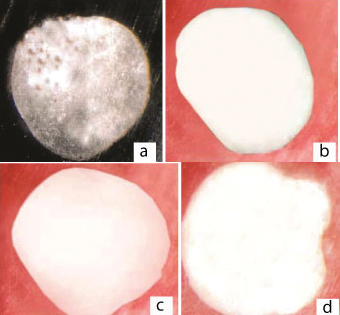
Degrees of dye penetration for apical microleakage in group-1 using MTA at each mm sections of cavities 4 mm in depth. a) MTA section-1, score-1 b) MTA section-2, score-0 c) MTA section-3, score-0 d) MTA section4, score-0
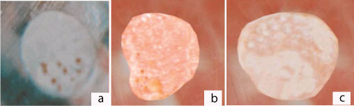
Degrees of dye penetration for apical microleakage in group-1 using RMGIC at each mm sections of cavities 3 mm in depth.
a) RMGIC-section-1, score-2 b) RMGIC-section-2, score-1 c)RMGIC-section-3 Score-0
Degrees of dye penetration for apical microleakage in group-1 using IRM at each mm sections of cavities 2 mm in depth; a) IRM-section-1, score-4; b) IRM-section-2, score-3
Comparison of microleakage between different thicknesses of materials in Group-1 with cavity in depth of 4 mm.
| Materials | Median | Mean±SD | H* Value Significance | Significant Pairs** |
|---|
| Depth | 1mm | 2 mm | 3 mm | 4 mm | 1 mm | 2 mm | 3 mm | 4 mm |
|---|
| MTA | 1 | 0 | 0 | 0 | 0.57±0.53 | 0.00±0.00 | 0.00±0.00 | 0.00±0.00 | 13.5, p<0.01 VS | 1 and 2, 1 and 3, 1 and 4 |
| RMGIC | 1 | 0 | 0 | 0 | 1±1.15 | 0.57±0.79 | 0±0.00 | 0±0.00 | 9.337, p<0.05 S | 1 and 2, 1 and 3, 1 and 4 |
| IRM | 2 | 2 | 1 | 0 | 2±1.53 | 1.43±1.13 | 0.71±0.76 | 0.00±0.00 | 10.35, p<0.05, S | 1 and 3, 1 and 4, 2 and 4, 3 and 4 |
VS- Very significant, S-significant, * Kruskal Wallis Test, ** Mann-Whitney U Test p<0.05 significant difference. p<0.01 very significant difference. p>0.05 not significant difference
Comparison of microleakage between different thicknesses of materials in Group- 2 with cavity in depth of 3mm.
| Materials | Median | Mean±SD | H* value,Significance | Significant Pairs** |
|---|
| Depth | 1 mm | 2 mm | 3 mm | 1 mm | 2 mm | 3 mm |
|---|
| MTA | 1 | 0 | 0 | 0.67±0.82 | 0.00±0.00 | 0.00±0.00 | 9.33, p<0.05 S | 1 and 2, 1 and 3 |
| RMGIC | 1 | 0 | 0 | 1.17±1.33 | 0.40±0.50 | 0.00±0.00 | 7.02, p<0.05 S | 1 and 3 |
| IRM | 2 | 1 | 1 | 1.67±1.37 | 1.00±0.89 | 0.50±0.84 | 3.49, p>0.05, NS | ---------- |
S- Significant, NS- Non-significant, * Kruskal-Wallis Test, ** Mann-Whitney U Test p<0.05 significant difference. p<0.01 very significant difference. p>0.05 not significant difference
Comparison of microleakage between different thicknesses of materials in Group-3 with cavity in depth of 2 mm.
| Materials | Median | Mean±SD | H* Value,Significance | SignificantPairs** |
|---|
| Depth | 1 mm | 2 mm | 1 mm | 2 mm |
|---|
| MTA | 1 | 0 | 0.60±0.55 | 0.00±0.00 | 7, p<0.01 VS | 1 and 2 |
| RMGIC | 1 | 0 | 0.80±0.84 | 0.57±0.84 | 49.5, p>0.05, NS | ----------- |
| IRM | 2 | 1 | 2.2±1.79 | 1.2±1.30 | 42.5, p>0.05, NS | ----------- |
Discussion
The apical sealing ability of root end filling material has important implication in the success of root canal treatment. The placement of retrograde filling materials after root-end resection and preparation is to establish an effective barrier between the root canal system and the periapical tissues [10-12]. Various materials have been used as retrograde filling materials. Dye leakage is one of the most frequently used methods to assess the sealing ability of root-end filling materials inspite of its limitations. Use of MTA in comparison with existing material like RMGIC, IRM there is a very limited scientific data available in terms of different thickness used to assess microleakage and the thickness of material used is of paramount importance for the success of treatment. Therefore, the results obtained may not be conclusive.
So, the present study has given specific insights of sealing ability of materials in relation to use of different thickness like 4 mm, 3 mm, 2 mm, 1 mm respectively. In the present study, the serial transverse sections of each specimen were measured in degrees of dye leakage, which allowed a more reliable assessment of the sealing ability of the materials.
Chong BS et al., showed less dye penetration in RMGIC than amalgam and reinforced zinc oxide-eugenol cement [11]. However, in the present study RMGIC showed more dye penetration when compared to MTA but less than IRM because MTA has better sealing ability than RMGIC and IRM has least sealing ability. Pereira et al., study showed similar results with MTA, Vitremer, Super Ethoxybenzoic Acid (EBA) and amalgam in a dye penetration test [13]. The findings of this in vitro study suggested 3 and 4 mm were equally effective; whereas 1 mm, 2 mm were not effective and less effective respectively, when RMGIC (Vitremer) was used as a root end filling material. Thicknesses evaluated here suggested 3 mm thickness is more effective in preventing apical microleakage. Glass Ionomer Cements have been reported to have several advantageous properties such as adhesiveness to tooth structure, fluoride release and antimicrobial activity [13]. RMGIC called as Tri-cure Glass Ionomer system has three distinct curing reactions.
Intermediate Restorative Material (IRM) is a Zinc Oxide Eugenol material reinforced by addition of 20% polymethacrylate by weight to the powder [4]. IRM has added advantages of being readily available, inexpensive and easy to manipulate, but shows more solubility and disintegration properties in the presence of tissue fluids [4]. In this study IRM showed more leakage than MTA and RMGIC and (Vitremer) the results are similar to published literature [14-16]. However, the present study results showed that 1 mm, 2 mm and 3 mm deep cavities of IRM were less effective when compared to 4 mm in preventing apical microleakage. Thicknesses evaluated here suggested 4 mm thick IRM is more effective when used as a root end filling material.
MTA is a biocompatible material with numerous clinical applications in endodontics [14,15]. Mangin C et al., showed similar results with Super EBA, Amalgam, Glass Ionomer Cements and MTA in a study on sealing ability [16]. MTA has been used as root end filling material, direct pulp capping, perforation repairs, apexification and resorptive defects [17-19]. Hydration of powder results in a colloidal gel that solidifies in approximately three hours. MTA has a pH of 12.5 after setting, similar to calcium hydroxide [18]. MTA showed the best sealing ability [14,18]. The successful ex-vivo and in vivo tests supported its use in various clinical applications since its approval by the U.S. Food and Drug Administration [20]. In this study, results showed that 1mm deep cavities of the MTA were less effective in preventing apical leakage compared to 2, 3 and 4mm which are equally effective when the MTA was used as root end filling material. This is because of its hydrophilic nature and expansion when cured in a moist environment [10,14,18].
Limitation
This was performed in vitro conditions; which may not mimic the exact situation of the oral cavity as blood or moisture contamination may affect the properties of sealant which are sensitive to moisture like RMGIC. Long term in vivo studies are required to check the sealing ability at different thickness for establishment of protocols for routine clinical usage. Further long term studies may be required in order to evaluate the sealing ability of various materials for retro filling at different thicknesses using interstitial fluid as well as methods such as bacterial leakage.
Conclusion
Apical leakage was seen in all groups with 1 mm depth root end cavities irrespective of the materials used. MTA with 2 mm thickness, RMGIC of 3 mm thickness and IRM with 4 mm thickness was effective to prevent apical microleakage. The apical leakage decreased with an increase in the thickness of root end filling materials. Our findings suggest MTA is a promising alternative in comparison with several existing materials as root end filling materials.
VS- Very significant, S-significant, * Kruskal Wallis Test, ** Mann-Whitney U Test p<0.05 significant difference. p<0.01 very significant difference. p>0.05 not significant difference
S- Significant, NS- Non-significant, * Kruskal-Wallis Test, ** Mann-Whitney U Test p<0.05 significant difference. p<0.01 very significant difference. p>0.05 not significant difference