The anteversion angle (declination) of the femur neck is the angle formed by the femoral condyles plane (bicondylar plane) and a plane passing through the center of the neck and femoral head [1]. The orientation of bicondylar plane to femoral neck axis differentiates between anteversion and retroversion. This bicondylar plane passing posterior to femoral neck axis indicates anteversion while this plane passing anterior to neck axis indicates retroversion [2]. Femoral neck anteversion is highest at birth (36°) and progressively decreases with age to an adult value of about 16°; the value of regression being about 1.5 (0.2-3.1) degree/year [3].
It is important to know the true value of these anthropometric parameters of proximal femur in our population and its relationship to values obtained by various other methods in different studies. Anteversion values are important from clinical point of view as well as these give us an insight into various possible pathologies like hip and knee osteoarthritis and labral hip pathology [4].
Many methods are available for measuring femoral neck anteversion which include clinical examination, fluoroscopy, radiography, Ultrasonography, CT and MRI [1,5]. Due to the wide variation in health infrastructure in our country, it may not always be possible to measure femoral neck anteversion by CT and MRI.
Hence, in the present study two methods of femoral neck anteversion have been used and their results evaluated-direct measurements from dry bone and measurements on digital radiography of patients. The present study aimed to compare femoral neck anteversion of proximal femur as measured by digital radiography with that of dry bones using direct measurements in sub himalayan population of north west India and to evaluate any variations of these measurements attributable to age and gender.
Materials and Methods
This prospective hospital-based study was conducted at a tertiary care institute after due ethical approval, over a period of one year (July 2015-June 2016). Patients in age group 20-60 years presenting to Department of Orthopaedics, who gave written consent to participate in the study were included. Patients with age less than 20 years, fracture proximal shaft of femur, fracture of neck head of femur, old operated patients with above mentioned fractures, patients with deformity in the hip and osteoarthritis of hip and knee were excluded.
Group A Digital Radiography Method
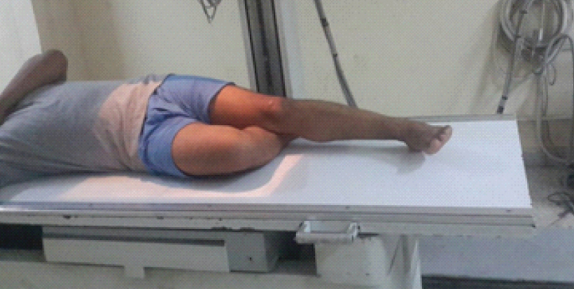
Eighty nine consecutive patients (89 paired femora) were analysed for femoral neck anteversion on digital radiographs. Patients were enrolled and subjected to detailed history and clinical examination. Biplanar X-ray method described by Ogata K et al., was used to calculate the femoral neck anteversion [6]. Femoral condylar axis was made parallel to the table by placing the patient supine with the knees flexed to 90° over the edge of the table and the legs suspended down. X-ray tube was centred over femoral neck and an Anteroposterior (AP) film was taken with beam perpendicular to table [Table/Fig-1].
Patient positioning for radiography proximal hip and femur AP view.

Subsequently, hip was flexed to 90° and rotated externally till the entire lateral aspect of the leg touched the table and lateral view of femoral neck taken. This position rotated the femur to 90° on its long axis and the condylar axis became perpendicular to the table [Table/Fig-2]. Neck axis was defined by marking two points each at widest diameter of head and base of neck; and their mid points marked. The central axis of the neck was located on each film by a line connecting these two mid points [Table/Fig-3].
Patient positioning for radiography proximal hip and femur Lateral view.
Radiograph proximal hip and femur-AP view.
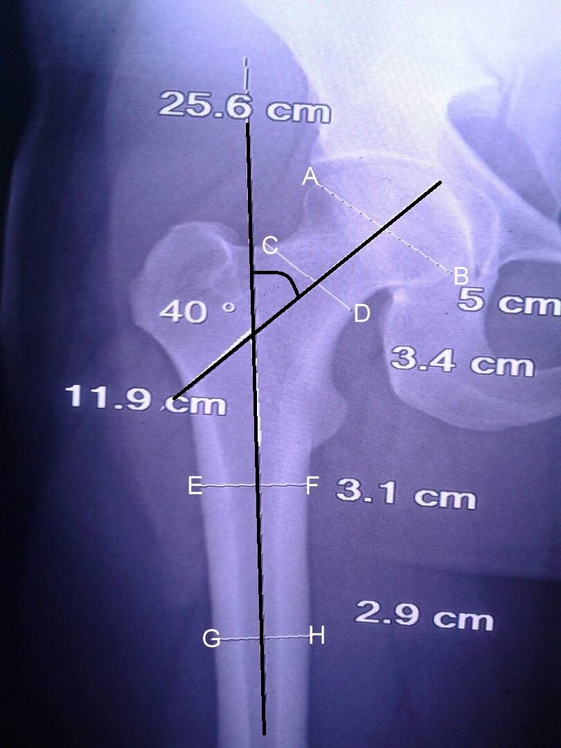
Two points were taken at outer cortex of shaft femur on both sides; just inferior to the lesser trochanter and 10 cm distal to the lesser trochanter. With reference to these points, the midpoint was measured at these two levels. The line joining these two midpoints represented the axis of the femoral shaft [Table/Fig-4].
Radiograph proximal hip and femur-lateral view.
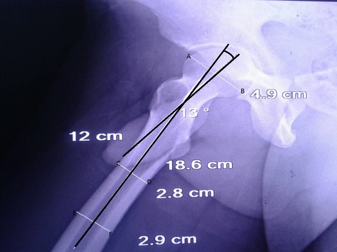
Angle was measured between the shaft axis and the neck axis on AP and Lateral films [Table/Fig-3,4]. Trignometric calculations were used to determine Femoral Neck Angle (FNA=tan of angle in lateral view/tan of angle in AP view) [7].
Group B Dry Bone Method
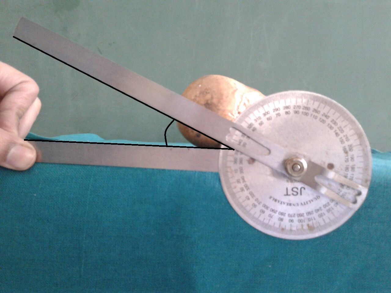
Ninety three unpaired cadaveric dry bone femora (50 Right and 43 Left) available with Department of Anatomy of our institute were analysed using Kingsley Olmsted method [8]. Age and sex of these femora were noted from Anatomy department records of our institute. Femora of unknown age and sex and those showing a significant bony or arthritic deformity were excluded from the study. The Kingsley Olmsted method was followed to determine the angle of femoral torsion in our study. Dried cadaveric femur was placed on a table with the posterior surface of its condyles and greater trochanter touching a smooth horizontal surface. Retrocondylar axis was represented by the horizontal surface against which anteversion was measured with the help of the axis of the head and neck of femur. The horizontal limb of a goniometer was fixed at the edge of the experimental table. The vertical limb was held along the axis of the head and neck of the femur. The angle between two-axis was recorded as femoral anteversion angle [Table/Fig-5].
Measurement of FNA using goniometer.
Statistical Analysis
Data obtained from digital radiography and dry bone measurements was separately analysed for values of femoral neck anteversion using SPSS trial version 23. The data was presented as frequency, percentage, and mean±SD. Difference between continuous and categorical variables was analysed using student’s t-test and chi-square test respectively. A p-value less than 0.05 were considered significant.
Results
In the present study, femoral neck anteversion in sub himalayan population presenting to a teritiary care institute was analysed using two different methods-digital radiography and dry bone measurements. Eighty nine patients (89 paired femora) were studied by digital radiography (Group A) while dry bone measurements (Group B) were used in 93 unpaired dry cadaveric femora. Mean age of the patients in Group A and Group B was 37.31±10.11 years (range: 30 to 60 years) and 46.64±7.71 years (range: 20 to 56 years) respectively with a male/female ratio of 3/1(Group A) and 5/1(Group B). It was observed that the mean femoral neck ante version was higher in digital radiography group (14.70±2.26) when compared with dry bone group (14.57±2.67) (p>0.05). Also, femoral neck anteversion dimensions were higher in males when compared with females in both groups, though statistically insignificant [Table/Fig-6]. The mean femoral neck anteversion angle in degrees for right femur was 14.60±2.67 and for left femur 14.35±2.9. The difference of means of femoral neck anteversion angle between the right and left femora was statistically non-significant (p>0.05).
Femoral neck anteversion mean males and females in digital radiography and dry bone (degrees).
| Age Group (Years) | Male | Female | p-value |
|---|
| Digital Radiography (n=66)a | Dry Bone (n=77)b | Digital Radiography (n=23)c | Dry Bone (n=16)d |
|---|
| 21-30 | 14.13±1.12 | 14.1±3.1 | 14.25±3.19 | 14±2.97 | pab=0.12;pac=0.64pcd=0.41;pbd=0.131 |
| 31-40 | 14.75±3.12 | 14.28±3.75 | 14.32±3.05 | 14.75±3.53 | pab=0.31;pab=0.53pcd*=0.07;pbd=0.151 |
| 41-50 | 15±4.12 | 14.74±3.19 | 14.77±3.72 | 14.83±2.89 | pab=0.061;pab=0.63pcd=0.41;pbd=0.71 |
| 51-60 | 15.28±3.14 | 14.96±3.07 | 15.1±2.97 | 14.92±3.54 | pab=0.171;pab=0.031pcd=0.47;pbd=0.66 |
Discussion
Measurement of femoral neck anteversion is important as abnormal femoral neck anteversion is known to be a predisposing factor to various pathological conditions like intoeing of gait, hip and knee osteoarthritis and femoroacetabular impingment. Some conditions like osteoarthritis of hip have been associated with increased as well as decreased femoral neck anteversion [2], while decreased anteversion neck femur has been associated with femoroacetabular impingment [9]. Increased femoral anteversion causes anterior displacement of the femoral head in the acetabulum and a decrease in congruity of the hip joint. Thus, improvement in the congruity of the hip joint may occur with excessive internal rotation of the hip but resulting valgus alignment at knee can cause anterior cruciate ligament injury [10].
Moreover, careful assessment of femoral neck anteversion measurement is important for surgical planning for orthopaedic procedures like derotation osteotomy of femur and total hip replacement [11]. Failure to recognise the abnormally anteverted or retroverted hip during total hip replacement may lead to abnormal range of motion and an unstable hip [12].
Variations in the values of femoral neck anteversion measurements have been reported in literature from across the globe. Therefore, analysis of anteversion femoral neck in a population assumes importance.
A multitude of methods like physical examination, radiology, CT, MRI, ultrasound and dry bone measurements are available to measure femoral neck anteversion. Measurements by physical examination are dependent on various parameters like musculoskeletal condition, dorsal/ventral decubitus attitude of patient (dorsal or ventral decubitus) and position of knee during examination of patient.
Hence, instead of measurements by physical examination, greater reliability in measurements has been reported with alternative methods [13].
Computed tomography and MRI have greater accuracy in determination of femoral neck anteversion with MRI having advantage of no radiation exposure and being able to provide greater insight into underlying pathology [14].
In the present study, two different methods were used to assess femoral neck anteversion-digital radiography and dry bone measurements. The mean femoral neck anteversion by digital radiography method was 14.70±2.26 while 14.57±2.67 by dry bone measurements.
Similarly, the dimensions of femoral neck anteversion were higher in males when compared with females, but statistically non-significant. Geographical factors like living on hilly terrain and day to day activities involving more of squatting and working in fields could be underlying factors for higher values of femoral neck anteversion in studied population.
Femoral neck anteversion with values of 13.68°±7.92° and 13° have been reported by some studies from Indian subcontinent [Table/Fig-7] [15,16]. While other studies have reported mean anteversion values of 11.50±5.90° (Males) and 11.4°±4.7° (Females) using radiography and 20.4° using CT [7,11,15,16,20].
Comparison of femoral neck anteversion among contemporary literature (Indian).
| Study | Population | Method | Measurements |
|---|
| Present Study | Indian (Sub himalyan) | Digital radiographyn=89Dry bone n=93 | Digital radiography 14.70±2.26Dry bone 14.57±2.67 |
| Jain AK et al., [7] | Indian (Delhi) | CT n=72X-ray n= 138Clinical n= 138Dry bone n=300 | X Ray 11.5°±5.4°CT 7.4°±4.6°Dry bone 8.1°±6.6°Clinical 13.1°±4.6° |
| Saikia KC et al., [17] | Indian (Guwahati) | n=92 | CT 20.4°±8.6° |
| Maheshwari AV et al., [16] | Indian (Delhi) | n=172 | CT 8.0°±4.7° |
| Srimathi T et al., [11] | Indian (Tamil Nadu) | n=164 | Dry bone 9.8° |
| Siwach RC and Dahiya S [15] | Indian | n=150 | Digital radiography 13.68°±7.92° |
Some authors have noted variations in femoral neck anteversion based on right and left side. Significantly, greater FNA was noted on left side in some studies while others reported a higher value on right side [15,16,19]. However, in our study the difference in mean values of anteversion between right and left side was insignificant.
Variability in values of femoral neck anteversion both in Indian and western studies could be due to different races, different methods of measurement and use of different anatomical landmarks while doing the measurements e.g., using transepicondylar axis rather than retrocondylar axis. The higher anteversion values can be due to persistent version due to abnormal postnatal sitting and sleeping postures [21]. Similarly, variable values for femoral neck anteversion ranging from 7° to 14° have been reported from western literature also [Table/Fig-8] [21-24].
Comparison with contemporary literature (other countries).
| Study | Population | Method | Measurements |
|---|
| Umebese PF et al., [21] | Nigerian | X-ray n=118 | X-ray 28±5° |
| Toogood PA et al., [22] | American | Dry bone n=375 | Dry bone 9.73° (-14.63 to 35.90) |
| Bargar WF et al., [18] | American | CT n=46 | CT 13.8±7.9° (-6.1 to 32.7) |
| Koerner JD et al., [23] | American | CT n=328 | CT T:8.84±9.66°;M: 8.70±9.44°; F: 9.51±10.72° |
| Yun HH et al., [24] | Korean | CT n=112 | CT T: 9.0±8.1° L: 9.0±7.4°R: 9.0±8.8° |
In our study, digital radiography was used instead of conventional radiography and measurements were done using Picture Archieving and Communication System (PACS) software provided with digital radiography, thereby increasing accuracy of measurements. Slightly higher values of femoral neck anteversion on digital radiography as compared to dry bone measurements in our study could be attributed to variations in patient positioning, rotations of the limb and magnification error on digital radiography.
Assessment of femoral neck anteversion is important in reconstructive surgery such as total hip arthroplasty and planning osteotomy about the hip. In a reconstructive surgery of the hip, especially the femoral stem replacement in hemiarthroplasty surgery, evaluation of contralateral anteversion of other hip is required for preoperative planning.
Limitation
Limitation of this study is its relatively small sample size. Much larger studies, preferably multicenter ones, would be required to expand the database of the studied population. This is a basic science study and further studies would be required to assess clinical relevance of this data in various disease conditions e.g., pathogenesis of osteoarthritis hip and also to validate it in relation to various orthopaedic surgical procedures (total hip replacement, osteotomies about the hip).
Conclusion
The study highlights the variations between the anteversion angles of femur of the sub himalayan north west Indian population with that from other parts of the globe. The data from this study will be helpful in planning of osteotomies about the hip and procedures like total hip replacement.