Vaginal agenesis is a rare congenital malformation of female genital tract. Its incidence is stated to be 1:4000-5000 female live birth [1]. Mayer Rokitansky Kuster Hauser (MRKH) syndrome is by far the most common manifestation in majority of them [2]. MRKH is often associated with renal (34%), skeletal (12%) abnormalities [3]. Renal abnormalities like unilateral renal agenesis, ectopic kidney(s), and horse shoe shaped kidney and skeletal abnormalities like fused vertebrae may be present along with vaginal agenesis [4]. Uterus and fallopian tubes are either absent or rudimentary with normal ovaries [5]. Isolated vaginal agenesis or absence of complete vagina with a normally functioning uterus is a very rare occurrence. These cases generally present with primary amenorrhea in late pubertal or early adolescent age when the patient, more often her mother gets worried about delay of the menarche. They are phenotypically female with well-developed secondary sexual character and 46 XX karyotype [6-8]. There are several non-surgical and surgical methods for treatment of vaginal agenesis. The purpose of the treatment is to create an adequate passageway for penetration and to facilitate satisfactory sexual intercourse and the McIndoe (Abbe-McIndoe-Reed) technique is the most frequently mentioned procedure in literature [9-11]. McIndoe’s technique with different modification has been mentioned in the literature. Putting a mould with graft material in the newly created neovagina is essential part of the treatment. Various graft materials have been used like split thickness skin graft, buccal mucosa and amnion etc., similarly, different material has been used to prepare the mould [12]. The study was carried out to evaluate the success of vaginoplasty in vaginal agenesis in terms of adequacy of neovaginal canal and satisfaction during sexual intercourse and to assess the acceptability and comfort level of soft mould in the postoperative period, by the patient.
Materials and Methods
This prospective study included nine patients aged 14 to 29 years underwent McIndoe’s vaginoplasty for vaginal agenesis, in Maharishi Markandeshwar Institute of Medical Sciences and Research, Mullana, Haryana, India, between January 2013 to June 2016. Approval of the institutional ethical committee was obtained before starting the study. Informed and written consent was taken from each patient in the study after explaining the procedure and their likely complications. All the patients presented with primary amenorrhea as their chief complaint. After taking a written consent from all patients a thorough general physical examination was done. They were found to have normal secondary sexual characters. Local examination revealed normal external genitalia but with blind vagina. Further evaluation was done with Ultrasound of abdomen and pelvis (USG), Magnetic Resonance Imaging (MRI) of abdomen-pelvis and Intravenousurography (IVU) when indicated. We present an innovative method of preparing mould from foam rubber [Table/Fig-1a-c] which is soft, pliable and gives excellent results. We also used roller bandage put in a condom lubricated with ointment [Table/Fig-2] for the post care to keep the neovagina patent, after removal of the first mould with skin graft; instead of hard acrylic mould which is uncomfortable to the patient during the postoperative period. All clinical parameters are depicted in [Table/Fig-3]. All the patients were explained about the surgical procedure and their complications. They were particularly briefed about the postoperative care of the neovagina. A team of gynaecologist and reconstructive plastic surgeon were involved in the surgical management. Preoperative preparation included preparation of skin graft site and operative field, bowel preparation and a pre-anaesthetic work up. Patients were given general anaesthesia.
(a) Picture of sponge slab from which the mould is taken. (b) Picture of the sponge mould with condom over it. (c) Picture of the sponge mould with skin graft over it.

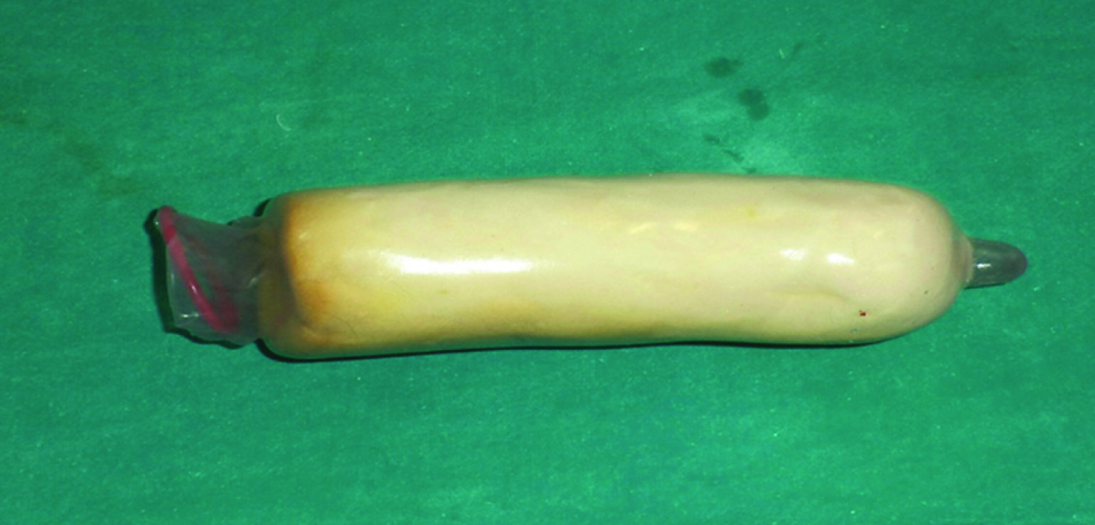
Picture of the soft mould made from roller gauze used following removal of first mould with skin graft.
Depicts clinical findings of each patient.
| Case 1 | Case 2 | Case 3 | Case 4 | Case 5 | Case 6 | Case 7 | Case 8 | Case 9 |
|---|
| Age | 18 years | 24 years | 28 years | 14 years | 26 years | 24 years | 22 years | 24 years | 23 years |
| Marital status | Unmarried | Unmarried | Married | Unmarried | Married | Unmarried | Unmarried | Unmarried | Unmarried |
| Complaint | Primary amenorrhea | Primary amenorrhea | Primary amenorrhea and Vaginismus since marriage | Primary amenorrhea and cyclical abdominal pain | Primary amenorrhea | Primary amenorrhea | Primary amenorrhea | Primary amenorrhea | Primary amenorrhea |
| Secondary sexual characters | Well developed | Well developed | Well developed | Well developed | Well developed | Well developed | Well developed | Well developed | Well developed |
| Gynecological examination | Blind vaginaNormal external genitalia | Blind vaginaNormal external genetilia | Blind vaginaNormal external genitalia | Blind vaginaNormal external genitalia | Blind vaginaNormal external genitalia | Blind vaginaNormal external genitalia | Blind vaginaNormal external genitalia | Blind vagina Normal external genitalia | Blind vaginaNormal external genitalia |
| UltrasoundMRIIVU | Uterus- HypoplasticRight ovary and kidney normalLeft ovary & kidney -absent | Uterus- HypoplasticOvaries-normalRight kidney-ectopicLeft kidney - absentIVU- Normally functioning Right ectopic kidney | Uterus RudimentaryOvaries-normal single right ectopic pelvic kidneyMRI- Confirm USG findings | Haematometra of 10 X 6 cmOvaries and kidneys normal. | Uterus-HypoplasticOvaries and kidneys normal | Uterus -Hypoplastic with normal ovaries and kidneys | Uterus -RudimentaryOvaries and kidneys normalIVU-Normal study | Uterus –Hypoplastic. Ovaries-normal Kidneys-horse shoe shapedIVUhorse shoe shaped kidney with normal function | Uterus-Hypoplastic. Ovaries and kidneys normal |
| Breast development | Tanner 4 | Tanner 4 | Tanner 5 | Tanner 3 | Tanner 4 | Tanner 4 | Tanner 4 | Tanner 4 | Tanner 4 |
| Length of neovaginaat 3 months follow up | 10 cm | 10 cm | 12 cm | 8 cm | 9 cm | 10 cm | 9 cm | 10 cm | 10 cm |
Creating neovaginal space: Operations were performed by first putting the patients in lithotomy position. After preparation of the part, bladder was catheterised with Foleys catheter [Table/Fig-4a] and a final assessment of local area and per rectal examination was done under anaesthesia. A circumlunar incision was given at the vaginal vestibule in the sub-urethral region. Space was created between the urethra and bladder anteriorly and rectum posteriorly on either side of median raphae by using progressively increasing size hegers dilator [Table/Fig-4b]. Finger was put in the rectum from time to time to ascertain the correct space between the fingers in the rectum posteriorly and Foleys in the urethra and bladder anteriorly. After creating the space of 10-12 cm, median raphae were incised [Table/Fig-4c]. An adequate diameter was created [Table/Fig-5a]. Haemostasis was ensured by using sutures and cautery. The space was packed with adrenaline saline (1:200000) soaked pack.
(a) Preoperative picture of blind vagina. (b) Intraoperative picture of neovagina with median raphae. (c) Intraoperative picture of neovagina after excising median raphae.

(a) Intraoperative pictures after creating the neovaginal space. (b) The mould (covered with the skin graft) placed into the neovagina.

Taking the graft and preparation of the mould: Lateral aspect of the thigh was prepared and a split thickness skin graft taken with a Humby’s knife. Graft site was covered with paraffin gauge dressing and bandaged. The skin graft was wound around a preprepared autoclaved sponge mould and the margins were stitched over it. The sponge mould was prepared from a commercially available sponge slab piece [Table/Fig-1a]. The mould with the skin graft wrapped over it [Table/Fig-1c] was introduced inside the created neovagina [Table/Fig-5b]. Free margins of the graft were secured by a few stitches with the labial skin to maintain the graft and mould in position. Edges of labia were stitched over the mould to secure it in its place. A ’T’-bandage was applied over the dressing at the operation site. Foley’s catheter was kept for seven days and patients were kept on IV fluid for one day then a liquid diet for another five days to avoid soiling of operation site with urine or stool. Prophylactic antibiotics were given to all cases. Dressing was changed on 7th postoperative day and the mould was removed from the newly created vagina. Graft was retained and was well taken up in all the cases. Thorough vaginal douching was given to remove the debris. A soft mould was prepared putting a condom over a 12 cm roller bandage [Table/Fig-2]. The soft lubricated mould was introduced in the vagina and dressing done with ’T’ bandage. Dressing and mould were changed on alternate days with cleaning and douching of the vaginal cavity each time. Patients were explained the method of preparing the mould, cleaning the vagina and self-introducing the mould inside the vagina. Within a few days when the patients learned the procedure themselves, they were discharged and reviewed fortnightly. Vaginal cavity was inspected for any infection, unusual discharge and condition of the graft during their follow up visits. This opportunity was utilized to assess how well the patient has learned the use of the mould, because proper use of mould is the cornerstone of the success of this surgery. Use of soft mould made from roller bandge was found to be well acceptable to the patients during this initial period. Sexual intercourse was allowed after three months if married. For unmarried women, dilator use was advised till they get married and start sexual intercourse. They were followed up for 1-2 years.
Results
Total of nine cases of vaginoplasty done from January 2013 to June 2016 are presented in this paper. Details of each case are presented in [Table/Fig-3]. Two of the patients were married and rest of them were unmarried at the time of going for the surgery. Average age of the patients was 21 years. All of them presented with primary amenorrhoea as their chief complain. The married patients also had coital difficulty as one of the major complaints. Secondary sexual characters were well developed and external genitalia was essentially normal in appearance in all the cases. Ultrasonography (USG) and MRI revealed hypoplastic or rudimentary uterus in all cases except one girl, aged 14 years (case no 4) who was found to be a case of complete vaginal agenesis with functional uterus as evident by the presence of haematometra of 10 X 6 cm size in USG and MRI. She also had complaints of cyclic dysmenorrhoea every month. All of them had normal ovaries and four of them had renal abnormalities.
Surgical procedure remained without any significant difficulty or intraoperative complication. Average operation time was 1½ to 2½ hours (mean two hours). Immediate postoperative period remained uneventful and graft were well taken in all cases. No postoperative complication was encountered in any of the cases. During their follow up visit at three month neovagina measured 8 to 12 cm, an average of about 10 cm. The married patients resumed intercourse after three months. Five of the unmarried girls got married in four to six months with initiation of sexual intercourse. One of them got divorced and came for follow up with vaginal stenosis. Case no 4 also had narrowing of neovagina when she reported for follow up. Both the cases had stopped regular use of dilator. Proper counselling about daily and regular use of vaginal dilator and its significance was reemphasised to the patients. Following resumption of regular use of vaginal dilator, desirable length and girth of vagina re-established in both of them. Cases were followed up for 1- 2 years and their vagina was found to be well formed in size and shape [Table/Fig-6a,b]. Those who are married are currently experiencing satisfactory sexual intercourse. Those who are not yet married are using dilator regularly.
(a&b) Picture of neovagina during the follow-up visit showing adequate Neovagina.

Discussion
Vaginal agenesis is one of the most common causes in patients presenting with primary amenorrhea and it is associated most commonly with Mayer-Rokitansky- Küster-Hauser syndrome [13]. The management of vaginal agenesis remains a challenge irrespective of the technique used to correct the abnormality. Historically, various non-surgical and surgical procedure have been applied by various workers to correct vaginal agenesis. Non-surgical techniques like Frank’s method and various surgical techniques like the McIndoe procedure, sigmoid vaginoplsty, Williams’ vulvogainoplasty, Vecchietti procedure, Davydove procedure etc. have been described [14].
However, McIndoe vaginoplasty is the most frequently applied surgical technique [11,15,16]. Advantages of the McIndoe technique are feasibility, high success rates and low morbidity. We also preferred this technique because the procedure is comparatively simple with a lower complication rate and gives excellent result in terms of patient’s coital satisfaction. Possibility of partial or total obliteration of vagina is its disadvantage [9-11]. Complications usually encountered during this procedure are failure of take up of skin graft, bleeding, urethrovaginal fistula, perforation of rectum, rectovaginal fistula. However, if the surgeon is careful and cautious during dissection, serious complications are few and final result is good [4]. We did not encounter any serious intraoperative complication except bleeding from the newly created vaginal bed which was successfully tackled by use of suture, cautery and adrenaline saline (1:200000) soaked packing.
Vaginoplasty operation needs a meticulous postoperative care and follow up for the neovagina to maintain its desirable shape and size. The patients are instructed to continue using the mould in the neovagina. Initially, this appears cumbersome and painful. However, with time it becomes less painful. The neovagina has a strong tendency to shrink at any stage of life in both diameter and length, in the absence of regular sexual intercourse or alternatively artificial dilatation of the neovagina [17]. Hence, a neovagina requires a lifelong maintenance in order to retain its benefits. Cooperation of the patient is of paramount importance in maintaining the neovagina in functional patency. Two of the cases in our series had stenosis of newly created vagina due to irregular use of dilator in one and non-resumption of dilatation following cessation of intercourse in another. However, they improved with resumption of regular use of dilator after proper counselling.
Many workers have used soft mould made up of various materials with good results. Sushree Jagadeb et al., has used mould made by sponge and condom [18]. Sujata Swain et al., used rubber mould and Pawan Tiwari et al., has used cotton pad as soft mould with good outcome [19,20]. Postoperative discomfort with soft mould is much less than the hard acrylic mould. Saraf S and Saraf P, also reported success with soft mould made of foam material covered with condom [4]. We have used a soft mould made of roller bandage put in a condom, for use in postoperative period. This was well acceptable by the patients and gave good result in terms of surgical outcome. The advantage of the soft mould we have used is easy availability of roller bandage in our stores and ease of preparing it even by the patients.
Limitation
It is not a comparative study. A Further larger comparative study is required with large sample size.
Conclusion
McIndoe’s vaginoplasty for treatment of vaginal agenesis is a safe and technically simple procedure with satisfactory long-term outcome. Good compliance with persistent use of the vaginal dilatation after surgery is the corner stone of success of this operation for maintaining satisfactory as well as functional neovagina. Use of soft mould in the postoperative period is a useful option.