Introduction
According to Revised National Tuberculosis Control Programme (RNTCP) annual report, one-fourth of the global TB burden is seen in India. In 2015, 28 lacs cases were estimated and 4.8 lacs people died from TB in India [1].
Various new modalities like Cartridge Based-Nucleic Acid Amplification Techniques (CB-NAAT), Microscopically Observed Drug Susceptibility (MODS), Loop Mediated Isothermal Amplification (LAMP), Line Probe Assay (LPA), Xpert MTB/RIF are employed in the diagnosis of TB [2]. But they are costly; consumables are not easily available and require technical expertise which can limit the utilisation of automated methods. Also the introduction of automated methods does not mean to replace the need for culture as mycobacterial culture is still needed for [1]. Surveillance of drug resistance as part of control programme performance [2] follow up of patients who fail on treatment regimens to check for drug resistance for drug susceptibility and identification tests which include both genotypic and phenotypic tests [3].
In developing countries like India, LJ medium is the most commonly used medium for the culture of M. tuberculosis. Growth of M. tuberculosis is slow, appear in two to six weeks and negative culture report cannot be given before eight weeks [4]. However few research papers published such as Drancourt M and Raoult D, Satti L et al., Coban AY et al., suggest that BA can also be used for the isolation of M. tuberculosis [5-7].
In the present study, an attempt has been made to assess the utility of BA for isolation of M. tuberculosis with LJ medium and BACTEC MGIT 960. To make it a selective medium, antibiotics and antifungal drugs were added. Further identification of the isolates recovered from BA were confirmed by Genotype MTBDR plus assay (Hains Lifescience, Nehren, Germany) targeting 23rRNA, which was available at the STDC-IRL.
If this study succeeds in proving the M. tuberculosis culture on BA as a possible modality in TB diagnostics, then it will be a path breaking finding in this field.
Materials and Methods
In this prospective study, samples were collected from the initial screened 100 clinically suspected TB cases attending Designated Microscopy Centre (DMC) of Government. General and Chest Hospital based on certain inclusion and exclusion criteria as per RNTCP guidelines [3] and were further processed in the STDC-IRL.
Inclusion Criteria
The study group includes patients with more than any two symptoms out of the following: 1. Fever and cough with expectoration >2 weeks; 2. Abnormal findings in chest radiograph; 3. Gradual weight loss; 4. HIV status (positive/negative); 5. Contacts of smear positive TB patients having cough of any duration. A patient with extra pulmonary TB may have general symptoms like weight loss, fever with evening rise and night sweats. Other symptoms depend on the organ affected [3].
Exclusion Criteria
Cases already on Anti Tuberculous Treatment (ATT);
Cases already confirmed as TB by other microbiological methods [3].
It included 95 sputum, 3 pleural fluid, 1 pus and 1 lymph node sample. Two sputum samples were collected from each patient, on the spot and early morning sputum sample. The extra pulmonary samples were collected by the treating physician under strict aseptic conditions. The informed consent of the patients was taken before the collection of the specimens.
The samples were then decontaminated and concentrated by using N-acetylcysteine Sodium Hydroxide method [3] for sputum samples and sulfuric acid [8] for extra pulmonary samples. If any delay in the processing, the samples were stored in the refrigerator at 4°C. A microscopic examination (ZN staining) followed by the parallel inoculation on 7% Sheep BA, LJ and BACTEC MGIT 960 (Becton, Dickinson and Company) was done. LJ medium were prepared as per RNTCP guidelines [3]. BA slants were prepared according to Mathur ML et al., instead of using petri dishes; McCartney bottles were used for BA to avoid desiccation [4]. About 50 μL of concentrated sample was inoculated on: (i) two plain LJ slants and one LJ slant with Para Nitro Benzoic (PNB) acid (500 μg/mL); (ii) two BA slants and one BA slant with PNB and 0.5 mL of the inoculum was added to MGIT tubes in duplicate. Growth obtained on solid medium and MGIT tubes were further tested for the presence of AFB by ZN staining and four biochemical tests, i.e., Niacin, Nitrate, Heat resistant catalase test and PNB test to differentiate the growth into M. tuberculosis and NTM. Standard controls Mycobacterium smegmatis (ATCC700084) and M. tuberculosis H37Rv (ATCC19977) were used in the study.
An isolate was designated to be that of M. tuberculosis, if it grew slowly (taking more than 7 days), showing no growth on medium containing PNB and which showed positive niacin and nitrate reduction tests and a low catalase activity. Any growth seen within seven days of incubation or which did not meet the above criteria was considered as NTM [3].
The genotype MTBDR plus assay, a multiplex PCR DNA strip test assay (Hains Lifescience, Nehren, Germany) was done to detect the molecular genetic identification of M. tuberculosis complex from BA culture positive samples. Step by step procedure was done as instructed by the manufacturers (DNA extraction, multiplex amplification with biotinylated primers and reverse hybridisation) [9]. The study was approved by the Ethics Committee, Osmania Medical College, Koti, Hyderabad, India.
Statistical Analysis
OpenEpi: Open Source Epidemiologic Statistics for Public Health, Version. www.OpenEpi.com, updated 2013/04/06 was used for statistical analysis. The Yates corrected Chi-square test was used to analyse the given data. A p-value <0.05 was considered significant.
Results
A total of 100 cases were included in the study, 70 were males and 30 females. The positivity rate of ZN stain was found to be maximum in the 20-40 age group (11 of 21 smear positives). Out of 100 cases, 14 were HIV positive of which five were smear positive for AFB. About 21 were smear positive for AFB of which 19 showed growth on BA, 16 on LJ medium and 21 on BACTEC MGIT 960. Out of the 79 smear negative cases, 15 showed growth on BA, 14 on LJ and 17 on MGIT 960. M. tuberculosis was isolated from 39 samples overall, sensitivities being 87.17% (34 out of 39) for BA, 76.9% (30 out of 39) for LJ and 97.3% (38 out of 39) for BACTEC MGIT 960.
About 39 M. tuberculosis isolates were obtained from any of the 3 culture media. NTM isolates were 3 on BA, 2 on LJ and 2 on MGIT 960 and the rate of contamination was 12% in MGIT 960, 6% on LJ and 2% in BA [Table/Fig-1].
Performance of different culture types.
| Total No. of Samples (n=100) | BA (n=34) | LJ (n=30) | MGIT (n=38) | Any culture positive (n=39) |
|---|
| Origin of Samples |
| Sputum (n=95) | 32 | 29 | 36 | 37 |
| Pleural fluid (n=3) | 1 | 1 | 1 | 1 |
| Pus (n=1) | 1 | 0 | 1 | 1 |
| Lymph node (n=1) | 0 | 0 | 0 | 0 |
| Smear positive (n=21) |
| Number of culture positive | 19 | 16 | 21 | 21 |
| Contaminated samples | 0 | 0 | 0 | 0 |
| Non Tuberculous Mycobacteria | 0 | 0 | 0 | 0 |
| Smear Negative (n=79) |
| Number of culture positive | 15 | 14 | 17 | 18 |
| Contaminated samples | 2 | 6 | 12 | - |
| Non Tuberculous Mycobacteria | 3 | 2 | 2 | - |
| Sensitivity | 87.17% | 76.9% | 97.3% | - |
In our study, the specificity, positive predictive value, and negative predictive value of BA were 97.06%, 92.42% and 86.84% when compared to BACTEC MGIT 960. The specificity, positive predictive value, and negative predictive value of BA when compared to LJ was observed to be 100%, 100%, and 94.29%, respectively [Table/Fig-2].
Specificity, positive predictive value, negative predictive value of Blood agar when compared to Lowenstein Jensen and BACTEC MGIT 960.
| Variables | Blood agar when compared to Lowenstein Jensen medium | Blood agar when compared to BACTEC MGIT 960. |
|---|
| Specificity | 100% | 97.06% |
| Positive predictive value | 100% | 92.42% |
| Negative predictive value | 94.29% | 86.84% |
In the present study, a significant difference (p-value < 0.001), was observed to detect M. tuberculosis culture positivity on BA when compared to LJ [Table/Fig-3]. Thirty isolates were culture positive on both BA and LJ medium, in addition BA could isolate four cases which were negative on LJ medium [Table/Fig-3].
Comparison of results of culture between Blood agar and Lowenstein Jensen medium.
| Variables | Blood agar culture positive | Blood agar culture negative |
|---|
| LJ culture positive | 30 | 0 |
| LJ culture negative | 4 | 66 |
χ2= 79.04; p value = <0.001.
In the present study, a significant difference (p-value <0.001) was observed to detect M.tuberculosis culture positivity on BA and MGIT 960. MGIT could isolate additional 5 M. tuberculosis which were culture negative on BA. But, there was one isolate which was negative by BACTEC MGIT 960 but positive for culture on BA [Table/Fig-4].
Comparison of results of culture between Blood agar and BACTEC MGIT 960.
| Variables | Blood agar culture positive | Blood agar culture negative |
|---|
| BACTEC MGIT 960 positive | 33 | 5 |
| BACTEC MGIT 960 negative | 1 | 61 |
χ2 = 72.51; p value= <0.001
The minimum time taken by M. tuberculosis to grow was eight days on BA, 21 days on LJ and six days on MGIT 960. The maximum time taken was 18 days on BA, 36 days on LJ, and 16 days on MGIT 960 [Table/Fig-5].
Duration of incubation for isolation of Mycobacterium tuberculosis on Blood Agar, Lowenstein Jensen and MGIT 960.
| Duration of incubation (days) | Blood agar – No. of isolates | LJ – No. of isolates | BACTEC MGIT 960-No. of isolates |
|---|
| 1 – 7 | - | - | 2 |
| 8 – 14 | 25 | - | 30 |
| 15 – 21 | 9 | 2 | 6 |
| 22 – 28 | - | 17 | - |
| 29 – 35 | - | 10 | - |
| 36 – 42 | - | 1 | - |
| Total | 34 | 30 | 38 |
Out of the 37 culture positive cases on BA, 34 samples showed TUB band in the Genotype MTBDR plus assay version 2.0, thus confirming the isolates belonging to MTB complex and remaining three showed no band confirming it as NTM.
Discussion
Robert Koch isolated M. tuberculosis from freshly crushed pulmonary tubercles using heat coagulated sheep and beef serum medium after 10 days of incubation in tubes [10]. Isolates were recovered using BA, but were replaced by an egg based agar which became the recommended medium for isolation of M. tuberculosis in 1920’s as the egg based medium is easy to sterilize [10]. Drancourt M et al., were the first to report the growth of M. tuberculosis colonies on BA [10]. The Indian studies conducted by Mathur ML et al., Rajdev S et al., Palange P et al., also have positively evaluated the utility of BA for primary isolation as well as Drug Susceptibility Testing (DST) of M. tuberculosis [4,11,12].
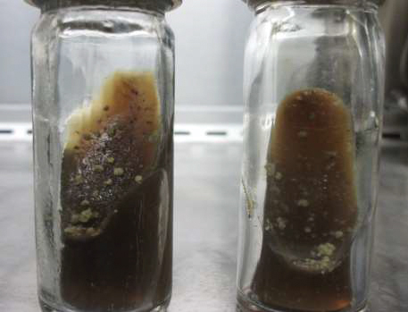
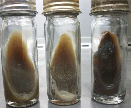
In our study, BA showed sensitivity of 87.17% as comparable to findings of Mathur ML et al., 94.2%, Satti L et al., 90%, Palange P et al., 89.3%, Shidiki A and Pokhrel N 98.9% [4,6,12,13]. LJ medium and MGIT 960 showed sensitivity of 76.9% and 97.3% in the present study. The mean duration of isolation of M. tuberculosis on BA was 13.2 days which are similar to findings done by Mathur ML et al., 13.6 days and, Satti L et al., 14 days [4,6], Palange P et al., 18.4 days and Shidiki A and Pokhrel N reported 13.6±5.2 days on BA [12,13]. The mean duration of isolation in LJ was 26 days and in MGIT 960 was 11.8 days in our study. The colonies on BA are pinpoint in size, light gray, glistening and easily recognized when examined under bright light, gradually increase in size, become irregular, warty, or cauliflower like colony [Table/Fig-6]. Growth of NTM also observed on BA [Table/Fig-7]. But the limitation is that Mycobacteria species are morphologically indistinguishable on BA.
Blood agar showing growth of Mycobacterium tuberculosis.
Growth of Non Tuberculous Mycobacteria on Blood Agar.
The cost to prepare one BA bottle was Rs 11/-, while one LJ medium was Rs 9/- and MGIT cost Rs 190/- per bottle. Also, it is easy to prepare in comparison to LJ.
In our study, BA showed good results when compared to gold standard LJ medium in sensitivity being 87.17%, mean duration of isolation being 13.2 days and least contamination rate being 2% while on LJ medium, sensitivity was 76.9%, mean duration of isolation was 26 days and contamination rate was 6%. Apart from our study, there are other 3 studies which compared BA with automated system for isolation of M.tuberculosis. Drancourt M et al., compared BA with BACTEC 9000 MB broth and reported superior performance of BA (98.9% versus 92.6%) over BACTEC 9000 MB broth and duration of isolation was 26 days on BACTEC and 19 days on BA [5]. Mathur ML et al., reported isolation of M. tuberculosis took 9 days by MGIT and 13.6 days on BA [4]. Coban AY et al., reported 10 days on MGIT and 14 days on BA [7]. In our study, the contamination rate on BA was 2%, preferably least when compared to LJ and MGIT 960 which were 6% and 12%. Mathur ML et al., observed a contamination rate of 1.6% on BA and 7.8% on LJ medium [4]. Satti L et al., reported contamination rates of 2.8% and 4.2% on BA and LJ medium and Palange et al., showed 7.2% on BA and 4.8% on LJ medium [6,12]. No contamination was observed in BA, LJ or MGIT 960 supporting NTM growth.
Few studies like Coban AY et al., [14] and Shidiki A and Pokhrel among others have also gone to a further extent of using BA for (DST) of M. tuberculosis [13]. Though our study has not been involved in DST of M. tuberculosis but it can suggest that BA can support the growth of M. tuberculosis, the contamination rate was lower and would be at least as good as, if not better than, LJ medium for recovering M. tuberculosis.
Limitation
In the present study, the sample size was low. Growth of M. tuberculosis is morphologically indistinguishable from other Mycobacterial species on BA.
Conclusion
Based on the above findings, it can be said that BA can provide as a good substitute over LJ medium in terms of rate of isolation, duration of isolation and sensitivity. The contamination rate on BA was also low as compared to LJ medium. Although the sample size was less and BA lacked a little behind over MGIT 960 in terms of sensitivity and duration of isolation but the contamination rate was higher in MGIT as compared to BA. Also MGIT 960 requires sophisticated equipment, expensive kits and skilled manpower while BA is easy to prepare, inexpensive and requires only an incubator, basic equipment in clinical laboratory provided appropriate microbiological safety measures are in place to counter the highly infectious nature of M. tuberculosis.
χ
2= 79.04; p value = <0.001.
χ
2 = 72.51; p value= <0.001
[1]. Govt. of India. Ministry of Health and Family welfare. Revised National TB Control Programme-annual status report: TB India; 2017;page 173 [Google Scholar]
[2]. Sharma K, Appannanavar SB, Goyal K, Sharma A, Recent advances in the diagnosis of tuberculosisJ Postgrad Med Edu Res 2013 47(4):181-87.10.5005/jp-journals-10028-1083 [Google Scholar] [CrossRef]
[3]. Revised National TB Control Programme Training Manual for Mycobacterium tuberculosis Culture and Drug susceptibility testing. Central TB Division, Directorate General of Health Services. Printed April 2009. Accessed: 25th March,2014: http://tbcindia.nic.in/WriteReadData/l892s/6995271860Training%20manual%20M%20tuberculosis%20C%20DST.pdf [Google Scholar]
[4]. Mathur ML, Gaur J, Sharma R, Solanki A, Rapid culture of Mycobacterium tuberculosis on blood agar in resource limited settingDan Med Bull 2009 56:208-10. [Google Scholar]
[5]. Drancourt M, Raoult D, Cost-effectiveness of blood agar for isolation of mycobacteriaPLoS Negl Trop Dis 2007 1(2):e8310.1371/journal.pntd.000008318060087 [Google Scholar] [CrossRef] [PubMed]
[6]. Satti L, Ikram A, Coban AY, Martin A, Rapid direct testing of susceptibility of Mycobacterium tuberculosis to Isoniazide and Rifampicin on Nutrient and Blood agar in Resource limited settingsJ Clin Microbiol 2012 50(5):1659-62.10.1128/JCM.00013-1222357498 [Google Scholar] [CrossRef] [PubMed]
[7]. Coban AY, Akqunes A, Durupinar B, Evaluation of Blood agar medium for the growth of mycobacteriaMikrobiyol Bul 2011 45(4):617-22. [Google Scholar]
[8]. Koneman EW, Allen SD, Janda WM, Schrenkenberger PC, Winn WC, Procop GW, Color Atlas and Textbook of Diagnostic Microbiology 2006 6th edPhiladelphiaLippincott Williams and WilkinsChapter 19, Mycobacteria; pp.1066-1124 [Google Scholar]
[9]. Hains Life Sciences. Genotype MTBDR plus – Manual of Standard Operating Procedures [Google Scholar]
[10]. Drancourt M, Carrieri P, Gévaudan MJ, Raoult D, Blood agar and Mycobacterium tuberculosis: the end of a dogmaJ Clin Microbiol 2003 41(4):1710-11.10.1128/JCM.41.4.1710-1711.200312682165 [Google Scholar] [CrossRef] [PubMed]
[11]. Rajdev S, Patel A, Mulla S, Use of blood agar along with Lowenstein Jensen media for rapid isolation of Mycobacterium tuberculosis for early drug susceptibility testingJ Acad Clin Microbiol 2014 16(2):100-103.10.4103/0972-1282.144737 [Google Scholar] [CrossRef]
[12]. Palange P, Narang R, Kandi V, Evaluation of culture media for isolation of mycobacterium species from human clinical specimensCureus 2016 8(8):e75710.7759/cureus.757 [Google Scholar] [CrossRef]
[13]. Shidiki A, Pokhrel N, A comparision of blood and egg based media for the rapid isolation and drug susceptibility testing of Mycobacterium TuberculosisResearch Journal of Pharmaceutical, Biological and Chemical Sciences 2013 4(1):736-43.:0975-8585. [Google Scholar]
[14]. Coban AY, Blood agar validation for susceptibility testing of isoniazid, rifampicin, ethambutol, and streptomycin to Mycobacterium tuberculosis isolatesPLoS ONE 2013 8(2):e55370Available from https://doi.org/10.1371/journal.pone.005537010.1371/journal.pone.005537023405140 [Google Scholar] [CrossRef] [PubMed]