Introduction
Microleakage is a major problem beneath deep Class II composite restorations where little or no enamel remains. One technique to overcome this problem is sandwich restoration in which an intermediate liner is sandwiched between tooth and composite restoration.
Aim
This study evaluated and compared gingival microleakage at tooth restoration interface in deep Class II composite closed sandwich restorations (mesio-occlusal) using different liners like Resin Modified Glass Ionomer Cement (RMGIC), Biodentine, Theracal LC.
Materials and Methods
Standardised conventional mesio-occlusal cavities were prepared on 40 extracted maxillary first premolars with dimension 2 mm buccolingually and the gingival seat placed at the level of cementoenamel junction. Teeth were divided into four groups (n=10). In Group 1 (control group) cavities were restored with composite (Filtek 250) using self etch bonding agent (scotch bond). In Group 2, 3 and 4, 0.8 mm thick liner of RMGIC, Biodentine and Theracal LC were applied respectively on the axial wall of the cavity. All the cavities were then restored with composite using self-etch bonding agent similar to Group 1. The specimens were then immersed in 0.5% aqueous solution of rhodamine B dye for 24 hours, sectioned and observed for the extent of dye penetration using confocal laser scanning microscope. Statistical analysis was performed using Kruskal-Wallis test for intergroup comparison followed by Mann-Whitney test for intragroup comparison.
Results
Microleakage scores indicated that use of a liner beneath deep Class II composite restorations significantly reduced microleakage. Among the liners used, Biodentine and Theracal LC showed better results than RMGIC.
Conclusion
This study concluded that use of a liner beneath deep Class II composite restoration reduced microleakage and Theracal LC performed similar to Biodentine and better than RMGIC, when used as a liner in deep Class II composite closed sandwich restorations.
Introduction
The substitution of dentine and enamel, that is lost either by caries or as a result of mechanical removal, with a restorative material is always a challenge to the clinician. A successful restoration should protect and support the tooth from further compromise and also prevent pulpal damage [1].
Composites are the commonly used restorative materials because of their superior aesthetic qualities, minimal removal of tooth structure and also they can be directly placed without any lab procedure [2]. But, the most undesirable characteristic of composite resin is its polymerisation shrinkage and bonding to the deeper dentine which leads to gap formation and microleakage over time [3]. Microleakage can lead to postoperative pain, recurrent caries and even pulpal damage. Several techniques were tried to overcome the problems associated with polymerisation shrinkage and one among that is the open sandwich technique where a liner is placed beneath the composite restoration. One concern with open sandwich technique is that the liner is exposed to the oral environment. To compensate this closed sandwich technique was introduced where a layer of composite is placed over the liner [4].
An ideal liner required for deep dental caries requiring pulp capping should have the ability to kill bacteria, induce mineralisation, establish a tight seal, be less soluble, pulpal protection, healing and good bonding to dentine and overlying composite restoration [5].
RMGIC has been successfully used as a liner in sandwich technique since many years because of its definite advantages like it bonds well to the tooth, its coefficient of thermal expansion is similar to dentin with the property to command set. It shows superior mechanical properties, less dissolution rate and good sealing ability as compared to conventional GIC. One of the main disadvantage of RMGIC was again shrinkage, due to resin component and technique sensitivity. Also, the monomers that leach out of RMGIC are said to have noxious effects on the pulp [6].
In deep carious lesion, liners which can promote dentin deposition are preferred. These materials should serve the purpose of a liner as well as an indirect pulp capping agent. Some of the materials used as liners under restorations in deep cavities include calcium hydroxide, mineraltrioxide aggregate, Biodentine and Theracal LC.
Calcium hydroxide is the gold standard pulp capping agent for many years. It is antibacterial and can neutralise the acidic bacterial byproducts. The high pH creates an environment conducive to the formation of reparative dentin. However, it has some disadvantages like is high solubility, it gets lost during acid etching procedure done prior to placement of composite and the dentin bridge formed by calcium hydroxide has also shown tunnel defects which can be a source for bacterial reinfection [7].
A calcium silicate cement (MTA) introduced by Dr. Mahmoud Torabinejad has been used as a material for pulp capping, because of its good sealing ability preventing bacterial leakage and the ability to stimulate cementum, bone and dentin. These properties have helped MTA unsurp the position of a gold standard in pulp capping, previously held by calcium hydroxide. However, it has some disadvantages, like long setting time (2 hours 45 miutes), low compressive strength, staining of teeth etc., which lead to the development of Biodentine [7,8].
Biodentine, developed by Septodont could overcome most disadvantages of MTA. It has a shorter setting time of 12 minutes. Its mechanical properties such as compressive strength and modulus of elasticity is similar to natural dentine and has sufficient strength to withstand occlusal loading. Only problem is that it is little difficult to handle and there is no direct bonding with composite restoration [8,9].
Theracal LC is a new light cured resin-modified calcium silicate-filled base/ liner material designed for pulp capping. The resin consists of a hydrophobic component (comprising hydrophobic monomers) such as Urethane Dimethacrylate (UDMA), Bisphenol A- Glycidyl Methacrylate (BisGMA), Triethylene Glycol Dimethacrylate (TriEDMA or TEGDMA) and a hydrophilic component (containing hydrophilic monomers) such as Hydroxyethyl Methacrylate (HEMA) and Polyethylene Glycol Dimethacrylate (PEGDMA) [9]. Theracal LC has the advantage of command set and it bonds directly to composite [10].
As there are no published data evaluating the microleakage of Theracal LC as liner, the aim of the study is to compare the gingival microleakage of Theracal LC, RMGIC and Biodentine in posterior deep Class II closed sandwich composite restoration.
Materials and Methods
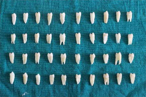
This in vitro study was carried out in the Department of Conservative dentistry and Endodontics of Dayananda Sagar College of Dental Sciences, Bengaluru, Karnataka, India. Forty intact human maxillary first premolars indicated for extraction for orthodontic or periodontal reason were selected for the study and stored in saline [Table/Fig-1]. Carious, restored, hypoplastic and fractured teeth were avoided. The teeth were mounted with adjacent teeth in an artificial dental jaw model. Standardised conventional mesio-occlusal preparations were made on these teeth using No. 245 bur with an airotor hand piece. The dimensions of these preparations were 2 mm buccolingually and the gingival seat placed at the level of CEJ and 1.5 mm deep axially, verified using a periodontal probe. No. 245 bur was changed after every five preparations. Tofflemire matrix band retainer was adapted to the preparation to prevent gingival overhang of the restoration.
Forty premolars specimen.
Restorative Procedure
Group1: Self-etch bonding agent (Scotch bond universal) was applied with an applicator tip to the entire cavity and light cured for 20 seconds. Cavities were then restored with composite resin and light cured for 30 seconds.
Group 2: About 0.8 mm thick liner of RMGIC was applied on the axial wall. Self etch bonding agent (Scotch bond universal) was applied to the entire cavity including the liner and light cured for 20 seconds followed by restoration with composite resin as in Group 1.
Group 3: About 0.8 mm thick liner of Biodentine (Septodont) was applied on the axial wall. Self etch bonding agent (Scotch bond universal) was applied to the entire cavity including the liner and light cured for 20 seconds followed by restoration with composite resin as in Group 2.
Group 4: About 0.8 mm thick liner of Theracal LC was applied on the axial wall. Self etch bonding agent (Scotch bond universal) was applied to the entire cavity including the liner and light cured for 20 seconds followed by restoration with composite resin as in Group 3.
Preparation for Microleakage Test
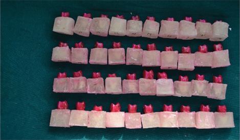
After the restorations were completed, excess proximal flash was removed with a sharp hand scaler. The apical foramen was sealed by embedding the teeth in acrylic resin blocks and two coats of nail varnish applied to the entire tooth, except for the restoration and 1 mm around it [Table/Fig-2]. The specimens were then subjected to thermocycling at temperatures of 5±1°C and 55±1°C for 1000 cycles with 30 seconds dwell time to simulate oral conditions.
Mounted specimens after insertion in dye.
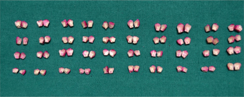
The specimens were then stored in humidor and immersed in 0.5% aqueous solution of rhodamine B dye for 24 hours. The dye was then rinsed off with water to remove excess dye. The portions of the root embedded in acrylic blocks were severed and the specimens were sectioned mesiodistally through the centre of restoration with a diamond disc under copious water spray [Table/Fig-3]. Buccal half of the tooth section was retained and the lingual section was discarded. The entire tooth section was polished with aluminium oxide paste to obtain high degree of flatness. The tooth restoration interface was observed for the extent of dye penetration using confocal laser scanning microscope (ZEISS LSM) of 10X magnification and scored according to the study done by Tredwin CJ et al., [11].
Scores (C J TREDWIN)
0 No dye penetration
1 Dye penetration upto1/3rd gingival seat axially
2 Dye penetration 1/3rd to 2/3rd gingival seat axially
3 Dye penetration in excess of 2/3rd gingival seat axially
4 Extensive dye penetration at the entire gingival seat upto axial wall.
Statistical Analysis
The scores obtained were statistically analysed by Kruskal-Wallis test. Later to find significant differences between different groups Mann-Whitney was carried out. SPSS version 22.0 software was used for the analysis. Level of significance is set at p-value <0.05.
Results
Higher mean microleakage was recorded in Group 1 (no liner) followed by Group 2 (RMGIC), Group 4 (Theracal LC) and Group 3 (Biodentine) respectively. The difference in mean microleakage among the groups was found to be statistically significant [Table/Fig-4].
Mean microleakage recorded in the groups.
| Group | Mean | Standard deviation | Standard error of Mean | 95%Cl for Mean | Kruskal-Wallis Chi -Square | p-value |
|---|
| Lower bound | Upper bound |
|---|
| Group 1 | 4.00 | 0.00 | 0.00 | 4.00 | 4.00 | 26.551 | <0.001* |
| Group 2 | 3.20 | 0.79 | 0.25 | 2.64 | 3.76 |
| Group 3 | 2.10 | 0.32 | 0.10 | 1.87 | 2.33 |
| Group 4 | 2.30 | 0.67 | 0.21 | 1.82 | 2.78 |
*=significant difference
Group 1-No liner; Group 2-Resin modified glass ionomer cement; Group 3-Biodentine; Group 4: Theracal LC
In order to find out between which pair of groups there exist a significant difference, Mann-Whitney U test was applied for pair-wise comparisons.
The difference in mean microleakage was found to be statistically significant between Group 1 and Group 2 (p<0.01), Group 1 and Group 3 (p<0.001), Group 1 and Group 4 (p<0.001), Group 2 and Group 3 (p<0.001) as well as between Group 2 and Group 4 (p<0.05). No significant difference was observed between Group 3 and Group 4 (p>0.05) [Table/Fig-5].
Mean microleakage difference between groups.
| Groups | Mean Difference | z | p-value |
|---|
| Group 1 | Group 2 | 0.800 | -2.814 | 0.005 * |
| Group 3 | 1.900 | -4.264 | <0.001* |
| Group 4 | 1.700 | -3.873 | <0.001* |
| Group 2 | Group 3 | 1.100 | -3.104 | 0.002* |
| Group 4 | 0.900 | -2.466 | 0.014 * |
| Group 3 | Group 4 | -0.200 | -0.669 | 0.503 |
*=significant difference
Group 1-No liner; Group 2-Resin modified glass ionomer cement; Group 3-Biodentine; Group 4: Theracal LC
Discussion
Deep carious lesions cause pulpal inflammation (i.e., pulpitis); if not managed, they may result in pulp necrosis and involvement of the periradicular tissues, with possible pain requiring endodontic treatment or extraction [12]. One of the major disadvantage of root canal therapy is the loss of proprioception which can lead to excessive transmission of occlusal load and can ultimately lead to catastrophic fractures. So, it is always preferred to maintain the vitality of teeth whenever possible [13]. The choice of restorative materials for deep dental caries depends on many factors like location, remaining dentin thickness etc.
The increase in demand for tooth coloured restorations along with the concerns regarding mercury toxicity has lead to a dramatic decrease in the use of amalgam restorations. Resin composites represent the materials most commonly used as an alternative to amalgam. It is largely due the aesthetic results, requirement of little to no preparation, acceptable longevity, and relatively low costs. However, it has relatively high surface roughness, low polishability, poor resistance to staining and poor bonding at the tooth restoration interface, which, in combination with polymerisation shrinkage resulted in margin degradation and microleakage [14].
As microleakage is a major drawback in composite restorations, over years researchers have been working on ways or methods to reduce polymerisation shrinkage, hence microleakage. Diverse materials and techniques have been developed to decrease the shrinkage like using non shrinking resins, modifying filler particles, using low elastic modulus liners, various layering techniques and altering the degree of conversion of monomer systems by varying curing source and technique [15].
Another technique used to counteract the contraction stresses is “sandwich technique” where a liner is used between the dentin and the composite restoration. They are of two types-closed sandwich and open sandwich technique. In a closed sandwich, liner is completely covered by restorative material, whereas in open sandwich the liner is exposed to the oral environment [16].
The liners commonly used in deep dental caries with remaining dentin thickness less than 0.5 mm are calcium hydroxide and calcium silicate cements like MTA, Biodentine and Theracal LC [17].
In this study, dye penetration in each sample was scored according to the scores given by Tredwin CJ et al., [11]. In the present study, the depth of gingival seat was 1.5 mm of which 0.8 mm was occupied by liner and 0.7 mm filled with composite (Group 2,3 and 4) and entire 1.5 occupied by composite in Group 1. Tredwin’s Score ‘0’ indicates no dye peneteration, Score ‘1’ indicates dye peneteration upto 1/3rd gingival seat axially, which in this study is 0.5 mm marginal leakage in the gingival seat. So, leakage is completely beneath composite restoration in Score ‘1’. Similarly, Score ‘2’ indicates dye peneteration 1/3rd to 2/3rd gingival study axially which is about 0.3 mm marginal leakage in the gingival seat beneath the liner in this study. Tredwins Score ‘3’ denotes dye peneteration in excess of 2/3rd gingival seat which means leakage more than 0.3 mm in the gingival seat beneath the liner in our study. Score ‘4’ indicates dye peneteration at the entire gingival seat upto the axial wall which in this study indicates leakage beneath entire 0.8 mm of liner in gingival seat and extending to the axial wall.
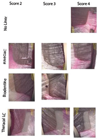
On evaluating the results of the study, the highest marginal leakage was seen in composite restoration placed directly on the dentine without any liner. All the specimens showed leakage score ‘4’ [Table/Fig-6]. Thus, the use of liner reduces microleakage in cervically placed deep Class II composite restorations.
Confocal microscopic images of all groups.
Lower scores in other Groups (Group 2,3,4) were attribuited to the presence of a liner. Liners are less rigid and could have a modulus of elasticity 20-30% lower than conventional hybrid composites. Liners overcome polymerisation shrinkage concept of elastic cavity wall [18].
Also, application of liners reduces the volume of composite in the cavity. By decreasing the bulk amount of resin used, volumetric shrinkage will be less which reduces the stress generated as well as microleakage [19].
Liner also prevents the composite resin from bonding with the dentin. The reduction in bonded composite surfaces decreases the configuration factor (C-factor) of the cavity. As a result, the tensile stress generated by the polymerisation contraction of the composite is also reduced [20].
Among the groups RMGIC showed statistically significant higher leakage values as compared to Theracal LC and Biodentine when used as a liner beneath closed sandwich restorations. About 40% of the samples with RMGIC liner showed Score ‘4’, next 40% showed Score ‘3’ and the remaining 20% showed Score ‘2’. So here majority of samples exhibited more leakage beneath RMGIC.
Some researchers state that, RMGIC bonds get distrupted with dentin, mainly in the initial stages of GIC maturation due to contraction forces which occur within polymerising composite resin. So, the polymerisation stress leads to pulling away of RMGIC from dentin and cementum during polymerisation of composite resin layer [21].
Monomers may diffuse through tubules and can have direct, toxic effects on pulpal cells [22]. Resin content in RMGIC also increases polymerisation shrinkage leading to increased risk of microleakage beneath these liners [23].
Stability, setting time is short enough to complete the whole procedure in single appointment unlike other calcium silicate based cements and has good compressive strength to withstand occlusal forces when overlaid by composite resin. Good marginal integrity of sandwich restorations filled with Biodentine and Theracal LC is also likely due to the ability of the calcium silicate materials to form hydroxyapatite crystals at the surface, when formed at the interface between the restorative material and the dentin walls, these crystals may contribute to the sealing efficiency of the material [24]. This explains the reason for lower leakage values seen beneath Biodentine and Theracal LC in Class II closed sandwich restoration in this study. Biodentine performs better even when margins are located in cementum. It forms tag like structures at the interface which is an advantageous property over RMGIC which is sensitive to moisture [25].
Among the calcium silicate cements used in the present study, Biodentine showed less leakage compared to Theracal but the difference was not statistically significant. Nearly, 90% of Biodentine samples showed Score ‘2’ leakage that is less than 0.3 mm microleakage beneath the liner. Remaining 10% showed Score ‘3’. Whereas, in Theracal LC 80% of samples showed Score ‘4’, 10% exhibited Score ‘3’ and the remaining 10% showed Score ‘4’ leakage respectively which means marginal leakage below the entire 0.8 mm liner gingivally and even extending to the axial wall was also seen with Theracal LC [Table/Fig-6]. This might be due to the presence of a resin matrix in Theracal LC which might have modified some of its properties [9]. Polymerisation shrinkage due to its resin content might be a contributary factor as well.
Within the limitations of the present in vitro study, calcium silicate cements proved to be a better option as a protective liner beneath deep Class II closed sandwich restoration.
Limitation
The major limitation of this study is that it is an in vitro study and the effect of various factors in the oral environment on the liner cannot evaluated.
Theracal LC can be considered as a liquid apatite at your finger tips and because of its ease of handling, command set property and possibility of direct bonding to composite, it is gaining more popularity as a protective liner beneath deep restorations, over other calcium silicate cements [26]. However, long term in vivo studies are required to investigate the clinical implications of this material like the quality of calcific bridge formed, solubility, mechanical strength, its durability as well as the bonding to substrate and overlying restoration.
Conclusion
It can be concluded that application of a liner below a deep Class II composite restoration decreased microleakage. The microleakage scores were significantly lesser in those teeth where Biodentine, Theracal LC were used as a liner as compared to RMGIC. Theracal LC when used as a liner demonstrated similar microleakage scores compared to Biodentine. It can be concluded that Biodentine and Theracal may be considered as the material of choice as a liner in deep Class II cavities requiring pulp capping procedure. However, none of the materials and techniques were able to completely eliminate microleakage from gingival margins of Class II composite restorations.
*=significant difference
Group 1-No liner; Group 2-Resin modified glass ionomer cement; Group 3-Biodentine; Group 4: Theracal LC
*=significant difference
Group 1-No liner; Group 2-Resin modified glass ionomer cement; Group 3-Biodentine; Group 4: Theracal LC
[1]. Thompson V, Craig RG, Curro FA, Green WS, Ship JA, Treatment of deep carious lesions by complete excavation or partial removalJ Am Dent Assoc 2008 139(6):705-12.10.14219/jada.archive.2008.025218519994 [Google Scholar] [CrossRef] [PubMed]
[2]. Wahab F, Abu-Tabra IT, Wala MA, An in vitro study of micro leakage of different types of composites with respect to their matrix compositionsBr J Med Med Res 2014 4(9):190810.9734/BJMMR/2014/7790 [Google Scholar] [CrossRef]
[3]. Ferracane JL, Resin composite-state of the artDentMater 2011 27:29-38.10.1016/j.dental.2010.10.02021093034 [Google Scholar] [CrossRef] [PubMed]
[4]. Bausch JR, de Lange K, Davidson CL, Peters A, de Gee AJ, Clinical significance of polymerization shrinkage of composite resinsJ Prosthet Dent 1982 48:59-62.10.1016/0022-3913(82)90048-8 [Google Scholar] [CrossRef]
[5]. Alvarez-Gayosso C, Barcelσ-Santana F, Guerrero-Ibarra J, Sαez-Espνnola G, Canseco-Martνnez MA, Calculation of contraction rates due to shrinkage in light- cured compositesDent Mater 2004 20:228-23.10.1016/S0109-5641(03)00097-6 [Google Scholar] [CrossRef]
[6]. Koubi S, Raskin A, Dejou J, About I, Tassery H, Camps J, Effect of dual cure composite as dentin substitute on the marginal integrity of Class II open- sandwich restorationsOper Dent 2010 35(2):165-71.10.2341/08-104-L20420059 [Google Scholar] [CrossRef] [PubMed]
[7]. Pathak SD, Bansode PV, Wavdhane MB, Khedgikar S, Birage PP, Advances in Pulp Capping Materials: A ReviewJournal of Dental and Medical Sciences 2017 16(2):31-37.10.9790/0853-1602073137 [Google Scholar] [CrossRef]
[8]. Qureshi A, Soujanya E, Kumar N, Kumar P, Hivarao S, Recent advances in pulp capping materials: An overviewJ Clin Diagn Res 2014 8(1):316-21.10.7860/JCDR/2014/7719.398024596805 [Google Scholar] [CrossRef] [PubMed]
[9]. Camilleri J, Hydration characteristics of biodentine and theracal used as pulp capping materialsDent Mater 2014 30(7):709-15.10.1016/j.dental.2014.03.01224793199 [Google Scholar] [CrossRef] [PubMed]
[10]. About I, Murray PE, Franquin JC, Remusat M, Smith AJ, The effect of cavity restoration variables on odontoblast last cell numbers and dental repairJ Dent 2001 29:109-17.10.1016/S0300-5712(00)00067-1 [Google Scholar] [CrossRef]
[11]. Tredwin CJ, Stokes A, Moles DR, Influence of flowable liner and margin location on microleakage of conventional and packable class II resin compositesOper Dent 2005 30(1):32-38. [Google Scholar]
[12]. Bjorndal L, Dentin and pulp reactions to caries and operative treatment: biological variables affecting treatment outcomeEndod Topics 2002 Jul 1 2(1):10-23.10.1034/j.1601-1546.2002.20102.x [Google Scholar] [CrossRef]
[13]. Gonzaga CC, Campos EA, Baratto-Filho F, Restoration of endodontically treated teethRSBO 2011 8(3):33-46. [Google Scholar]
[14]. Burke FT, Amalgam to tooth-coloured materials—implications for clinical practice and dental education: governmental restrictions and amalgam-usage survey resultsJ Dent 2004 32(5):343-50.10.1016/j.jdent.2004.02.00315193781 [Google Scholar] [CrossRef] [PubMed]
[15]. Kaup M, Dammann CH, Schäfer E, Dammaschke T, Shear bond strength of Biodentine, proroot MTA, glass ionomer cement and composite resin on human dentine ex vivoHead Face Med 2015 11(1):110.1186/s13005-015-0071-z25908430 [Google Scholar] [CrossRef] [PubMed]
[16]. Nunes MC, Franco EB, Pereira JC, Marginal microleakage: critical analysis of methodologySalusvita 2005 24(3):487-502. [Google Scholar]
[17]. Alani AH, Detection of micro leakage around dental restorations A reviewOper Dent 1997 22:173-85. [Google Scholar]
[18]. Schneider LF, Cavalcante LM, Silikas N, Shrink age stresses generated during resin- composite applications: a reviewJ Dent Biomech 2010 (1):110.4061/2010/13163020948573 [Google Scholar] [CrossRef] [PubMed]
[19]. Simi B, Suprabha BS, Evaluation of microleakage in posterior nano composite restorations with adhesive linersJournal of Conservative Dentistry 2011 14(2):17810.4103/0972-0707.8263121814362 [Google Scholar] [CrossRef] [PubMed]
[20]. Arora V, Kundabala M, Parolia A, Thomas MS, Pai V, Comparison of the shear bond strength of RMGIC to a resin composite using different adhesive systems: An in vitro studyJ Conserv Dent 2010 13(2):8010.4103/0972-0707.6671620859480 [Google Scholar] [CrossRef] [PubMed]
[21]. Sawani S, Arora V, Jaiswal S, Nikhil V, Comparative evaluation of microleakage in Class II restorations using open vs. closed centripetal build-up techniques with different lining materialsJ Conserv Dent: J Conserv Dent 2014 17(4):34410.4103/0972-0707.13645025125847 [Google Scholar] [CrossRef] [PubMed]
[22]. Beriat NC, Nalbant D, Water absorption and HEMA release of resin-modified glass- ionomersEur J Dent 2009 3(4):267-72.10.1055/s-0039-1697443 [Google Scholar] [CrossRef]
[23]. Lim HN, Kim SH, Yu B, Lee YK, Influence of HEMA content on the mechanical and bonding proper ties of experimental HEMA-added glass ionomer cementsJ Appl Oral Sci 2009 17(4):340-49.10.1590/S1678-7757200900040001319668995 [Google Scholar] [CrossRef] [PubMed]
[24]. Malkondu Ö, Kazandağ MK, Kazazoğlu E, A review on biodentine, a contemporary dentine replacement and repair materialBiomed Res Int 2014 16:01-10.10.1155/2014/16095125025034 [Google Scholar] [CrossRef] [PubMed]
[25]. Koubi S, Elmerini H, Koubi G, Tassery H, Camps J, Quantitative evaluation by glucose diffusion of microleakage in aged calcium silicate-based open-sandwich restorationsInt J Dent 2011 12:1-6.10.1155/2012/10586322194747 [Google Scholar] [CrossRef] [PubMed]
[26]. Gandolfi MG, Siboni F, Prati C, Chemical–physical properties of Theracal, a novel light–curable MTA–like material for pulp cappingInt Endod J 2012 45(6):571-79.10.1111/j.1365-2591.2012.02013.x22469093 [Google Scholar] [CrossRef] [PubMed]