Eradicating infections and preventing reinfections are two most critical steps for the long-term success of endodontic treatment [1]. While shaping the root canal with a greater taper, endodontic instruments are likely to remove more amount of infected dentin at canal orifice and create space for the irrigants and antibacterial medicaments to eradicate bacteria [2]. Vertical root fractures, which could be caused due to excessive removal of dentin, are catastrophic events that often result in tooth extraction [3]. However, mechanical instrumentation with large tapered files may remove excessive dentin and thus exert more stress on the canal walls [4,5]. This further generates dentinal cracks. On application of an external force, dentinal defects act as high-stress concentration areas and eventually propagate to the root canal surface [6].
To overcome the disadvantages of NiTi rotary instruments, reciprocating instruments were introduced. Torsion and flexion are two main reasons for instrument fracture which occur in continuous rotating NiTi instruments while preparing root canals. To prevent this, reciprocating movement was proposed [7], which mimics balanced force technique introduced in 1985 for preparation of root canal. Reciprocating movement allows maintenance of the original canal shape in curved root canals during the preparation. However, with introduction of reciprocating system, decreased cutting efficiency has been noticed due to their peculiar feature having small and equal clockwise (CW)/Counterclockwise (CCM) angles, thus making progression into canal more tedious [8]. It was also thought that reciprocation might decrease the incidence of dentinal cracks formation, however this speculation is still controversial. Moreover, Bürklein S and Schäfer E stated that debris transportation towards the apex enhances with reciprocal motion [9], thus, torsional forces might increase [10].
Thus, considering the available literature, the main aim of this systematic review was to examine the dentinal defects in endodontically treated teeth using NiTi rotary instruments and comparing it with reciprocating instruments.
Materials and Methods
Focussed Question
What was the effect of kinematics i.e. continuous rotation and reciprocation on dentinal defects of the root dentin on human teeth treated for root canal using motorized endodontic file systems?
Eligibility Criteria
A literature search was performed for in vitro studies reported on comparison of incidence of dentinal defects induced after rotary and reciprocating endodontic file system in studies from 1st January 2005 to 31st December 2016.
Studies published in English or those having detailed summary in English were considered.
Studies showing the evaluation of dentinal defects in endodontically treated teeth using greater tapered instruments and motion kinematics were included.
Evaluation of the specimens was done with Cone Beam Computed Tomography (CBCT), Scanning Electron Microscope (SEM), Stereo microscope and Synchrotron Radiation-Based μCT (SRμCT).
Data sources: A thorough literature search was done on PubMed - MEDLINE, The Cochrane Library, Science Direct, Wiley Online Library and Google Scholar. A total of four combinations of the following keywords: NiTi file systems, Rotary instruments, Reciprocating file systems, Dentin microcracks, Dentinal defects, Dentinal craze lines, Endodontics, motion kinematics were used to gather the required literature using appropriate filters.
Screening and Selection
The papers were independently scanned by Reviewer 1 (R1) and Reviewer 2 (R2), first by the title and abstract. Case reports, letters and narrative/historical reviews were not included in the search. If the search keywords were present in the title and/or the abstract, the papers were selected for full-text reading. Papers without abstracts but with titles suggesting that they were related to the objectives of this review were also selected to screen the full text for eligibility. After selection, full-text papers were read in detail by two reviewers (R1 and R2). Those papers that fulfilled all of the selection criteria were processed for data extraction. The reviewers (R1 and R2) hand searched the reference lists of all selected studies for additional relevant articles. Disagreements between the two reviewers were resolved by discussion. If a disagreement persisted, the judgment of a third Reviewer (R3) was considered decisive.
Data Extraction
From the collection of papers that met the eligiblity criteria, data was extracted with respect to the dentinal defects in endodontically treated tooth using greater tapered instruments as evaluated with CBCT, SEM, stereo microscope and SRCT were considered.
Results
Search and Selection Results
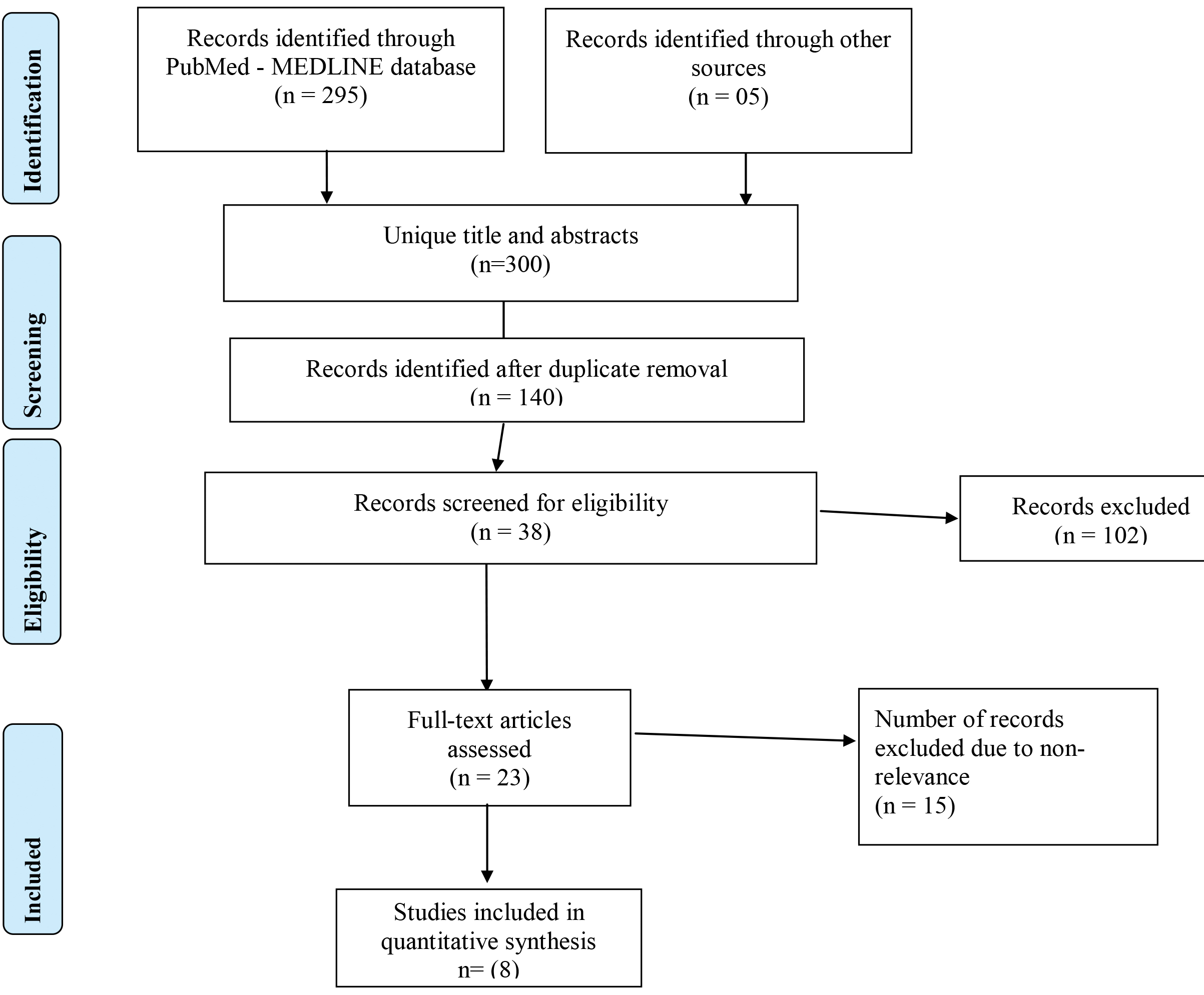
Preliminary screening identified 300 unique records, out of which 23 articles were selected by title and abstract [Table/Fig-1]. After full-text reading, 15 records were excluded. This exclusion resulted in eight full-text articles which were processed for data extraction. Additional hand searching of the reference lists of the selected studies yielded no additional records. An overview of the selected studies and their characteristics are presented in [Table/Fig-2] [11-18].
Flowchart summarizing the article selection process.
Overview of the studies processed for data extraction [11-18].
| StudyID | Authors, year of publication | Kinematics | Tooth type | Sample size (n) | Evaluation technique | Assessmentof subjects | Results | Conclusion |
|---|
| 1 | Priya NT et al.,2014 [11] | Hand, Rotary, Reciprocating | Extracted mandibular central incisors | n=100 Negative control Grp*=10, Control Grp*=10, Grp PT†=10, Grp PT† rec‡=10 Grp PTN§=10, Grp PTN§ rec‡=10 Grp OS||=10, Grp OS|| rec‡=10 Grp Reciproc=10, Grp Reciproc rec=10 | Stereomicroscope. | Dentinal micro cracks were assessed. | Percentage of defects in each Group-Grp-Positive control Grp=0%, Negative control Grp=5%, Grp PT=80%, Grp PT rec=75%, Grp PTN=36%, Grp PTN rec=38%, Grp OS=70%, Grp OS rec=50%, Grp Reciproc=65%, Grp Reciproc rec=40% | Endodontic Files in reciprocating motion showed less cracks than rotary files. |
| 2 | Dhingra A et al., 2015 [12] | Rotary and Reciprocating | Extracted mandibular first molars | n=60 Grp WO††=20 Grp OS||=20 GrpReciproc=20 | Pre instrumentation and post instrumentation scans were done using CBCT** | Canal transportation, Cross-sectional area, cervical dentin thickness | Mean values at different cross sectional levels | Reciprocating motion better than rotary. |
| Group WO | Group OS | Group Reciproc |
| 1 mm=0.571,2 mm=1.4660,3 mm=3.0150,5 mm=5.355,7 mm=5.6370,9 mm=1.5160 | 1 mm=3.3212 mm=2.56803 mm=4.72005 mm=6.9267 mm=6.79909 mm=3.5250 | 1 mm=0.66662 mm=1.35603 mm=2.95505 mm=5.5977 mm=5.59659 mm=1.4760 |
| 3 | Monga P et al., 2015 [13] | Rotary and Reciprocating | Extracted mandibular premolars | Grp A, n=30 (negative control) | Digital Stereo microscope | Dentinal defects in the form of craze lines, cracks and fracture were assessed. | Number of teeth showing cracks/Percentage showing cracks | Continuous rotating instruments produces dentinal crack formation. |
| Grp B, n=30 (hand instrumented) | Coronal third-nil | Middle third-nil | Apical third-nil |
| Grp C, n=30 (ProTaper) | Coronal thirdScore 0-22 (73.3%)Score 1-6 (20%)Score 2-1 (3.3%)Score 3-1 (3.3%) | Middle thirdScore 0-24 (80%)Score 1-3 (10%)Score 2-2 (6.7%)Score 3-1 (3.3%) | Apical third-nil |
| Grp D, n=30 (K3XF) | Coronal thirdScore 0-25 (83.3%)Score 1-3 (10%)Score 2-2 (6.7%)Score 3-nil | Middle thirdScore0-29 (96.7%)Score 1-1 (3.3%)Score 2-nilScore 3-nil | Apical third-nil |
| Grp E, n=30 (WaveOne) | Coronal thirdScore 0-28 (93.3%)Score 1-2 (6.7%)Score 2-nilScore 3-nil | Middle thirdScore 0-27 (90%)Score 1-2 (6.7%)Score 2-1 (3.3%)Score 3-nil | Apical third-nil |
| 4 | Zhou X et al., 2015 [14] | Rotary and reciprocating | Extracted mandibular premolars and molars | n=280Control Grp (n=40), Grp WO†† (n=60), Grp PT† (n=60), Grp TF‡‡ (n=60), Grp TFA§§ (n=60) | Scanning Electron Microscopy | Dentinal cracks and craze lines were assessed | Teeth showing cracks at 3,6,9 mm sections. Control Grp=0/40, WO=14/60, PT=15/60, TF=8/60, TFA=10/60. | Rotary and reciprocating NiTi|||| systems are more likely to induce dentinal and apical cracks. |
| 5 | Kansal R et al., 2014 [15] | Rotary and reciprocating | Extracted mandibular premolars | n=120Control grp (n=30) Grp WO†† (n=30) Grp PT† (n=30) Grp PT† (n=30) | Digital Stereomicroscope | Dentinal microcracks | The control group and WaveOne, single F2 ProTaper in reciprocating motion, and continuous ProTaper groups caused cracks in 0%, 15%, 26%, and 53% of samples. | Dentinal cracks formation is less with instruments working in reciprocating motion comparedwith those working in continuous rotation. |
| 6 | Pop I et al., 2015 [16] | Rotary and reciprocating | Fourteen extracted maxillary and mandibular molars | n=14 Grp PT† (n=6) Grp WO†† (n=6) Grp control (n=2) | Synchrotron radiation-based μCT (SRμCT) | Dentine microcracks | Mean and Standard deviation of length of cracks in micrometres in the pre and post-instrumentation experimental groups (based on all slices). | Reciprocating and rotary instrumentationare similar in terms of effect. |
| measurements | preinstrumentation | Post instrumentation |
| Grp A: (n=6) | 22.18 (77.24) | 58.06 (124.37) |
| Grp B: (n=6) | 26.44 (74.78) | 59.58 (127.75) |
| Grp C: (n=2) | 68.18 (64.24) | 70.21 (66.32) |
| Total samples | 24.63 (75.86) | 58.88 (126.20) |
| 7 | Liu R et al., 2013 [17] | Three single-file systems and the ProTaper system | One hundred mandibular incisors | n=100 Grp Control (n=20) Grp ProTaper† (n=20) Grp OneShape|| (n=20) Grp SAF††† (n=20) Grp Reciproc‡ (n=20) | Stereo microscope | Dentin cracks. | Number of teeth with cracks observed at different positions | The self-adjusting file and Reciproc files caused less cracks than the ProTaper and OneShape files. |
| Group | n | Apicalsurfaceonly | Sectiononly | Both | Total (%) |
| Control | 20 | 0 | 0 | 0 | 0 |
| ProTaper | 20 | 2 | 6 | 2 | 10 (50) |
| OneShape | 20 | 0 | 6 | 1 | 7 (35) |
| SAF | 20 | 0 | 0 | 0 | 0 |
| Reciproc | 20 | 0 | 1 | 0 | 1 (5) |
| Total | 100 | 2 | 13 | 3 | 18 (18) |
| 8 | Jalali S et al., 2015 [18] | Reciproc, ProTaper Universal and Mtwo | One hundred extracted mandibular premolars | n= 100 Grp control (n=25)Grp Mtwo (n=25) Grp ProTaper Universal† (n=25)Grp Reciproc‡ (n=25) | Stereo microscope | dentinal crack and crazelines | Groups | Specimens N (%) | Total | All three engine-driven systems created dentinal defects. Reciproccaused less cracks than Mtwo and ProTaper Universal. |
| Defected | No defect |
| ProTaper | 6 (24%) | 19 (76%) | 25 (100%) |
| Mtwo | 6 (24%) | 19 (76%) | 25 (100%) |
| Reciproc | 1 (4%) | 24 (96%) | 25 (100%) |
| Control | 0 (0%) | 25 (100%) | 25 (100%) |
Grp* = Group, PT† = ProTaper, rec‡ = reciprocating, PTN§ = ProTaper NEXT, OS|| = One Shape, CBCT** = Cone beam computed tomography, WO†† = WaveOne, TF‡‡= Twisted Files, TFA§§ = Twisted file adaptive, Niti||||= Nickel titanium, SAF††† = Self adjusting file
Study Characteristics
All the included studies had comparable study groups. All the selected teeth had similar characteristics in their group. Characteristics such as teeth type, root length, width and curvature were similar. Adequate root length was selected to ensure that the dentinal cracks occurred due to instrumentation and not due to morphology. In these studies, slices for detection of dentinal cracks were made at adequate length for accurate detection of dentinal cracks away from the root apex. Two studies compared ProTaper with WaveOne systems [15,16]. Rest all studies compared a combination of rotary and reciprocating file systems. The control groups were unprepared in all studies. The studies were published in the time frame of 2013–2016.
Quality of the Study
The information integrated and summarised by each researcher was assessed. The risk of bias was ascertained. The specimens were randomly distributed among the groups in each of the included study. A single operator performed the instrumentation to minimise any risk of operator bias. Post-instrumentation blind assessment of the dentinal slices was done by two independent reporters who were unaware of the study outcome.
Discussion
Summary of Evidence
Almost all rotary systems produce dentinal cracks, they are inevitable [16]. Greater taper instruments are prone to remove more root canal dentin than conventional root canal instrumentation. Various comparative studies have been conducted in the past to evaluate the propagation of dentinal cracks formed post-instrumentation using continuous rotary and reciprocating file systems [11-18].
It has been reported that endodontic file systems when set in reciprocating motion induce less dentinal defects when compared to full rotary motion. According to a study, full sequence file systems showed less cracks than single file systems [11]. Endodontic file systems ProTaper Universal and OneShape showed 80% and 70% dentinal defects in rotary motion, whereas in reciprocating motion ProTaper showed 75% dentinal defects which might be attributed to the greater taper and the file design of ProTaper files that generates more stress on the root canal walls than any other file systems. Hand instrumentation do not cause much damage to the root canal walls which could be because of the less aggressive movements of the hand files in the canal compared with engine operated files [19].
On comparing three file systems i.e. Reciproc, OneShape and WaveOne on their effect on cervical dentin wear, it was inferred that reciprocating motion is better than rotary motion. OneShape presented more effective wears in the cervical third, when compared to reciprocating [12]. Similar findings were reported in other studies [8,20].
In a study, it was stated that instruments used in continuous rotating motion produce more dentinal cracks compared to reciprocating motion, thus making them a better option [13]. This is in accordance with most of the studies [15,16,18]. Another study compared WaveOne, ProTaper Universal system, Twisted File (TF), Twisted File Adaptive (TFA) and their influence on dentinal crack propagation [14]. ProTaper Universal in full rotary motion showed highest crack formation (15/60) than other reciprocating file systems and it was concluded that any greater tapered instruments are capable to create dentinal defects and craze line regardless to their kinematics used. Similar findings were reported in 2013 where ProTaper system showed highest crack formation (50%) [17]. In a study comparing reciprocating and rotary file systems with TFA systems, it was concluded that TFA system produced significantly less cracks than the full sequence rotary or reciprocating file systems [14]. Dentinal crack formation with greater tapered instruments is more due to high levels of stress concentrations in root canal walls. In accordance to the present literature, NiTi engine driven systems remove greater volume of root canal dentin when compared to hand filing, which may lead to formation of the defects. These defects may extend to the external surface thus breaching the intact root dentin. However, it is essential to balance this against the better efficiency of motor driven systems in cleaning and shaping the root canal [21]. Reciprocating movement reduces the torsional and flexural stresses, thus increasing the canal centering ability within the root canal [11]. It also has advantages to create less invasive root canal preparations [22].
Thus, on reviewing the articles it is evident that kinematics play a very important role in certain areas during root canal instrumentation procedures. Propagation of dentinal aberrations/defects is seen more in greater tapered instruments used in continuous rotary motion.
Implications for Future Research
Introduction of rotary instruments has been a boon in the field of endodontics. It reduces the treatment time as well as operator and patient fatigue. However, literature shows that there are inherent disadvantages like dentinal damage and microcrack formation. Due to crown-down technique of instrumentation, greater tapered endodontic instruments are very popular amongst dental practitioners. This systematic review highlights the drawbacks of using greater taper instruments indiscriminately, as well as, suggests that alteration in kinematics of instrumentation may reduce the dentinal damage. This may have an influence on the longevity of dental tissues and a positive impact on prognosis of the treatment rendered. Hence, there is tremendous scope for further research on the changing kinematics of endodontic instrumentation and their effects on dental tissues.
Limitation
Although, the major databases were used for the literature search, papers might have been missed because they might not be listed in these sources. The present review encompasses articles published in English language, which may have excluded potentially valuable evidence. Extensive literature can be found related to the ProTaper file system. However, there is a dearth of literature in evaluating the new generation of file systems. Most of the literature found compared a number of mechanised root canal instrumentation systems with each other. Few studies showed inconclusive results due to lack of standardisation with regard to methods used for evaluation and assessment of the dentinal defects and cracks. The technique which was used for sectioning and decoronation of samples using diamond coated high speed burs was inappropriate leading to dentinal cracks and craze lines.
Conclusion
Greater taper instruments have a tendency to generate apical microcracks and dentinal defects. Rotary endodontic systems generated more dentinal cracks and defects as compared to reciprocating systems. Regardless of the kinematics used, majority of mechanised instruments tend to create dentinal defects.