Introduction of Another Approach of Ultrasound-Guided Maxillary Nerve Block via Pterygopalatine Fossa
Ying Ying Lyu1, Dong Ping Du2
1 Attendant, Department of Pain Management, Shanghai Jiao Tong University, Shanghai Sixth People’s Hospital, Shanghai, China.
2 Director, Department of Pain Management, Shanghai Jiao Tong University, Shanghai Sixth People’s Hospital, Shanghai, China.
NAME, ADDRESS, E-MAIL ID OF THE CORRESPONDING AUTHOR: Dr. Dong Ping Du, No.600, Yishan Road, Pain Management Center, Shanghai Jiao Tong University, Shanghai Sixth People’s Hospital, Shanghai, China.
E-mail: dudp@mail.sjtu.edu.cn
Dear editor,
We have read the article “Recognition of the Lateral Pterygoid Muscle and Plate during Ultrasound-Guided Trigeminal Nerve Block” published in your journal [1]. We would like to mention about another approach via Pterygopalatine Fossa (PPF), in the maxillary never block in management of trigeminal neuralgia and atypical facial pain. Blockades of maxillary nerve by this approach were used in two cases and both of them had their trigeminal neuralgia relieved effectively.
The fossa is a small space deep between pterygoid process and maxillary tuberosity [2]. Main part of its medial boundary is perpendicular plate of the palatine bone. The roof of the fossa is formed by the sphenoid bone at the union between the body and the great wing. The posterior boundary is the pterygoid process with foramen rotundum at its root. Its lateral side is pterygomaxillary fissure which is the opening of PPF communicated with infratemporal fossa.
Maxillary nerve is located just under the roof of PPF and very close to perpendicular plate of the palatine bone according to the location of foramen rotundum. To perform a successful block of maxillary nerve, the appropriate place of needle tip is as close as to the superior and medial part of PPF. The fossa is so narrow and deep that it is hard for the needle to reach maxillary nerve because of the obstruction of bones around PPF. To avoid hitting the bones, we inserted the needle under ultrasound guided between maxillary bone and coronoid process instead of between coronoid and condylar process, as Dr. Chang described [1]. The needle was parallel to mandibular rami and aimed at the pterygomaxillary fissure to get into the pterygopalatine fossa. By this approach, it was easy to handle the needle to avoid the bones and reach the maxillary nerve easily [Table/Fig-1].
Showing the patient’s position and approach.
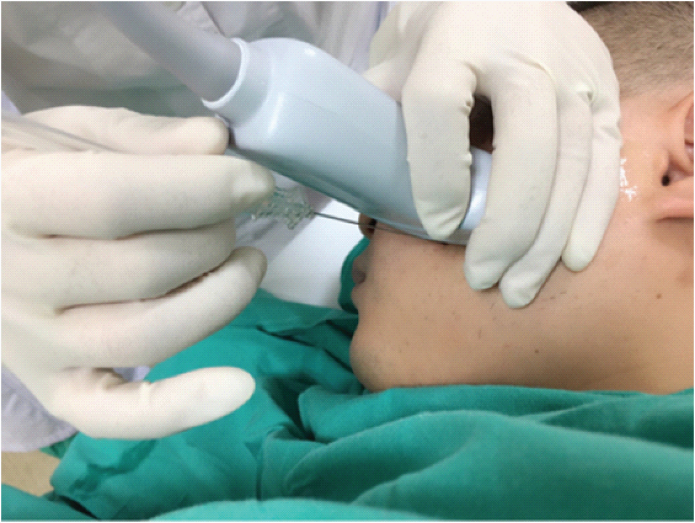
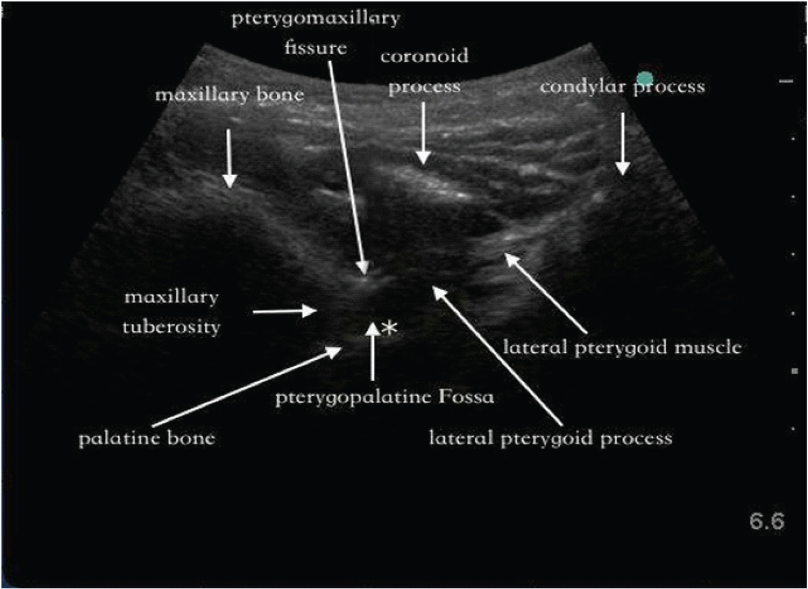
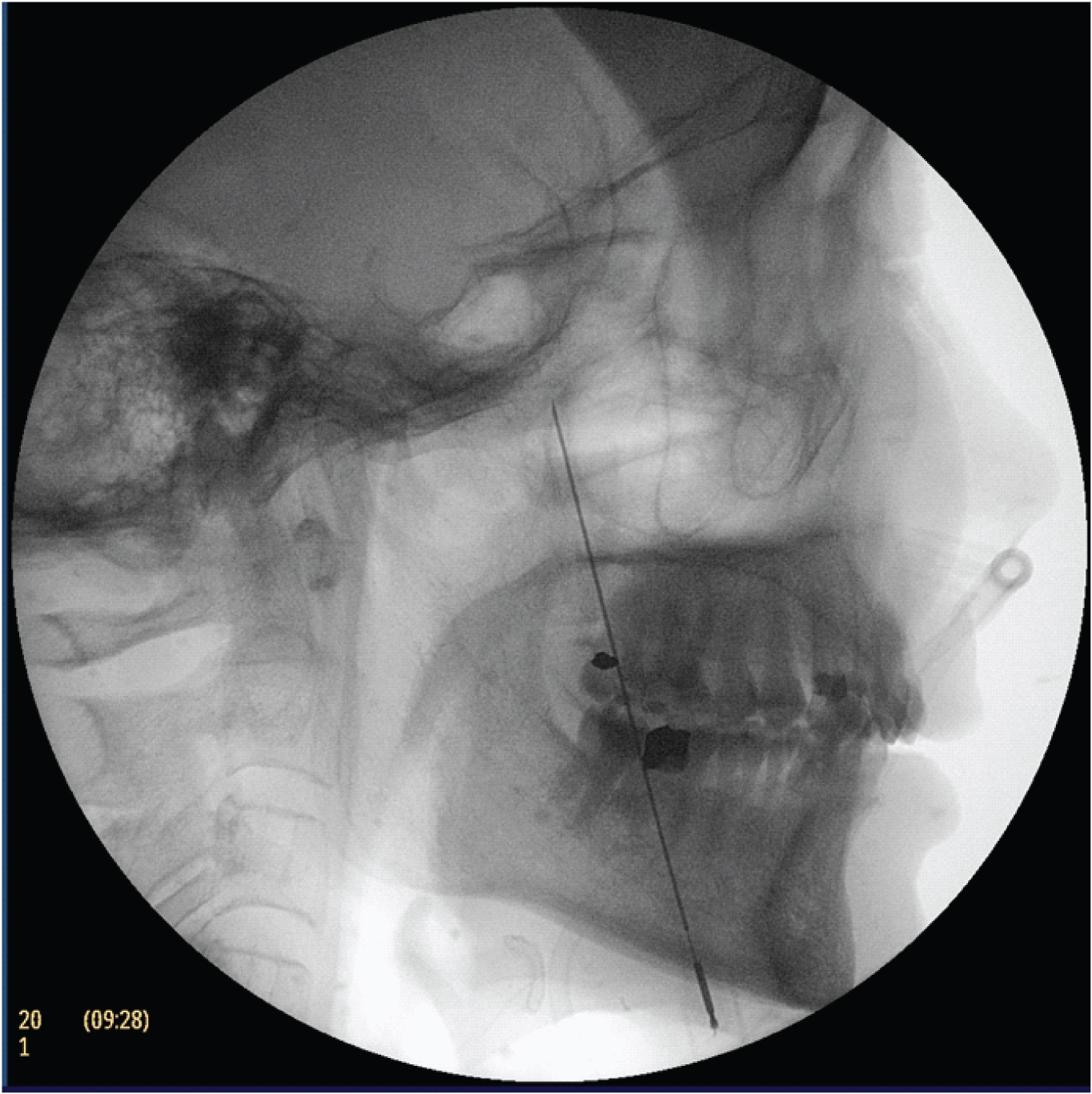
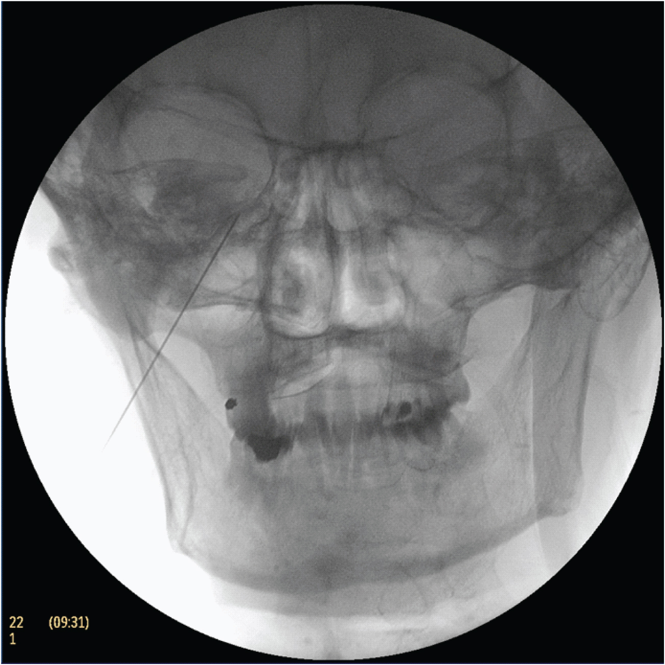
Curvilinear transducer was applied transversely to the face under the zygomatic arch [Table/Fig-1] and the beam was slightly cranial so that maxillary bone, coronoid process and condylar process were visualized [Table/Fig-2]. Moreover, pterygpalatine fossa could be identified among the maxillary tuberosity, lateral pterygoid process and perpendicular plate of the palatine bone. The needle was inserted out of plane approach and head for the upper and medial part of pterygopalatine fossa. Finally, fluoroscopy was used in order to confirm precise location of the needle tip [Table/Fig-3,4].
Ultrasonic image of PF and the needle approach.
The image of the patient demonstrating the direction of needle by lateral radiograph under fluoroscopy.
The image of the patient illustrating the situation of needle by anteroposterior radiograph under fluoroscopy.
[1]. Ke-Vin Chang, Lin CS, Lin CP, Wu WT, Özçakar L, Recognition of the lateral pterygoid muscle and plate during ultrasound-guided trigeminal nerve blockJ Clin Diagn Res 2017 11(5):UL01-UL022. [Google Scholar]
[2]. Drake Vogl Mitchell Gray’s Anatomy for Students 2014 3nd Edition:914-920. [Google Scholar]