A systematic approach of the pituitary region is very important because sometimes findings are very subtle. For complete assessment of pituitary gland, we should be aware of its normal anatomy with the physiological variations in its size and shape in different age groups in both males and females. Changes are seen in size, shape and signal intensity of pituitary gland which reflects the changes in complex hormonal physiology of the gland. Frequently there are borderline pituitary abnormalities encountered such as physiological hypertrophy of gland, subtle microadenoma, increased lobulated margins, inflammatory diseases and empty sella. Measurements of the normal pituitary gland for various age ranges are helpful to diagnose such cases. MRI has proved to be an accurate diagnostic modality for the assessment of pituitary gland. Necessity of the study arises from the lack of previous instances of measurement of pituitary height and volume in such a wide variety of age groups and for both sexes in a designated Indian population. The only evidence of similar study that was carried out in Indian population was by Sanjay SC et al., i.e., ‘Variation in size and shape of normal adult female pituitary gland: A Radiological Survey’ [1]. The study showed that the most common shape of pituitary gland was flat and the height of pituitary gland decreases as the age progresses except for the age group 40-49 years [1]. This study did not took into consideration volume of the gland and was limited to female cases only. Therefore, the present study was conducted to study the size and shape of normal pituitary gland in different age groups of both genders with MRI and to evaluate mean normal volume of the pituitary gland in relation with age and gender.
Materials and Methods
This was a retrospective observational study done in department of radiology at Dr. DY Patil Medical College, Hospital and Research Institute, Pune, Maharashtra, India. Study was conducted after institutional ethics committee clearance was obtained. The study period extended from November 2015 to December 2016 in which 500 patients were included in the study (261 males and 239 females, ranging from one year up to 80 years of age) who underwent brain MRI in the Radio-diagnosis department and the patients who were suffering from significant endocrine disorder; head injury or any genetic syndrome; those patients who were pregnant or breast feeding at the time of examination. Empty sella and patients having sella which was completely filled with cerebrospinal fluid with pituitary gland having height of less than 2 mm were regarded as empty sella and were not included in this study and subsequent analysis. All the patients who were included in the study were divided into 6 age groups (1 to 10 years, 11 to 20 years, 21 to 30 years, 31 to 40 years, 41 to 50 years, 50 years above) for both genders.
MRI examinations were performed using Siemen’s Mangnetom® Avanto 1.5 Tesla scanner. The coronal and sagittal views were displayed using the midline plane of both T1-weighted sagittal spin-echo and T2- weighted coronal spin-echo images. MRI protocol for sagittal scan was matrix 320x240, FOV was 230 mm and 5 mm slice thickness were taken. MRI protocol for coronal scan was matrix 320x196, FOV was 230 mm and 5 mm slice thickness were taken.
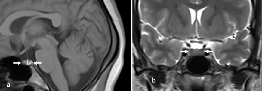
The mid-sagittal image T1 weighted image was used to obtain all pituitary gland height (cranio-caudal) and Anterior-Posterior (AP) dimensions [Table/Fig-1a]. Pituitary gland width (transverse) was measured on a coronal T2 weighted slice through the pituitary stalk [Table/Fig-1b]. Pituitary gland volume was estimated by using the formula: V=Antero-posterior dimension X Craniocaudal dimension X Transverse dimension X 0.52 (This factor is obtained from the sphere volume equation coefficient and cubic volume calculation: (4/3π)(r3)/(2r)3=3.1416/6=0.52).
a) Mid saggital T1W MR image. White arrows indicate the length (AP) and central line indicates the height (CC) of pituitary gland; b) Coronal T2W MR image. The white arrows indicate width (T) of pituitary gland.
Pituitary gland shape was obtained in the mid-sagittal T1WI. Shape of the superior surface of the gland was observed as flat, convex or concave.
Statistical Analysis
Statistical calculations were done by Software OpenEpi Version 3 for epidemiological and statistical calculations. Mean and Standard deviations of pituitary height were calculated in the scale of mm. Volume in different age groups were calculated in the scale of mm3. Relation between mean height and age and relation between volume and age were done by ANOVA test, Chi-square test and p-value <0.05 was considered as significant.
Results
The mean pituitary height in our study group was 6.4±2 mm. The mean pituitary height in the age group 1-10 years came out to be 5.3±1.4 mm. In the age group 11-20 years, mean pituitary height was 6.2±1.1 mm, in the age group 21-30 years 6.8±1.9 mm, 31-40 years age group 6.3±1.8 mm, in 41-50 years age group 6.5±1.5 mm and individuals above 50 years of age mean pituitary height was observed as 7±2.1 mm [Table/Fig-2]. A p-value for relation of pituitary height with age came out to be 0.17 (>.05) in between the male age groups which was not statistically significant, however p-value was 0.002 (<0.05) i.e., significant in between the females age groups. The mean value of pituitary height and volume in different age group and in both the sexes were given in the [Table/Fig-3].
Mean value of pituitary height and pituitary volume in different age group.
| Age Group | Mean Height in mm (SD) | Mean Volume in mm3 (SD) |
|---|
| 1-10 Years | 5.3 (±1.4) | 200 (±0.70) |
| 11-20 Years | 6.2 (±1.1) | 330 (±140) |
| 21-30 Years | 6.8 (±1.9) | 440 (±116) |
| 31-40 Years | 6.3 (±1.8) | 400 (±130) |
| 41-50 Years | 6.5 (±1.5) | 420 (±190) |
| 50 Years Above | 7.0 (±2.1) | 410 (±170) |
Mean value of pituitary height and pituitary volume in different age group and in both sex.
| Age Group | Sex | Mean Height in mm (SD) | Mean Volume in mm3 (SD) |
|---|
| 1-10 Years | Male | 5.4 (±1.5) | 210 (±0.73) |
| Female | 5.1 (±1.2) | 200 (±0.75) |
| 11-20 Years | Male | 6.2 (±1.7) | 340 (±127) |
| Female | 6.0 (±1.6) | 280 (±123) |
| 21-30 Years | Male | 6.6 (±1.5) | 430 (±116) |
| Female | 7.0 (±1.9) | 440 (±180) |
| 31-40 Years | Male | 6.3 (±1.4) | 380 (±140) |
| Female | 6.5 (±1.7) | 440 (±111) |
| 41-50 Years | Male | 6.1 (±1.5) | 400 (±159) |
| Female | 6.4 (±1.3) | 420 (±116) |
| 50 Years Above | Male | 6.0 (±1.6) | 410 (±168) |
| Female | 6.7 (±1.9) | 420 (±174) |
A p-value in relation with volume with age came <0.001 (<0.05) as statistically significant in between the groups for both males and females categories. Consequently, the mean height of pituitary gland in female patients of each age group was greater than that of male patients in the same age group except in the age group of 1-10 years and 11-20 years. However, representative differences in the mean pituitary heights were observed only in the 21-30 years old age group, 31-40-year-old age group and the 50-year-old and above age group. The height of pituitary gland in both males and females was less in the 1-10 years age group [Table/Fig-3,4], increasing in puberty [Table/Fig-5] and reached maximum in the 21-30 years age group, and declined in 30-50 years and above age groups, except in the 50 years above age group in women, in which again there was an increment in the mean pituitary height [Table/Fig-6].
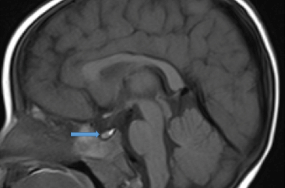
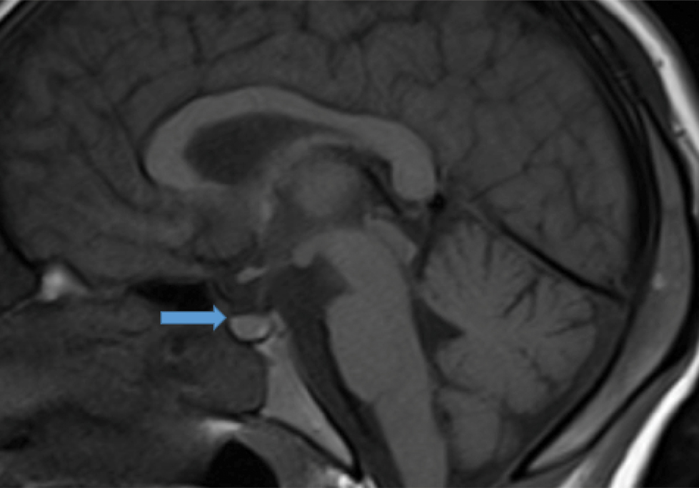
Pituitary gland in 1 year old male showed small size and upper border is concave in shape (arrow). Pituitary height measured 2.9 mm.
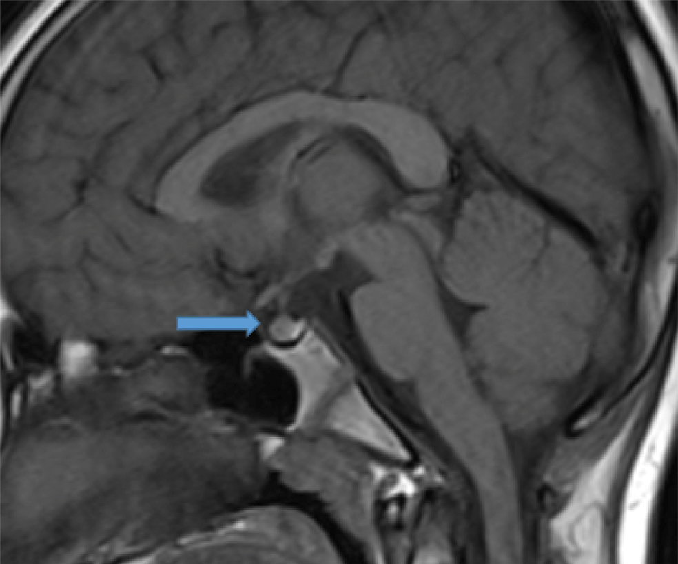
Pituitary gland in 10 year old female child. Upper border is convex in shape (arrow). Pituitary height measured 8.5 mm.
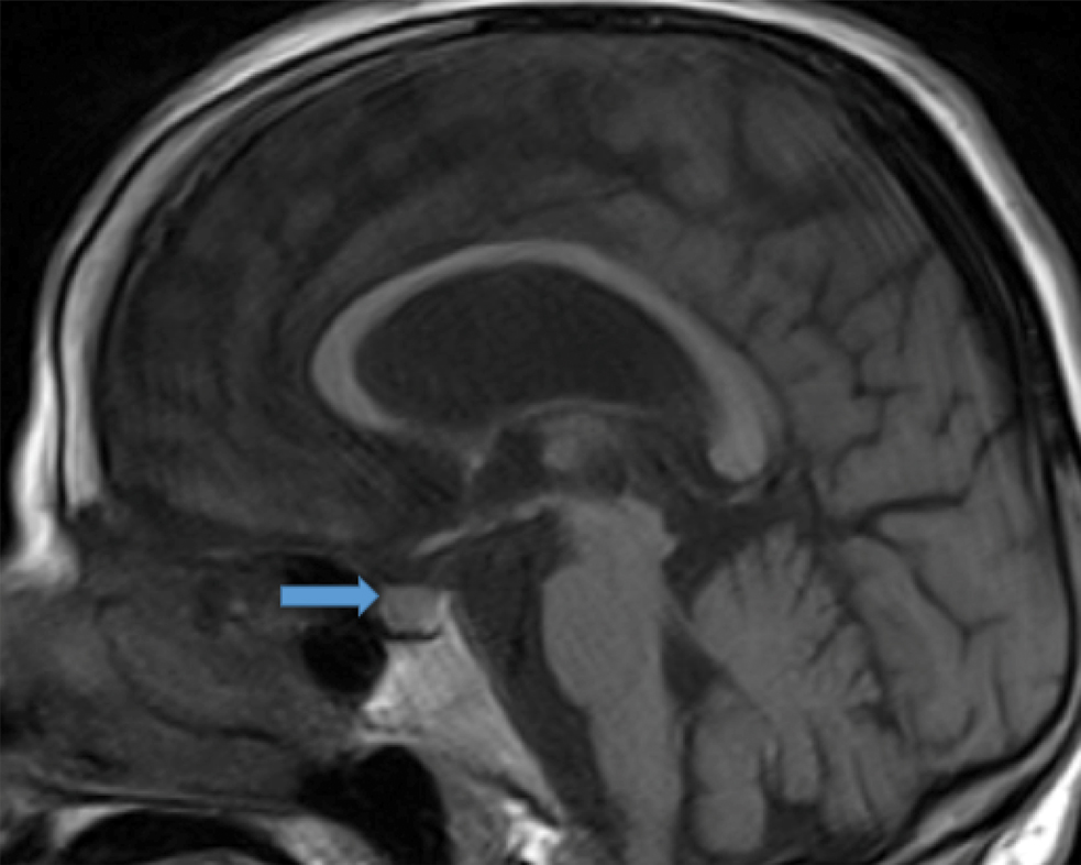
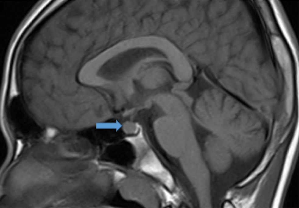
Pituitary gland in post menopausal age group also show mild increase in height measured 9.8 mm (arrow).
In all the age groups and both the sexes, the most common shape was flat which was seen in 46% of people (n=230) [Table/Fig-7], convex shape was seen in 31.2% (n=156) [Table/Fig-8] and concave shape was seen in (n=114) 22.8%.
Pituitary gland showing flat upper margin (arrow). Pituitary height measured 5.6 mm.
Pituitary gland is large in size showed convex upper border (arrow), pituitary height measures 10 mm.
Discussion
From the many result compounded during this study, the most striking result was that the height of the pituitary gland in females increases again in the 50-59-year-old age group. The first increase in pituitary height during puberty is well known [2-7], and the age-related decrease in height of pituitary gland is also documented in previous studies [3-5,7-9]. Studies done previously suggest that changes in the hormone levels may be the cause of such changes in the pituitary morphology. The above mentioned increase in pituitary height during puberty can be related to the increased production of Luteinizing Hormone (LH) during this time of growth. Also, relatively greater pituitary height in young patients, both the males and females, may be because of the physiological differences in the neuro- endocrinal hormones between younger and older subjects. The decreased in the pituitary height with age also shows the changes in the endocrine status with aging and also physiological atrophy of the pituitary gland.
The basal serum concentrations of gonadotropic hormones such as LH, Follicle Stimulating Hormone (FSH) decreased after puberty up to the fifth decade of life [10]. However, in females the concentrations of these hormones start to rise in their fifth and sixth decades, because of age-related decrease in circulating gonadal steroids hormones and an increase in gonadotropin releasing hormones [10]. As per the results in our study, females have a greater pituitary height in the same age group (i.e., 50 years and above). Previous studies [3] have also shown that the increase in the pituitary height is noted in the elderly subjects possibly due to compensatory hypertrophy after a significant decrease in gonadal steroid feedback effect.
Our study observed the noteworthy increase in the pituitary height in females of post-menopausal age. The association between the increased height of pituitary gland in young people is documented previously [2-5,7,8]. Studies done previously have also reported that height of pituitary gland reaches a maximum in the 10-19-year-old age group of both sexes [2,5,7], or in the 20-29 years old age group [4], which was also consistent with our findings.
Sanjay SC et al., concluded that mean height of pituitary gland was 6.27±0.56, the mean length was 9.10±0.78 and mean width was 11.22±0.82. They also observed that the height decreases as the age advances, however there was mild increase in height in 40-49 years age group [1].
The changes in pituitary size are basically due to changes in pituitary gland height, as there are no age related effects on gland length or width. As suggested by Lurie SM et al., future studies might reasonably use pituitary height alone with findings on mid-sagittal T1-weighted images serving as the single measure of pituitary size [11]. As seen in our study, the pituitary gland is often concave in its superior aspect, and this would not be reflected in a single, thick sagittal image, which may show only the higher lateral margins of the gland. This can cause errors in the measurements of the pituitary gland height. This can lead to decreased gland even though the pituitary height is increased (as it was measured from the lateral margins of the gland). The same problem is also seen in partially empty sellae where we can see the gland tissue extending upto the full height along its lateral margins which may lead to error in the measurements.
The results of our study were based on evaluation of pituitary size for 500 patients. As per the results of this study, height of pituitary gland reached a maximum in the 20 to 29 years of age group in both males and females, after which there was a decline in the pituitary height in the subsequent age groups.
A pituitary height greater than 9 mm in the 20 to 29 age group or 8 mm in the other age groups should be considered abnormal. In the females, the pituitary height showed a significant increase again in the 50 to 59 years age group which is due to the hypertrophy after a greater postmenopausal loss of gonadal steroid feedback mechanism. This data should help in the further evaluation of pituitary morphology in various neuro-endocrinal disorders.
Naik D et al., have done pituitary gland volume assessment in the normal Indian adolescent population and they concluded that there was moderate correlation between all pituitary measurements and age; however they have done the study only in adolescent age group [12]. They also found as in our study the most common shape of pituitary gland was flat.
Our study did provide a data on the normal pituitary height and volume in the Indian population in different age groups. Normal pituitary gland dimensions were influenced by age and gender. Pituitary height peaked in 21-30 years age group. Pituitary height more than 9mm in 21-30 years age group and more than 8mm in other age group was considered as abnormal. Perimenopausal women show increase in pituitary height again which might be due to loss of gonadal steroid feedback. This observation in the normal variation of pituitary size and shape is helpful in evaluation of pituitary morphology in neuroendocrine disorders.
Limitation
Limitation of our study was selection bias as expensive MRI test prohibited the normal volunteers for the study.
Conclusion
Pituitary size can be accurately determined by using MRI and should be correlated with the patient’s age and sex for further correlation. Pituitary gland size increased during puberty and it completely fills the pituitary fossa. Decline in gland size with age reflect the endocrinology of aging and physiological atrophy. In the cases where there is a borderline abnormality in the size and shape of pituitary gland, it should be further evaluated by dynamic contrast study on MRI. This study provides the normal range of height and volume of the normal pituitary gland in various age groups in both genders in Indian population.