Introduction
The restoration of structurally compromised endodontically treated teeth pose most challenging adhesive restorative procedure and controversies still exist about which material or technique are best for their restoration.
Aim
To evaluate the push-out bond strength of two fiber post systems luted with self-etch adhesive after phosphoric acid conditioning of the root dentin at two different storage periods.
Materials and Methods
Eighty mandibular premolars with single straight canals, decoronated at cementoenamel junction were endodontically treated and post spaces of 8 mm were prepared. Root samples were randomly divided into two groups; Groups 1 and 2 (n=40 each) according to the storage period and four subgroups (n=10 each) depending on the type of fiber post (subgroups A and C: glass fiber posts, B and D: carbon fiber posts) cemented. In subgroups A and B, fiber posts were luted with self-adhesive (Rely X U200) cement. In C and D subgroups, 37% phosphoric acid etching of the root dentin was done prior to cementation of the posts. After one month and 12 months storage period, uniform root slices were obtained for all the specimens with the help of a diamond saw and the slices were subjected to push-out test. The results were analysed with two-way ANOVA and Tukey’s multiple post-hoc test.
Results
The 37% phosphoric acid etching significantly improved the bond strength of fiber posts luted with self-adhesive resin (p<0.05) cement. Storage time significantly influenced the bond strength (p<0.05). The statistically significant difference was found between the type of posts tested, glass fiber posts exhibited better push-out bond strength compared to carbon fiber posts. Among the prepared post space regions lowest bond strength was observed in apical thirds.
Conclusion
Post space conditioning with 37% phosphoric acid prior to cementation could improve the bond strength of fiber posts luted with self-adhesive cements. Both storage time and location within the post space significantly influenced the push-out bond strength.
Endodontically treated teeth, Phosphoric acid, Self-etch adhesives, Universal testing machine
Introduction
Restoration of endodontically treated teeth using posts with a high modulus of elasticity causing irreversible root fractures have contributed to the increased use of Fiber-Reinforced Composite (FRC) posts along with resin luting cements resulting in homogenous stress distribution along the root dentin [1-3]. The basic requirement for the longevity of post retained restoration is to achieve an effective bond between the resin composite to root canal dentin and resin matrix of fiber post [4]. However, the fundamental goal is not only to achieve high retentive strength of fiber post but also to reduce microleakage along the post space to prevent degradation of the fiber post structure [5].
The most frequent clinical failure of these systems is adhesive or cohesive debonding of the post, which occurs at the dentin-adhesive cement and post interfaces [6]. For achieving a stable bond, several challenges have been demonstrated such as an unfavorable C-factor exceeding 200 contributing to high polymerization stress, the difficulty of controlling moisture during adhesive bonding procedure and the accessibility of the canal walls for treatment [7]. Further, the retention of post in the root canal may also be influenced by the degradation of the adhesive bond over a period of time due to aggressive etching of the canal walls that leaves the exposed collagen fibrils to metalloproteinases, accentuating the degradation of hybrid layer and adhesive interface [8,9].
Various luting agents using self-etching or etch and rinse adhesive systems have been proposed for bonding FRC posts to root canal dentin. In an effort to reduce the technique sensitivity self-adhesive resin cements have been developed with the advantage of not requiring dentin pretreatment [8,10].
The self-etching adhesive systems efficiently infiltrating the thick smear layer of root dentin forming a true hybrid layer is of great concern [11,12]. However, laboratory studies revealed superiority of self-adhesive cements when compared to conventional dual-cured resin cements [13,14]. Recently, it was suggested that etching the root canal dentine with phosphoric acid for 15 seconds can improve bond strength values of self-etch adhesive resin cements significantly [15].
Establishing an adequate displacement resistance between the post and the root canal is an important factor for clinical success. Among the several research protocols developed, the push-out test has been widely used to test the bond strength of fiber posts to root dentin with less variability in data distribution, more uniform values and with fewer premature failures [16]. The clinical condition is frequently simulated by thermal, mechanical loading or by ageing methods [13,17].
Based on these considerations the aim of this in-vitro study was to evaluate the bond strength of glass fiber posts and carbon fiber posts to root dentin that were luted with self-etch adhesive system, using thin slice push-out test. The null hypothesis tested in this study were: i) Post space conditioning with 37% phosphoric acid before the application of self-etching adhesive will not influence the bond strength of the fiber post to root dentin; ii) Bond strength will not differ relative to the storage time or the type of fiber; iii) There is no difference in push-out bond strength among different regions of the post space.
Materials and Methods
The research protocol of this in-vitro study was approved by the research committee of NTR university of health sciences and the ethical clearance was obtained from the Ethical Committee of GITAM Dental College and Hospital Visakhapatnam. Eighty single rooted, noncarious mandibular premolars with approximately similar dimensions were collected and were used within four months after extraction. Teeth were excluded if caries, restorations, cracks or craze lines were present on the crown or root surfaces. Using flexible diamond disk (Novo dental products, Mumbai) all the teeth were sectioned 1 mm above the Cementoenamel Junction (CEJ). The root of each tooth was standardized to a length of 12 mm as measured from the apex to the facial CEJ.
For all the root samples, coronal access was prepared using #245 bur (Mani Inc, Tochigi, Japan). After radiographic working length determination cleaning and shaping of the root canals was completed with ProTaper rotary NiTi files (Dentsply Maillefer, Switzerland) at a speed of 300 RPM using 3% sodium hypochloride irrigation. The root canals were obturated using gutta-percha and AH plus (Dentsply, Munchen, Germany) sealer, with lateral condensation technique and the orifices were sealed with interim restorative material (IRM, Dentsply). The filled roots were then stored in an incubator (Nuaire Us Autoflow, Model No:NU4500 E, Plymouth, USA) for seven days at 37°C and 100% relative humidity to allow the sealer to set completely.
After one week, post spaces were prepared using the drills provided by the manufacturer to a depth of 8 mm from CEJ leaving an apical seal of 4 mm approximately.
Among the 80 post space prepared teeth, in 40 root samples the Glass fiber posts (Reforpost /Glass fiber, No:2, Angelus, Brazil) and in another 40 samples the Carbon fiber posts (Reforpost/Carbon fiber, No:2, Angelus, Brazil) were cemented [Table/Fig-1].
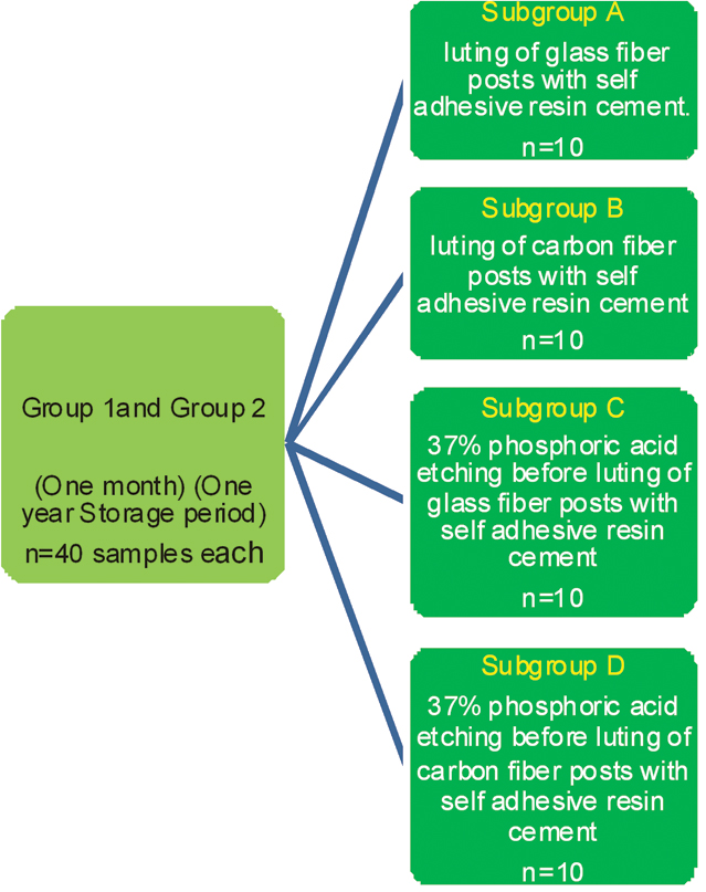
In both the groups, for subgroups A and B post spaces were flushed with normal saline and then gently air dried with an air syringe. In sub groups C and D, the root canals were etched with 37% phosphoric acid (3M ESPE, Scotch bond universal etchant, munchen, Germany) Then the luting agent RelyXTM U200 (3M ESPE, Deutschland, Germany) was mixed according to the manufacturer’s instructions and a thin layer of cement was coated onto the post space surfaces using a brush. Immediatly post was placed into the root canal and pressure was applied to hold it in position. Excess cement was removed and light cured for 40 seconds. To simulate the coronal restoration, all root samples were coated by a thin layer of resin composite Filtek Z350 (3M ESPE, Muunchen, Germany) that sealed the cervical surface and were light cured. The specimens were then stored in artificial saliva (Wet mouth, ICPA Health products. Ltd) with 100% humidity at 37oC for one month (group1) and for 12 months (group2). Artificial saliva was changed every week to avoid fungal growth.
After the appropriate storage periods (one month and 12 months), apical 4 mm portions of all the root samples were embedded in methacrylate resin blocks. Remaining portion of each root was then sectioned into 1 mm thick slices corresponding to cervical, middle and apical thirds of the post space with the help of a diamond saw (Leica Saw microtome, Nussioch, Germany) under water coolant. The thickness of each slice was measured with a digital caliper with an accuracy of 1±0.1 mm thick. To ensure that each inverted truncated root section could dislodge the fiber post from the root slice in an apico-coronal direction, each specimen was secured with cyanoacrylate glue on the push-out jig attached to the universal testing machine (Dak test bench, Mumbai). A compressive load was applied on the center of the fiber post at a cross-head speed of 0.5 mm/min, until it was completely dislodged from the root slice.
The bond strength values in Megapascals (MPa) was derived by dividing the load at failure (In Newtons) by the total surface area of the segment [18].
Statistical Analysis
The data was subjected to statistical analysis by using SPSS/PC version 16 software. Comparison of displacement resistance between two main groups offered at different root regions was done using t-test. Two-way analysis of variance (ANOVA) test was used to compare the bond strength between two groups and four subgroups. Tukey’s multiple post-hoc test was done for pair-wise Comparison of two groups and four subgroups with respect to bond strength.
Results
The mean push-out bond strength of both groups at different root regions is listed in [Table/Fig-2]. Of the two main groups, the push-out bond strength values were higher in group 1 with one month storage when compared to group 2 with the exception of subgroup B with carbon fiber posts where there is slight increase in bond strength in the coronal region. Both glass fiber posts and carbon fiber posts showed higher retentive bond strength values to root dentin after acid etching with 37% phosphoric acid (p<0.05).
Comparison of mean and standard deviation of push-out bond strength values in MPa/mm2 at different post space regions between two main groups and subgroups by t-test.
| Sub groups | Region | Group 1 | Group 2 | t-value | p-value |
|---|
| Mean | SD | Mean | SD |
|---|
| A | Coronal | 128.24 | 6.07 | 119.00 | 2.98 | 4.3251 | 0.0004* |
| Middle | 114.06 | 5.99 | 107.66 | 2.60 | 3.5821 | 0.0021* |
| Apical | 70.13 | 4.43 | 63.20 | 3.71 | 3.7957 | 0.0013* |
| B | Coronal | 116.07 | 5.04 | 122.54 | 9.41 | -1.0211 | 0.3207 |
| Middle | 110.06 | 3.99 | 106.44 | 8.93 | 0.8471 | 0.4081 |
| Apical | 63.20 | 8.50 | 56.42 | 3.35 | 2.3467 | 0.0306* |
| C | Coronal | 158.92 | 2.31 | 143.35 | 9.36 | 2.2011 | 0.0410* |
| Middle | 143.81 | 7.83 | 129.63 | 5.74 | 2.3941 | 0.0278* |
| Apical | 112.21 | 5.76 | 68.44 | 8.32 | 13.6780 | 0.00001* |
| D | Coronal | 145.51 | 5.50 | 128.55 | 6.71 | 6.1836 | 0.00001* |
| Middle | 131.64 | 7.24 | 117.45 | 6.07 | 4.7473 | 0.0002* |
| Apical | 100.19 | 6.33 | 69.36 | 4.31 | 6.2284 | 0.00001* |
*p<0.05- Statistically significant difference.
The highest push-out bond strength values were observed in the coronal third of the post space among all the groups, indicating a significant influence of the root regions on the bond strength (p<0.05). Among the regions, significant increase in bond strength values after acid etching was observed in apical region compared to the values without etching. Among the two types of fiber posts, lowest bond strength values were observed for carbon posts in the apical area without etching the root surface.
Discussion
Clinically, fiber posts have been used successfully and most of the studies have shown low failure rates [19-21]. Root fractures with fiber posts when they occur tend to be of favourable type when compared with metal posts that cause irreparable root fractures [22]. When retreatment is advised, fiber posts are relatively easy to remove by using an ultrasonic or rotary instrument by boring through the middle of the post [23]. The original carbon fiber posts were dark but they are indicated for posterior teeth where aesthetics is not that critical [19]. Therefore, in the present study comparison of bond strength was done for carbon fiber posts and glass fiber posts. Mandibular premolars were selected because of their delicate tooth morphology and also as the functional stresses extend on them simulate the clinical situation where the chewing forces are maximum [24].
The main goal of using adhesive luting cements is to reinforce the tooth by bonding the post to root canal dentin and to retain the coronal restoration. Adhesive resin luting cements have shown the compressive strength of around 200 MPa and lower elastic moduli (4 to 10 Gpa) [10].
The self-adhesive resin cements achieved better results than the conventional dual-cured resin cements mainly due to higher inorganic filler content that lowers its polymerization shrinkage [8]. However, some studies have shown that self-adhesive resin cements does not promote the formation of a hybrid layer as these adhesives are not able to dissolve the thick smear layer present on the canal walls after instrumentation for post placement [11,16].
The null hypothesis of the present study had to be rejected since the acid etching and 12 months storage period significantly affected the bond strength of fiber posts inside the root canal. The bond strength values were increased when acid etching was done with phosphoric acid before the cementation of posts to the root dentin. The thick secondary smear layer produced by post space preparation on the canal walls may be one of the reason why etching with phosphoric acid significantly improved the push-out strength of one-step self–etching systems. The methacrylated phosphoric esters present in RelyX U200 are not as effective as phosphoric acid in dissolving the thick smear layer in the root canal walls [15,16,25]. The results are in accordance with the results of other studies where prior etching of canal wall with 35% phosphoric acid enhanced the push-out bond strength values of fiber posts to root dentin [26]. After 12 months storage in 100% humidity, the push-out bond strength values were decreased for fiber posts and these values are statistically different when compared to one month storage period and the results are in accordance with the other studies [27]. The smear layer hybridized by self-etching adhesive is a weak bonding area with the top of the hybrid layer containing disorganized collagen fibrils that degrade over time. Contrary to these findings, few studies quoted that storage time has not influenced the push-out bond strength of the post within the root canal [8,13].
The mean bond strength values (with or without etching root dentin) were higher for glass fiber posts when compared to the carbon fiber posts though the difference is statistically not significant in coronal and apical regions. These results are in accordance with other previous studies where the glass fiber posts have presented highest bond strength values when compared to carbon fiber posts [10,28].
The results of this study revealed a decrease in bond strength values proceeding from the coronal to the apical third of the post space. Both the groups and all subgroups have shown highest bond strength values at coronal third and lowest at the apical third of the post space. Several studies are in accordance with this outcome [14,15,29]. Different clinical factors could be responsible for this phenomena, i.e., less visibility in deeper areas resulting less predictable post space cleaning, less distribution and density of dentinal tubules in the apical portion of root canal system, apical sclerosis, cavity configuration factor of the post space and difficulty of accessibility leading to restriction in adhesive cement flow to apical portion. In contrast with these results, some studies suggested that the root canal region does not influence adhesion of the post to root dentin [8,30]. These contradictory findings could be attributed to the different luting cements employed or the differences in the procedure of sample preparation.
Limitation
One of the limitations of the study is the dislodging forces exerted during push-out test cannot be correlated with functional forces during clinical service. In addition, debris accumulated during post space preparation could affect the bond strength. This may partially explain the high standard deviation that was observed within the same group.
Conclusion
Post space conditioning with 37% phosphoric acid before the application of self-etching adhesive could improve the bond strength of the fiber post. There will be a significant degradation of bond strength with aging probably due to collagenolytic activity in the root dentin.
It was reported that the collagenolytic activity can be prevented by the application of the agents like chlorhexidine, Dimethyl sulfoxides, Galardin, Tetracyclines, Benzalkoniumm chloride and Ethylenediaminetetraacetic Acid (EDTA). The use of any of these agents before the cementation of fiber post may decrease the bond degradation. Further research on this aspect is recommended to improve the longevity of post endodontic restorations.
*p<0.05- Statistically significant difference.
[1]. Qing H, Zhu Z, Chao Y, Zhang W, In-vitro evaluation of the fracture resistance of anterior endodontically treated teeth restored with glass fiber and zircon postsJ Prosthet Dent 2007 97(2):93-98. [Google Scholar]
[2]. Asmussen E, Peutzfeldt A, Sahafi A, Finite element analysis of stresses in endodontically treated, dowel-restored teethJ Prosthet Dent 2005 94(4):321-29. [Google Scholar]
[3]. Salameh Z, Qunsi HF, Aboushelib MN, Al-Hamdan R, Sandig W, Ferrari M, Effect of different onlay systems on fracture resistance and failure patterns of endodontically treated mandibular molars restored with and without glass fiber postsAm J Dent 2010 23(2):81-86. [Google Scholar]
[4]. Gu XH, Mao CY, Liang C, Wang HM, Kern M, Does endodontic post space irrigation affect smear layer removal and bonding effectivenessEur J Oral Sci 2009 117(5):597-603. [Google Scholar]
[5]. Breschi L, Mazzoni A, Ruggori A, Cadenaro M, Di Lenarda R, De Stefano Dorigo E, Dental adhesion review: Aging and stability of the bonded interfaceDent Mater 2008 24(1):90-101. [Google Scholar]
[6]. Coniglio I, Carvalho CA, Magni E, Cantoro A, Ferrai M, Post space debridement in oval-shaped canals the use of new ultrasonic tip with oval sectionJ Endod 2008 34:752-55. [Google Scholar]
[7]. Cheung W, A review of the management of endodontically treated teeth post, core and the final restorationJ Am Dent Assoc 2005 136:611-19. [Google Scholar]
[8]. Leme AA, Coutinho M, Insaurralde AF, Scaffa PM, Silva LM, The influence of time and cement type on push-out bond strength of fiber posts to root dentinOper Dent 2011 36(6):643-48. [Google Scholar]
[9]. Albashaireh ZS, Ghazal M, Kern M, Effects of endodontic post surface treatment, dentin conditioning, and artificial aging on the retention of glass fiber-reinforced composite resin postsJ Prosthet Dent 2010 103(1):31-39. [Google Scholar]
[10]. Calixto LR, Bandéca MC, Clavijo V, Andrade MF, Vaz LG, Campos EA, Effect of resin cement system and root region on the push-out bond strength of a translucent fiber postOper Dent 2012 37(1):80-6. [Google Scholar]
[11]. Yang B, Ludwig K, Adelung R, Kern M, Microtensile bond strength of three luting resins to human regional dentinDent Mater 2006 22(1):45-56. [Google Scholar]
[12]. Radovic I, Mazzitelli C, Chieffi N, Ferrari M, Evaluation of the adhesion of fiber posts cemented using different adhesive approachesEur J oral Sci 2008 116(6):557-63. [Google Scholar]
[13]. Bitter K, Meyer LH, Priehn K, Kanjuparambil JP, Neumann K, Kielbassa AM, Effects of luting agent and thermocycling on bond strength to root canal dentinInt Endod J 2006 39(10):809-18. [Google Scholar]
[14]. Onay EO, Korkmaz Y, Kiremitci A, Effect of adhesive system type and root region on the push-out bond strength of glass fiber posts to radicular dentinInt Endod J 2010 43(4):259-68. [Google Scholar]
[15]. Scotti N, Rota R, Scansetti M, Migliaretti G, Pasqualini D, Berutti E, Fiber Post adhesion to radicular dentin: The use of acid etching prior to a one step self-etching adhesiveQuintessence Int 2012 43(7):615-23. [Google Scholar]
[16]. Goracci C, Fabianelli A, Sadek FT, Papacchini F, Tay FR, Ferrari M, The contribution of friction to the dislocation resistance of bonded fiber postsJ Endod 2005 31(8):608-12. [Google Scholar]
[17]. Leitune VC, Collares FM, Werner Samuel SM, Influence of chlorhexidine application at longitudinal push-out bond strength of fiber postsOral Surg Oral Med Oral Pathol Oral Radio Endod 2010 110(5):e77-81. [Google Scholar]
[18]. Pala K, Demirbuga S, Gümüs HÖ, Arslan S, Zorba YO, Effect of different bonding techniques on the bond strength of two different fiber postsJ Res Dent 2014 2(1):32-36. [Google Scholar]
[19]. Williams C, Kumar M, Bajpai M, Agarwal D, Prosthodontic Management of Endodontically Treated Teeth: A Literature ReviewInt J Clin Prev Dent 2014 10(1):45-50. [Google Scholar]
[20]. Cagidiaco MC, Godoy FG, Vichi A, Grandini S, Goracci C, Ferrari M, Placement of fiber prefabricated or custom made posts affects the 3-year survival of endodontically treated premolarsAm J Dent 2008 21(3):179-84. [Google Scholar]
[21]. Saker S, El-Kholany N, El-Wassefy N, Effect of different surface treatments on push-out bond strength of glass fiber posts to resin composite core materialJ Dent App 2015 2(6):246-50. [Google Scholar]
[22]. Silva GR, Santos-filho PC, Simamoto-Junior PC, Martins LR, Mota AS, Soares CJ, Effect of post type and restorative techniques on the strain and fracture resistance of flared incisor rootsBraz Dent J 2011 22(3):230-37. [Google Scholar]
[23]. Goracci C, Ferrari F, Current perspectives on post systems: a literature reviewAust Dent J 2011 56(1):77-83. [Google Scholar]
[24]. Ferrari M, Vichi A, Fadda GM, Cagidiaco MC, Tay FR, Breschi L, A randomized controlled trial of endodontically treated and restored premolarsJ Dent Res 2012 91(7):72S-78S. [Google Scholar]
[25]. Saraiva LO, Aguiar TR, Costa L, Correr-Sobrinho L, Muniz L, Mathias P, Effect of different adhesion strategies on fiber post cementation: Push-out test and scanning electron microscopy analysisContemp Clin Dent 2013 4(4):443-47. [Google Scholar]
[26]. Zhang L, Huang L, Xiong Y, Fang M, Chen JH, Ferrari M, Effect of post-space treatment on retention of fiber posts in different root regions using two self-etching systemsEur J Oral Sci 2008 116(3):280-86. [Google Scholar]
[27]. Durate RM, DeGoes MF, Montes MA, Effect of time on tensile bond strength of resin cement bonded to dentine and low–viscosity compositeJ Dent 2006 34(1):52-61. [Google Scholar]
[28]. Ferrari M, Cagidiaco MC, Goracci C, Vichi A, Mason PN, Radovic I, Tay F, Long-term retrospective study of the clinical performance of fiber postsAm J Dent 2007 20(5):287-91. [Google Scholar]
[29]. Mumcu E, Erdemir U, Topcu FT, Comparison of micro push-out bond strengths of two fiber posts luted using simplified adhesive approachesDent Mater J 2010 29(3):286-96. [Google Scholar]
[30]. Wang VJ, Chen YM, Yip KH, Smales RJ, Meng QF, Chen L, Effect of two fibre post types and two luting cement systems on regional post retention using the push-out testDent Mater 2008 24(3):372-77. [Google Scholar]