Colonic Arteriovenous Malformation in a Child with Klippel Trenaunay Weber Syndrome
Suneel Mundkur1, Sandeep Kumar2, Shrikiran Aroor3, Adel Moideen4
1 Professor, Department of Paediatrics, Kasturba Medical College, Manipal, Udupi, Karnataka, India.
2 Assistant Professor, Department of Paediatrics, Kasturba Medical College, Manipal, Udupi, Karnataka, India.
3 Professor, Department of Paediatrics, Kasturba Medical College, Manipal, Udupi, Karnataka, India.
4 Senior Resident, Department of Paediatrics, Kasturba Medical College, Manipal, Udupi, Karnataka, India.
NAME, ADDRESS, E-MAIL ID OF THE CORRESPONDING AUTHOR: Dr. Sandeep Kumar, Assistant Professor, Department of Paediatrics, Kasturba Medical College, Manipal University, Manipal, Udupi-576104, Karnataka, India.
E-mail: bksandydoc@gmail.com
Angio-osteohypertrophy, Gastrointestinal bleed, Venous malformation
Dear editor,
Klippel-Trenaunay Weber Syndrome (KTWS) is a rare congenital vascular anomaly characterized by the presence of cutaneous capillary malformation, venous varicosities and hypertrophy of bone and (or) soft tissue. Visceral vascular malformations involving multiple organ systems are reported with this syndrome. However, the presence of gastrointestinal vascular malformations is relatively rare. We report a child who presented with transfusion dependent anemia and life-threatening hematochezia secondary to colonic arteriovenous malformation.
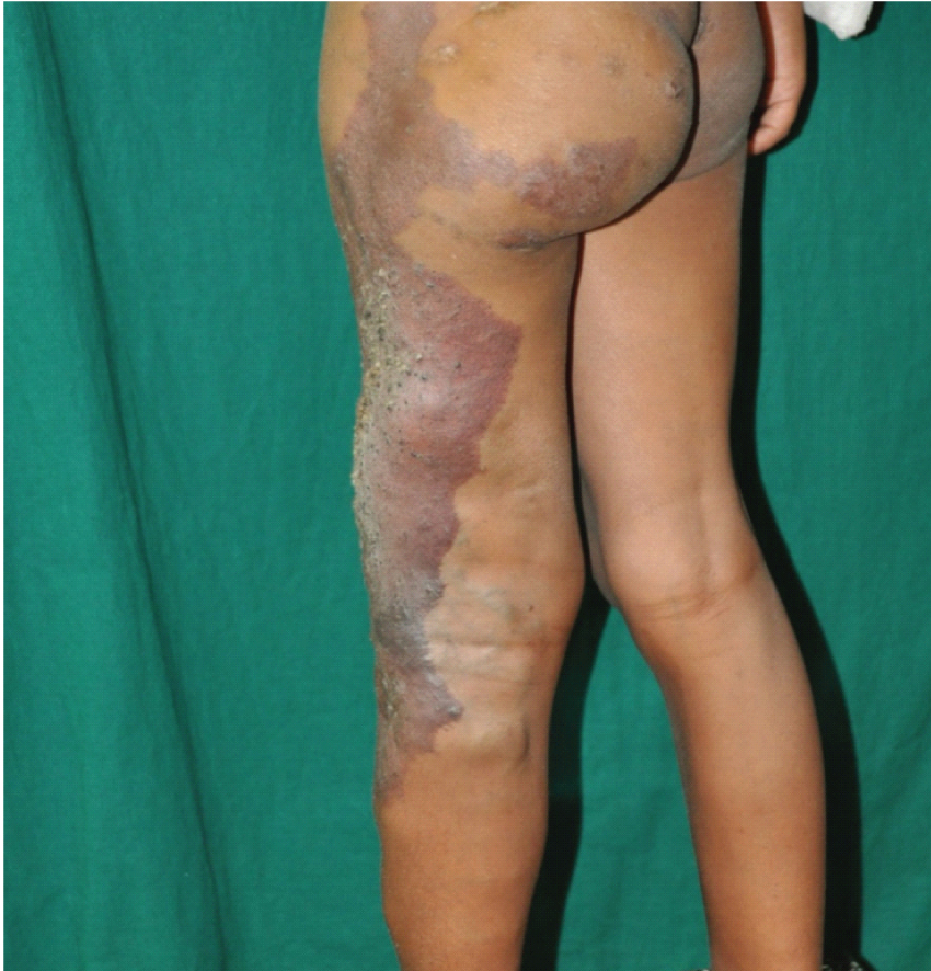
A 10-year-old girl born to non-consanguineous parents presented with hematochezia for two days. There was history of intermittent episodes of hematochezia requiring packed cell transfusions since infancy. Her birth, development and family history was normal. On examination there was severe pallor with large haemangioma over lateral aspect of left lower limb involving buttock with multiple verrucous lesions over left leg [Table/Fig-1]. Her heart rate was 110 beats per minute and blood pressure was 100/60 mmHg. Her systemic examination was unremarkable. Investigations showed severe anemia (haemoglobin 7.2 gm %) with normal platelet count, liver enzymes and normal coagulation profile. A Doppler USG revealed persistence of fetal lateral marginal vein of left lower limb with hypoplasia of the left common femoral, superficial femoral and popliteal veins. Anemia was corrected with packed cell transfusions. Colonoscopy revealed large internal hemorrhoids with venous malformations involving the descending colon and sigmoid colon [Table/Fig-2]. Open hemorrhoidectomy (Milligan-Morgan procedure) was done. The child was started on oral iron supplement. Currently she is under regular follow up with no further bleeding episodes.
Clinical photograph showing venous malformation over left lower limb with soft tissue hypertrophy.
Colonoscopy image showing venous malformations involving the descending colon.

Klippel-Trenaunay Syndrome (KTS) is a triad of capillary malformation, venous varicosity and combination of bony/soft tissue hypertrophy involving an extremity [1]. The International Society for the Study of Vascular Anomalies included the presence of lymphatic varicosities in addition to the above features [2]. The term KTWS is used by some authors in the presence of significant arteriovenous malformations. Varicosities may affect the superficial, deep, and perforating venous systems. The incidence of gastrointestinal involvement is relatively rare and reported in around 20% of patients [3]. Distal colon and rectum are the common sites affected. Rarely jejunum, oesophagus or whole gastrointestinal tract can be involved. The spectrum of the Gastro Intestinal bleeding may vary from asymptomatic occult bleeding to life-threatening haemorrhage.
Imaging studies such as contrast enhanced MRI, ultrasonography, and Doppler study are needed for proper diagnosis and to delineate the extent of lesion, thus planning the interventions if required [4]. Ideally contrast angiographic should be used for visualizing the vascular anatomy and determining the disease extent. Contrast angiography could not be performed in our patient. Endoscopic therapies have limited value in the management as KTS lesions are usually extensive and progressive in nature [4,5]. Active intervention is done only when there is localized lesion, or serious complication such as life threatening haemorrhage or congestive cardiac failure. Treatment options include surgery, sclerotherapy, and compression therapy [5]. Rarely resection of the segment of colon may be necessary to control bleeding.
Thus, gastrointestinal bleed secondary to colonic arteriovenous malformation is an uncommon presentation and can be life threatening. Routine screening of these children for arteriovenous malformations of gut is necessary for early intervention thus preventing fatal complications.
[1]. Oduber CE, van der Horst CM, Hennekam RC, Klippel-Trenaunay syndrome: Diagnostic criteria and hypothesis on etiologyAnn Plast Surg 2008 60(2):217-23. [Google Scholar]
[2]. Tetangco EP, Arshad HM, Silva R, Klippel-Trenaunay syndrome of the rectosigmoid colon presenting as severe anemiaACG Case Rep J 2016 3(4):161-71. [Google Scholar]
[3]. Wang ZK, Wang FY, Zhu RM, Liu J, Klippel-Trenaunay syndrome with gastrointestinal bleeding, splenic haemangiomas and left inferior vena cavaWorld J Gastroenterol 2010 16(12):1548-52. [Google Scholar]
[4]. Wilson CL, Song LM, Chua H, Ferrara M, Devine RM, Dozois RR, Bleeding from cavernous angiomatosis of the rectum in Klippel-Trenaunay syndrome: Report of three cases and literature reviewAm J Gastroenterol 2001 96:2783-88. [Google Scholar]
[5]. Schmitt B, Posselt HG, Waag KL, Müller H, Bender Steffen W, Severe hemorrhage from intestinal haemangiomatosis in Klippel-Trenaunay syndrome: Pitfalls in diagnosis and managementJ Pediatr Gastroenterol Nutr 1986 5(1):155-58. [Google Scholar]