Go-kart Accident - Induced Isolated Sternal Body Fracture
Onur Kaplan1, Ozgur Sogut2, Mehmet Yigit3, Mustafa Ozturk4, Muhamed Furkan Ozden5
1 Senior Resident, Department of Emergency Medicine, University of Health Sciences, Haseki Training and Research Hospital, Istanbul, Turkey.
2 Associate Professor, Department of Emergency Medicine, University of Health Sciences, Haseki Training and Research Hospital, Istanbul, Turkey.
3 Senior Resident, Department of Emergency Medicine, University of Health Sciences, Haseki Training and Research Hospital, Istanbul, Turkey.
4 Senior Resident, Department of Emergency Medicine, University of Health Sciences, Haseki Training and Research Hospital, Istanbul, Turkey.
5 Junior Resident, Department of Emergency Medicine, University of Health Sciences, Haseki Training and Research Hospital, Istanbul, Turkey.
NAME, ADDRESS, E-MAIL ID OF THE CORRESPONDING AUTHOR: Dr. Ozgur Sogut, Associate Professor, University of Health Sciences, Department of Emergency Medicine, Haseki Research and Training Hospital, Millet Street, Zip Code: 34096, Fatih/Istanbul, Turkey.
E-mail: ozgur.sogut@sbu.edu.tr
Sternal fractures can occur in isolation or with concomitant injuries, such as deceleration injuries and blunt anterior chest trauma. They are seen in 3%-8% of cases of blunt chest trauma, the most common cause of which is motor vehicle accidents. These cases account for 60%-90% of sternal fractures. With the mandatory use of seatbelts, the incidence of sternal fracture has increased. A go-kart (or go-cart) is an open-wheeled car that comes in all shapes and forms, from motorless models to high-powered racing machines. Here, we report on the case of a patient who was involved in a go-kart accident that resulted in an isolated sternal body fracture, with no evidence of concomitant injury on portable anteroposterior and lateral chest radiographs. The patient was treated conservatively with a sternum external fixation corset.
Blunt chest trauma, Diagnosis, Management
Case Report
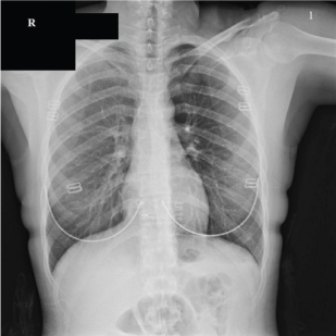
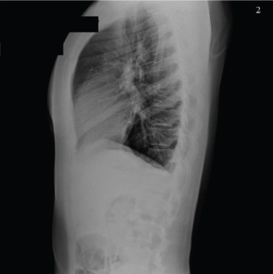
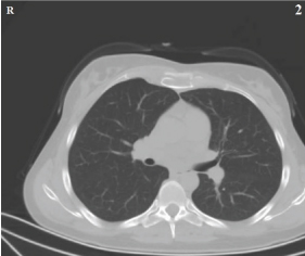
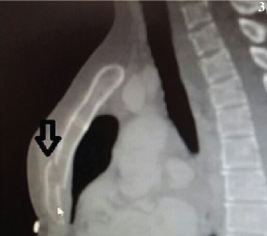
A 36-year-old woman who had been involved in a go-kart accident was admitted to the Emergency Department (ED) with persistent chest pain of one-hour duration. She stated that the seatbelt was fastened at the time of injury. On presentation to our ED, her blood pressure was 120/70 mm Hg, pulse rate was 76 beats per minute, respiratory rate was 14 breaths per minute, body temperature was 36.7°C, and oxygen saturation by pulse oximetry was 95% while breathing room air. On physical examination, the patient was conscious and oriented but she was extremely agitated because of conspicuous chest pain which increased with inspiration. She also complained of mild abdominal pain. Chest examination showed tenderness on palpation over the anterior part of the chest without swelling or bruising. Her heart sounds were normal and examination revealed no abnormal finding for the respiratory system. Abdominal examination revealed tenderness in the right upper quadrant. A Focused Abdominal Sonography for Trauma (FAST) was performed and revealed no intraperitoneal fluid or organ injury. Portable anteroposterior [Table/Fig-1] and lateral chest radiographs obtained on admission showed the normal upper mediastinum, mediastinal structures, and cardiothoracic ratio. A lateral chest radiograph demonstrated an inconspicuous fracture line and displacement [Table/Fig-2]. An axial lung-section image obtained by non-enhanced chest Computed Tomography (CT) with sagittal reformatting revealed a clearly displaced transverse fracture of the sternal body, with no concomitant thoracic injury [Table/Fig-3,4] respectively.
Antero-posterior chest radiograph obtained on admission showing the normal upper mediastinum, mediastinal structures, and cardiothoracic ratio.
Lateral chest radiograph obtained on admission showing a subtle fracture line and displacement.
Normal, non-enhanced chest computed tomographic image with axial reformatting, obtained on admission.
Thoracic computed tomographic image showing an obvious fracture line and slight displacement of the sternum (black arrow) in the sagittal plane.
A 12-lead Electrocardiogram (ECG) was obtained at the time of admission to rule out myocardial contusion but no dysrhythmia, conduction disturbance, or ST-segment change was noted. Laboratory results, including the patient’s haematocrit, white blood cell count, electrolytes, Creatinine Kinase (CK), CK myocardial band, and cardiac troponin-I values, were within the normal limits. The results of a second troponin I measurement obtained six hour later were also within the reference range.
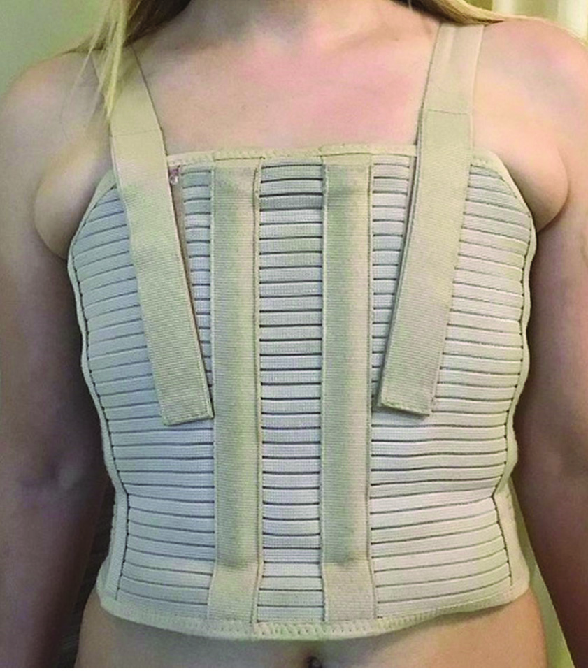
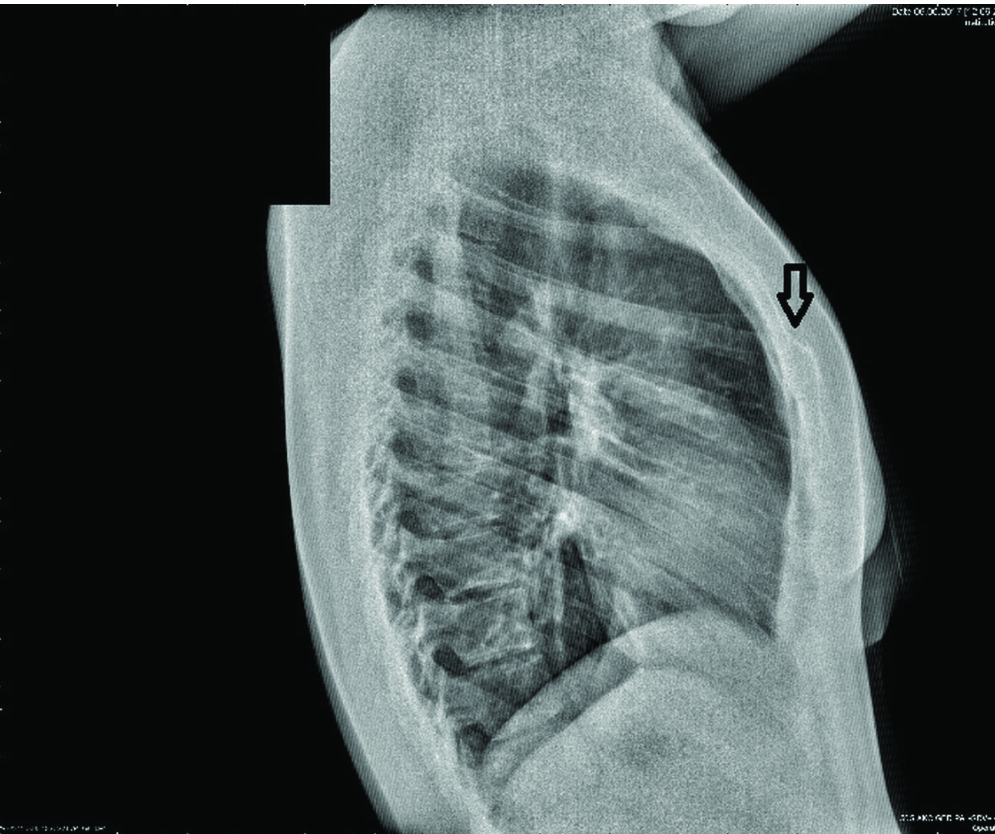
A single dose of fentanyl at a dosage of 100 micrograms (μg) was administered intravenously in the ED and the patient responded well. She was admitted to the thoracic surgery service for observation. The patient was placed in a supine position to minimize chest movement during hospitalisation. As she had no complication during the hospital course, she was discharged after two days with a sternum external fixation (Stern-E-Fix) [Table/Fig-5] corset. At the follow up examination three weeks later, she had no chest discomfort. Evidence of healing of the fracture line was detected on a follow up lateral chest radiograph [Table/Fig-6]. Written informed consent was obtained from patient for publishing the images.
View of patient with a sternum external fixation corset.
Lateral chest radiograph obtained at 3 weeks showing evidence of callus formation with complete bony union around the fracture site (black arrow).
Discussion
This index case focuses on the management of a patient with an uncommon isolated fracture of the sternal body sustained in a go-kart accident. She had no concomitant injury. An ECG and chest X-rays were normal, and she had no cardiac enzyme elevation.
Sternal fractures account for 3%-8% of all blunt chest wall traumas and comprise only a small percent of all injuries in patients with thoracic trauma who are treated in the ED [1]. In patients with sternal fractures, careful investigation should be conducted to detect injuries of the heart and other mediastinal structures [2], as the early identification of such injuries is of great importance. Concomitant intrathoracic injuries associated with sternal fracture include tension pneumothorax, massive haemothorax, open pneumothorax, flail chest, and cardiac tamponade [1,3]. Furthermore, sternal fractures may be associated with injuries of the great vessels and spinal cord, or acute myocardial contusion. The later is often clinically subtle and may thus be misdiagnosed [4,5].
The utility of ECGs and cardiac biomarker measurements in the investigation of myocardial contusion resulting from sternal fracture has been validated [1]. Wiener Y et al., suggested that patients with ECG changes or cardiac enzyme elevation be admitted for echocardiography and observation to rule out cardiac contusion [6]. Patients with normal admission ECGs, normal troponin I levels, and no concomitant injury can be discharged safely [7].
In our patient, cardiac contusion was evaluated based on the ECG results and cardiac enzyme levels. The combination of a normal ECG and a normal troponin I level at admission and six hour later ruled out a diagnosis of clinically significant blunt cardiac injury.
Although sternal fracture is rare, it may contribute significantly to the morbidity and mortality of patients with trauma, particularly when associated thoracic injuries are present. Mortality in these patients ranges from 0.7% to 19.2% [5,8]. The implementation of seatbelt legislation has led to an increase in the frequency of isolated sternal fractures sustained in road traffic accidents [5]. Most sternal fractures occur in drivers of older-model cars that use seat-belt which lack airbags and are due to the high pressure exerted by the belt against the rib cage at the time of injury [5,9].
Physical examination, lateral chest radiography, and thoracic CT are valuable in diagnosing sternal fracture [3]. Plain radiographs remain the gold-standard investigation in the diagnosis of this injury [3,4]. However, it may not be detected easily on frontal chest radiographs and may be relatively subtle on lateral chest radiographs [10]. An anteroposterior chest radiograph can be useful in determining the presence of concomitant injuries, which include rib fracture, pulmonary contusion, and haemothorax/pneumothorax, as well as mediastinal widening, which may be indicative of the presence of additional injuries [3]. Sagittal or coronal reconstructed thoracic CT images should be obtained when the suspicion of sternal fracture cannot be eliminated with conventional lateral chest radiographs [4,10]. Axial CT is inferior to radiography in the diagnosis of sternal fracture, as the CT slices may miss a transverse sternal fracture [10]. In our patient, no pathology was detected on the antero-posterior or lateral chest radiograph, but a transverse fracture line and slight displacement of the sternal body were visualised easily on a sagittal reformatted CT image.
The management of patients with isolated sternal fractures is usually conservative, with excellent outcomes achieved in those treated with Stern-E-Fix corsets and adequate pain control [3,9], typically with opioid analgesics and non-steroidal anti-inflammatory drugs [5]. Surgery is indicated in patients with severely displaced sternal fractures that result in flail chest, impairment due to severe pain, or limited ventilation [9].
Conclusion
It is of utmost importance for the emergency physician to anticipate the possibility of sternal fracture in a patient with blunt chest trauma. After the exclusion of other additional injuries patients with isolated sternal fractures can be managed conservatively and discharged home safely after 24 hour observation, provided that the ECG and cardiac enzyme (troponin at admission and 6 hour later) results are normal. As exemplified by our patient, because go-kart vehicles are equipped with seatbelts but not deployable airbags, the anterior chest may collide with the steering wheel or dashboard during an accident. We recommend that frontal airbag systems be used in go-kart vehicles to avoid sternal fracture and concomitant intrathoracic injuries.
[1]. Turhan K, Çakan A, Özdil A, Çağırıcı U, Traumatic sternal fractures: diagnosis and managementEge Journal of Medicine 2010 49:107-11. [Google Scholar]
[2]. Majercik S, Pieracci FM, Chest Wall TraumaThorac Surg Clin 2017 27(2):113-21. [Google Scholar]
[3]. Khoriati AA, Rajakulasingam R, Shah R, Sternal fractures and their managementJ Emerg Trauma Shock 2013 6(2):113-16. [Google Scholar]
[4]. Ayrik C, Cakmakci H, Yanturali S, Ozsarac M, Ozucelik DN, A case report of an unusual sternal fractureEmerg Med J 2005 22:591-93. [Google Scholar]
[5]. Karangelis D, Bouliaris K, Koufakis T, Spiliopoulos K, Desimonas N, Tsilimingas N, Management of isolated sternal fractures using a practical algorithmJ Emerg Trauma Shock 2014 7:170-73. [Google Scholar]
[6]. Wiener Y, Achildiev B, Karni T, Halevi A, Echocardiogram in sternal fractureAm J Emerg Med 2001 19:403-05. [Google Scholar]
[7]. Salim A, Velmahos GC, Jindal A, Chan L, Vassiliu P, Belzberg H, Clinically significant blunt cardiac trauma: Role of serum troponin levels combined with electrocardiographic findingsJ Trauma 2001 50(2):237-43. [Google Scholar]
[8]. Recinos G, Inaba K, Dubose J, Barmparas G, Teixeira PG, Talving P, Epidemiology of sternal fracturesAm Surg 2009 75(5):401-04. [Google Scholar]
[9]. Knobloch K, Wagner S, Haasper C, Probst C, Krettek C, Otte D, Sternal fractures occur most often in old cars to seat-belted drivers without any airbag often with concomitant spinal injuries: clinical findings and technical collision variables among 42,055 crash victimsAnn Thorac Surg 2006 82(2):444-50. [Google Scholar]
[10]. Collins J, Chest wall traumaJ Thorac Imaging 2000 15:112-19. [Google Scholar]