An Additional Visceral Impression on Spleen – A Rare Anatomical Scenario
Shweta Kumari1, Prabhat Goel2, Shikha Singh3, Vandana Mehta4, Rajesh Kumar Suri5
1 Senior Resident, Department of Anatomy, Vardhman Mahavir Medical College, New Delhi, India.
2 Assistant Professor, Department of Anatomy, Vardhman Mahavir Medical College, New Delhi, India.
3 Senior Resident, Department of Anatomy, Vardhman Mahavir Medical College, New Delhi, India.
4 Professor, Department of Anatomy, Vardhman Mahavir Medical College, New Delhi, India.
5 Professor, Department of Anatomy, Vardhman Mahavir Medical College, New Delhi, India.
NAME, ADDRESS, E-MAIL ID OF THE CORRESPONDING AUTHOR: Dr. Vandana Mehta, Room No. 111, Vardhman Mahavir Medical College, New Delhi-110029, India.
E-mail: drvandanamehta@gmail.com
An accessory spleen is a small mass of splenic tissue, found apart from main body. An unexpectedly rare location of accessory spleen in phrenicocolic ligament was encountered during the course of preclinical cadaveric dissection programme. The accessory spleen also created an oval impression measuring 2.5x2 cm on the visceral surface of the spleen. Precise knowledge of such anatomical variants is relevant to Radiologists and Surgeons in their clinical practice.
Abdominal mass, Accessory spleen, Phrenicocolic ligament
Case Report
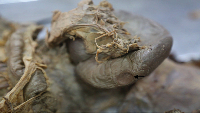
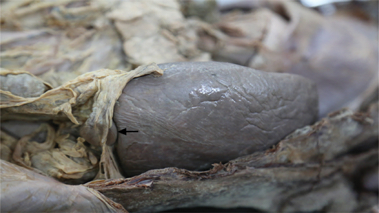
During the course of routine cadaveric dissection on 55-year-old female cadaver, by preclinical medical students, we encountered an additional visceral impression on the spleen which was created by an accessory spleen. The visceral surface of the spleen was carefully examined. An oval impression [Table/Fig-1] measuring 2.5x2 cm was noticed on the visceral surface near the inferior margin. This impression intervened between diaphragmatic surface and colic impression just adjacent to the inferior border of the spleen. The accessory spleen creating a visceral impression was found embedded in phrenicocolic ligament near the close proximity with the spleen [Table/Fig-2]. All the common sites i.e., splenic hilum, tail of the pancreas, gastrosplenic ligament, lienorenal ligament, and whole peritoneal cavity for the presence of accessory spleen were examined carefully, but there were no other accessory spleen found.
An additional impression over the visceral surface of the spleen (marked by black arrow).
An accessory spleen embedded in phrenicocolic ligament (marked by black arrow).
Discussion
An accessory spleen or supernumerary spleen refers to a small mass of splenic tissue encountered in addition to the normal spleen [1].
Development of spleen takes place as congregation of mesenchymal cells in the dorsal mesogastrium in the fifth week of embryonic life. The failure of fusion of these mesenchymal buds constitutes the embryological basis of accessory spleen formation [2].
Various splenic anomalies have been reported viz. lobulated spleen, accessory spleen seen at the splenic hilum, in the gastrosplenic ligament, in the lienorenal ligament, within the pancreas and along the splenic artery [3]. Verga I et al., found the various congenital variations like accessory spleen, wandering spleen, polysplenia, ectopic spleen [4]. In addition Weiand G and Mangold G also found accessory spleen present in 10-15% of cases [5]. This explains that accessory spleen is the commonest congenital anomaly.
Accessory spleens, although asymptomatic, can sometimes present as abdominal mass which might undergo cystic formation, hemorrhage, torsion or spontaneous rupture [6]. Thus, knowledge of presence of accessory spleens is important not only for Anatomists but also for Radiologists, Physicians, Pathologists and Surgeons for accurate diagnosis and management of conditions like acute abdomen with abdominal mass.
Accessory spleens are usually diagnosed incidentally during laparotomy or radiological examinations performed while investigating for other pathologies and are of no clinical significance in most cases. Sometimes however it may undergo cystic formation, haemorrhage, torsion or spontaneous rupture.
Notably, it is important to identify the accessory spleen because it may be confused with lymphadenopathy or a tumour of pancreas, kidney or adrenal gland [7]. Needless to mention that accessory spleen reasonably deserves to be considered in the differential diagnosis of abdominal mass. At the time of splenectomy surgeon has to take special care in removing accessory spleen as its persistence may lead to recurrence of disease [8,9]. Accessory spleen is most commonly seen in splenic hilum in 75% of cases, it can also be seen at the tail of pancreas in 25% of subjects. Other sites are gastrosplenic ligament, lienorenal ligament, wall of stomach or bowel, mesentery, greater momentum, it can be seen in pelvis or scrotum [10]. To the best of our knowledge accessory spleen in the phrenicocolic ligament with visceral impression on the main spleen have not been reported so far. So the uniqueness of the present investigation lies in the fact that accessory spleen in phrenicocolic ligament is located in very close proximity to the main spleen. The degree of proximity of accessory spleen is exhibited in the form of an additional oval impression created on the visceral surface of the spleen. This anatomical scenario could probably constitute an enigma to Radiologists while interpreting a Computed Tomography (CT) examination of upper abdomen. In view of the extremely rare location of a spleen as encountered in the current study case, the authors wish to recommend that Surgeons should also exercise extra caution in order to avoid recurrence of the disease.
We as Anatomists wish to highlight that the exclusiveness of the present paper is in the form of additional impression on the visceral surface of spleen. This unusual impression on the spleen could also possibly constitute a puzzle for the medical students in their preclinical curriculum. The present paper is a humble attempt to report an extremely rare location of an accessory spleen in the phrenicocolic ligament in close vicinity of spleen. The authors also aim to reinforce awareness of such rare position of accessory spleen among Surgeons, Radiologists and Anatomists.
Conclusion
Precise insight into anatomical variations of spleen is crucial to Surgeons and Radiologists in their clinical practice. The knowledge about variability in positional anatomy of accessory spleen is very important; for Surgeons during operative procedures and for Radiologists while interpreting the CT scans of upper abdomen.
[1]. Freeman JL, Jafri SZ, Roberts JL, Mezwa DG, Shirkhoda A, CT of congenital and acquired abnormalities of the spleenRadioGraphics 1993 13(2):579-610. [Google Scholar]
[2]. Seo T, Ito T, Watanabe Y, Umeda T, Torsion of an accessory spleen presenting as an acute abdomen with an inflammatory mass. US, CT, and MRI findingsPediatr Radiol 1994 24(7):532-34. [Google Scholar]
[3]. Grinbaum R, Zamir O, Fields S, Hiller N, Torsion of an accessory spleenAbdom Imaging 2006 31(1):110-12. [Google Scholar]
[4]. Verga I, Galfiova P, Adamkav M, Danisovic L, Polak S, Kubikova E, Congenital anomalies of the spleen from an embryological point of viewMed Sci Monit 2009 15(12):269-276. [Google Scholar]
[5]. Weiand G, Mangold G, Accessory spleen in the pancreatic tail - A neglected entity? A contribution to embryology, topography and pathology of ectopic splenic tissueChirurg 2003 74(12):1170-1177. [Google Scholar]
[6]. Development of the peritoneal cavity, gastrointestinal tract and its adnexae. In: Standring S, EditorGray’s anatomy–the anatomical basis of clinical practice 2008 LondonChurchil Livingstone Elsevier:1203-23. [Google Scholar]
[7]. Perez-Fontan FJ, Soler R, Santos M, Facio I, Accessory spleen torsion: US, CT and MR findingsEur Radiol 2001 11:509-12. [Google Scholar]
[8]. Mendi R, Abramson LP, Pillai SB, Rigsby CK, Evolution of the CT imaging findings of accessory spleen infarctionPediatr Radiol 2006 36:1319-22. [Google Scholar]
[9]. Hussein M, Hassan K, Yadav B, Usman N, Anatomical variation of spleen in north Indian population and its clinical significanceInnovative Journal of Medical and and Health Sciences 2013 3(4):190-192. [Google Scholar]
[10]. Yildiz AE, Ariyurek MO, Karcaaltincaba M, Splenic anomalies of shape, size, and location: Pictorial essayThe Scientific World Journal 2013 (2013):321810 [Google Scholar]