Anatomical Configuration of Roots and Canal Morphology of Primary Mandibular First Molars: A CBCT Study
Navin Hadadi Krishnamurthy1, Chikku Dani Jacob2, Umapathy Thimmegowda3, Jaya Agali Ramachandra4, Veena Arali5, Prasanna Kumar Bhat6
1 Reader, Department of Paedodontics, Rajarajeswari Dental College and Hospital, Bengaluru, Karnataka, India.
2 Postgraduate Student, Department of Paedodontics, Rajarajeswari Dental College and Hospital, Bengaluru, Karnataka, India.
3 Reader, Department of Paedodontics, Rajarajeswari Dental College and Hospital, Bengaluru, Karnataka, India.
4 Reader, Department of Paedodontics, Rajarajeswari Dental College and Hospital, Bengaluru, Karnataka, India.
5 Reader, Department of Paedodontics, Rajarajeswari Dental College and Hospital, Bengaluru, Karnataka, India.
6 Senior Lecturer, Department of Paedodontics, Rajarajeswari Dental College and Hospital, Bengaluru, Karnataka, India.
NAME, ADDRESS, E-MAIL ID OF THE CORRESPONDING AUTHOR: Dr. Navin Hadadi Krishnamurthy, No. 14, Ramohalli Cross, Mysore Road, Kumbalgodu, Bengaluru-560074, Karnataka, India.
E-mail: navina30@yahoo.co.in
Introduction
Root and canal morphology plays a vital role in improving the success rate of the endodontic treatment in primary teeth. The morphology of root canals in primary teeth leads to complications during pulp therapy. Studies done for assessing the root canal morphology and variations in primary teeth in Indian population are very scarce.
Aim
To evaluate the root and canal morphology of primary mandibular first molar (PMFMs) teeth using cone beam computed tomography (CBCT) in paediatric Indian population.
Materials and Methods
A total of 30 complete mandibular arch CBCT images of paediatric patients of the age group five to 10 years used in this study. The database was collected from Department of Oral Medicine and Radiology, Rajarajeswari Dental College and Hospital, Bengaluru. Sixty PMFMs from these 30 CBCT images were assessed for root and canal morphology and variation. Children of five to 10 years only were included in this study because the root completion of primary mandibular molars is around three years of age. This particular age group was chosen in order to ensure the integrity of the original morphology of the root canals of primary mandibular molars and also the amount of root resorption is minimal in this age. The study data was analyzed using SPSS version. 22 [IBM, Corp.,] for windows.
Results
Out of the 30 individuals enrolled with 60 PMFM’s majority of the primary mandibular first molars had two roots and three canals (68.3%), 20.0% had two roots and four canals and 11.7% had three roots and three canals. The bilaterally symmetrical incidence of variant 2 in this population was 70.4% variant 3 was – 18.5% and for variant 4 was 11.1%. There was no statistical difference between genders in the variance of root and canal number and morphology.
Conclusion
The results of this study indicated the PMFMs showed considerable variation in number and morphology of its root and its root canal. Very few studies have been conducted to find out the root canal morphology and anatomical variations in primary dentition. We as Paedodontists should be critical in evaluating root canal morphology in primary teeth for the overall success of the treatment plan.
Anatomical variation, Deciduous teeth, Radiographs
Introduction
Caries is the most common chronic disease in children. Primary teeth have a thinner layer of mineralized tissue between the external and internal surfaces, which leads to rapid involvement of the dental pulp [1]. The morphology of root canals in primary teeth usually leads to complications in root canal therapy [2]. Knowledge of the size, morphology and variation of the root canals are useful in visualizing the pulp cavity before, during and after endodontic treatment [3].
Premature loss of primary teeth, leads to changes in chronology and sequence of eruption of permanent teeth; thus, saving teeth in children is an important concept and frequently involves endodontic treatment [4]. Complexity of root canal systems dictates the parameters that affect the probability of success of root canal therapy especially in the primary mandibular first molars [5, 6].
Conventional radiography is the technique most frequently used for in vivo studies. The limitation with this technique is that the images of the roots and root canals always overlap, so it is difficult to see the buccolingual aspect [1]. Computed-based techniques have been used for studying root canal morphology and studies suggest that CBCT is a good option for studying root and canal morphology as it provides three dimensional information [1, 3, 5].
Digital radiography offers the benefit of less radiation exposure along with many advantages compared to conventional radiography. CBCT utility and its relevance to the practice of endodontics is reported with increasing frequency in the field of endodontics [6, 7]. However, there is hardly any study undertaken in the pre-evaluation and post-treatment using CBCT for pulp therapy in primary teeth.
Anatomically there are various root and canal configurations in case of primary teeth especially in case of primary molars but there is hardly any literature describing these variations unlike the various researches done on permanent teeth [8,9]. The PMFMs were taken into consideration in this study as it has a unique morphology as it does not have any resemblance to any other tooth in the oral cavity.
Therefore, the present study was undertaken to assess the variation in number and morphology of the root and canals of PMFMs and to study the applicability of CBCT in assessing the root and canal morphology.
Materials and Methods
A radiographic retrospective study was carried out on 60 CBCT images of PMFMs of children aged between five to 10 years obtained from the institution’s database of Department of Oral Medicine and Radiology of Rajarajeswari Dental College and Hospital, Bengaluru, India. The ethical clearance was obtained from the institutional ethical and research committee prior to the commencement of the study.
The sample size was estimated using the formula N=Z2(1-α) X PQ where Z(1-α) =1.96, P=0.50, Q=1-P, δ (Margin of Error) =0.15. δ2 The study was carried out between December 2016 to August 2017. The CBCT images of primary mandibular molars that were taken for valid diagnostic reasons such as developmental or congenital anomaly, pathological and traumatic lesions were collected and analyzed. The following teeth were selected, requiring endodontic treatment with or without periapical lesions and minimal root resorption in order to ensure the integrity of the original morphology of the root canals of PMFMs.
Radiographic Technique
The CBCT images were taken by a SCANORA® 3D machine at 90 kV and 12.5 mA, with a field of view of 60 mm and a voxel size of 0.1 mm. The slice thickness was 1 mm and the exposure time was 15 seconds. Scans were made according to the manufacturer’s recommended protocol and all of the images were taken by a licensed radiologist, with the minimum exposure necessary for adequate image quality. The lowest radiation dose and field were guaranteed.
Evaluation of the Image
The 60 CBCT images were analyzed with inbuilt software (On Demand) using a Dell Precision workstation and a 23 inch Dell LED screen in a dark room. The contrast and brightness of images could be adjusted using the image processing tool of the software to ensure optimal visualization. The evaluation criteria of the image was based on that given by Rang Yang et. al., in the year 2013 [1] [Table/Fig-1].
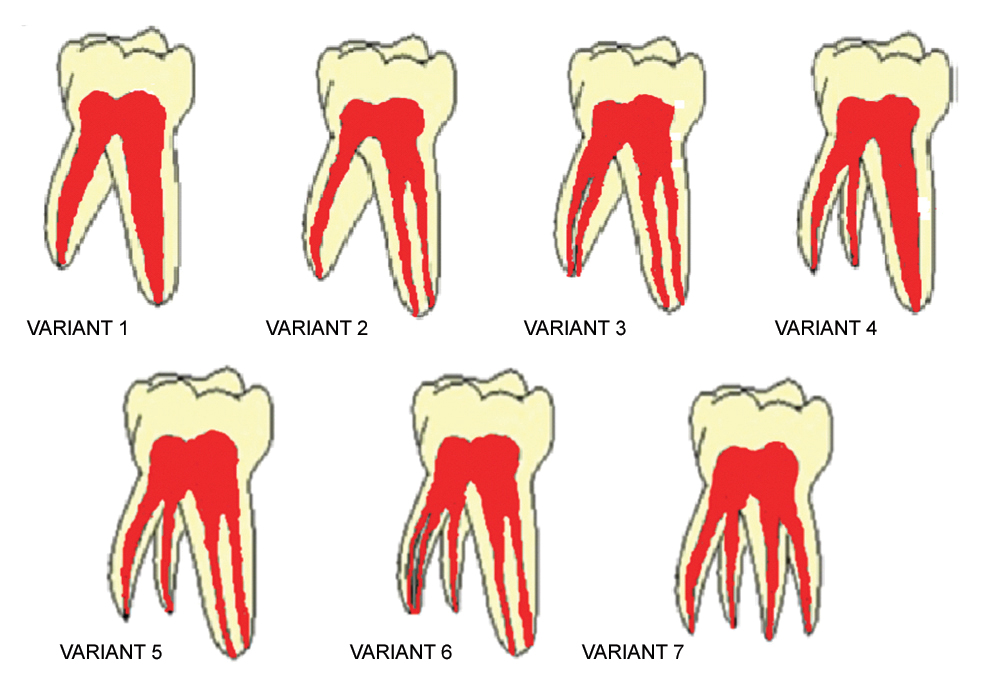
The three Variants which were Checked
Variant 2: Two separate roots, with two canals in the mesial root and one canal in the distal root.
Variant 3: Two separate roots, with two canals in the mesial root and two canals in the distal root.
Variant 4: Three separate roots a mesial, a distobuccal and a distolingual root with one canal in each root.
Statistical Analysis
The study data was analyzed using SPSS version 22.0 [IBM, Corp.,] for Windows. The experimental data obtained in this paper were presented as categorical variables. The frequency of the numbers of root and canals were determined and were compared by the Chi-square test, with a significance level of p<0.05.
Results
The Number of Roots and their Morphology
Sixty primary mandibular first molars CBCT images were examined. The mean age of the subjects included in the study was 6.8 years for the males (n=13) and 7.2 years for the females (n=17). 68.3% had two separate roots with three canals, 20.0% had two roots with four canals and 11.7% had three separate roots with one canal in each root. According to the gender wise distribution of total number of mandibular first molar teeth observed for different root and canal variants, there was no much variation observed [Table/Fig-2].
Gender wise distribution of total number of primary mandibular first molar teeth observed for different root and canal variants.
| Gender | Variant 2 | Variant 3 | Variant 4 | χ2-value | p-value |
|---|
| n | % | n | % | n | % |
|---|
| Males | 17 | 41.5% | 7 | 58.3% | 2 | 28.6% | 1.779 | 0.41 |
| Females | 24 | 58.5% | 5 | 41.7% | 5 | 71.4% |
| Total | 41 | 68.3% | 12 | 20.0% | 7 | 11.7% |
The Number and Morphology Variants of Canals
Gender wise comparison of the distribution of bilaterally symmetrical different root and canals showed that 70.4% had two separate roots and three canals, 18.5% had two separate roots and four canals and 11.1% had three separate roots and three canals [Table/Fig-3].
Gender wise comparison of the distribution of bilaterally symmetrical different root and canal variants.
| Gender | Variant 2 | Variant 3 | Variant 4 | χ2-value | p-value |
|---|
| % | % | % |
|---|
| Males | 66.7% | 25.0% | 8.3% | 0.682 | 0.71 |
| Females | 73.3% | 13.3% | 13.3% |
| Total | 70.4% | 18.5% | 11.1% |
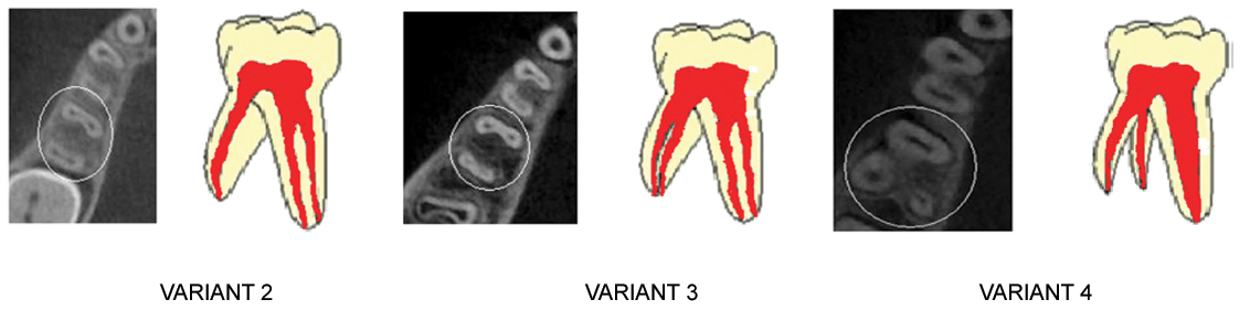
There were three variants found in the root canal morphology of the PMFMs [Table/Fig-4].
Illustrations showing the three variant.
Discussion
In the present study, we used CBCT to evaluate the number of roots and canals in 60 PMFMs of 30 individuals. The external morphology of PMFMs is unique and does not have any resemblance to any other tooth in the oral cavity [10,11]. When internal morphology is taken into consideration, mesiobuccal is the most prominent pulp horn followed by distobuccal, mesiolingual and distolingual [12,13]. An ex vivo study by Salama FS et al., examined 10 PMFMs and subjected half to a clearing technique and half to sectioning. Eight of these teeth had four canals and two had three canals present [14]. Another study conducted by Bagherian A et al., on studying 135 teeth, two types of variants were found. First variant had two roots with one canal in each (18.5%) and second variant had two roots with three canals, two in the mesial root and one in the distal root [15].
However, in our CBCT study, three types of root canal variations were determined, first variant was two separate roots along with two canals in the mesial root and one canal in the distal root (68.3%), second variant was two separate roots along with two canals in the mesial root and two canals in the distal root (20%) and the third variant was three separate roots, a mesial, a distobuccal and a distolingual root with one canal in each root (11.7%) which were statistically significant. Although, there are many case reports on the occurrence of three-rooted primary molars, there are very few studies on the prevalence of anatomical variants [8, 9].
CBCT can be considered when, it is determined that conventional radiographic techniques are yielding limited information and further details are required for diagnosis and treatment planning, while ensuring the radiation exposure as low as possible in patients. CBCT is an effective tool for the detection of the root and canal system of primary teeth. Two-rooted PMFMs occur frequently in this population which did not have relationship with gender predilection. If a PMFMs has an accessory root, there is a high probability that the PMFMs on the opposite side will also have one. The majority of PMFMs have three to four canals and the diversity of the root canal variants should be considered when performing clinical procedures [1, 13]. Our study provides information on canal variation types that can improve the success in the endodontics of primary teeth.
Until the year 1925, the study of root canal anatomy in primary molars were ignored, but Zurcher added an appendix to Hess’s work detailing his findings regarding the internal anatomy of primary dentition [14]. This study indicates that CBCT is helpful in evaluating the root and canal system, but CBCT cannot be used routinely because of over exposure to radiation risk. CBCT should only be considered when it is determined that conventional radiographic techniques are yielding limited information, ensuring that the patient’s radiation exposure is kept as low as possible.
Limitation
A limitation of this study was the sample size was less. Thus, further studies have to be conducted to identify more number of variants which could help paediatric dentists in improving their endodontic, diagnostic and treatment skills.
Conclusion
This study highlights the importance of various root canal variations in primary teeth, which is critical in the diagnosis as well as in the success of pulpal treatment in children. The majority of PMFMs have three to four canals and the diversity of the root canal variants should be considered when performing clinical procedures and can improve the success rate of endodontics in primary teeth. Thus, CBCT is an effective tool for the detection of the root and canal system of primary teeth accurately, but to be used with caution.
[1]. Yang R, Yang C, Liu Y, Hu Y, Zou J, Evaluate root and canal morphology of primary mandibular second molars in Chinese individuals by using cone-beam computed tomographyJ Formosan Medical Association 2013 112(7):390-5. [Google Scholar]
[2]. Gupta D, Grewal N, Root canal configuration of deciduous mandibular first molars - An in vitro studyJ Indian Soc Pedod Prev Dent 2005 23(3):134-7. [Google Scholar]
[3]. Zoremchhingi Joseph T, Varma B, Mungara J, A study of root canal morphology of human primary molars using computerized tomography: An in vitro studyJ Indian Soc Pedod Prev Dent 2005 23(1):7-12. [Google Scholar]
[4]. Fumes A, Versiani M, da Silva R, Root canal morphology of primary molars: A micro-computed tomography studyEur Arch Paediatr Dent 2014 15(5):317-26. [Google Scholar]
[5]. Wang Y, Chang H, Kuo C, Chen S, Guo M, Huang G, A study on the root canal morphology of primary molars by high resolution computed-tomographyJ of Dent Sci 2013 8(3):321-327. [Google Scholar]
[6]. Matherne RP, Angelopoulos C, Kulild JC, Tira D, Use of Cone-Beam Computed Tomography to Identify Root Canal Systems In VitroJ Endod 2008 34(1):87-9. [Google Scholar]
[7]. Gaurav V, Srivastava N, Rana V, Adlakha VK, A study of root canal morphology of human primary incisors and molars using cone beam computerized tomography: An in vitro studyJ Indian Soc Pedod Prev Dent 2013 31(4):254-9. [Google Scholar]
[8]. Liu JF, Dai PW, Chen SY, Huang HL, Hsu JT, Chen WL, Prevalence of three-rooted primary mandibular second molars among Chinese patientsPediatr Dent 2010 32(2):123-6. [Google Scholar]
[9]. Winkler MP, Ahmad R, Multi-rooted anomalies in the primary dentition of Native AmericansJ Am Dent Assoc 1997 128(7):1009-11. [Google Scholar]
[10]. Falk WV, Bowers DF, Bilateral three-rooted mandibular first primary molars: report of caseASDCJ DentChild 1983 50(2):136-7. [Google Scholar]
[11]. Zhang R, Yang H, Yu X, Wang H, Hu T, Dummer PM, Use of CBCT to identify the morphology of maxillary permanent molar teeth in a Chinese subpopulationInt Endod J 2011 44(2):162-9. [Google Scholar]
[12]. Curzon JA, Curzon ME, Congenital dental anomalies in a group of British Columbia childrenJ Can Dent Assoc (Tor) 1967 33(10):554-8. [Google Scholar]
[13]. Turner C, Christy G, Three-rooted mandibular first permanent molars and the question of American Indian originsAm J Phys Anthropol 1971 34(2):229-41. [Google Scholar]
[14]. Salama FS, Anderson RW, McKnight-Hanes C, Barenie JT, Myers DR, Anatomy of primary incisor and molar roor canalPediatric Dentistry 1992 14(2):117-8. [Google Scholar]
[15]. Bagherian A, Kalhori M, Sadhegi M, Mirhosseini F, Parisay I, An in vitro study of root and canal morphology of human deciduous molars in an Iranian populationJ Oral Sci 2010 52(3):397-03. [Google Scholar]