The elements of colour namely the hue, saturation, luminance has made mankind more aesthetic and fascinating. Extracts obtained from certain plants, insects and soil sources have good staining abilities. Numerous natural dyes are used in the diagnostic fields of histology, histochemistry and histopathology [1]. Turmeric is used as a dye for various purposes since centuries. Turmeric being an ecological compound is more readily available and non-toxic than synthetic stains. It belongs to the Zingiberaceae botanical group. The staining component of turmeric is curcumin [2]. It has affinity towards collagen fibres, cytoplasm, red blood cells and muscle cells [3]. In the present study, maceration and soxhlet methods were compared with respect to their staining efficacy along with the staining of turmeric extracts without and with mordant. TEM and eosin on normal and pathological tissues were also compared.
Materials and Methods
A prospective study was conducted on formalin fixed paraffin embedded blocks (normal and pathological tissues-OSCC) from archives of Department of Oral Pathology, Faculty of Dental Sciences, Ramaiah University of Applied Sciences, Bengaluru, Karnataka, India. The study was conducted for a period of six months. The ethical clearance was obtained from college Ethical Clearance Board (FDS/EC/2014-16/PG ST/05).
Turmeric was cut to slender pieces, dried and powdered. Maceration and soxhlet methods were employed to obtain its extracts [1, 2, 3].
Procedure of Turmeric Extraction
Maceration technique: About 15 g of finely ground turmeric powder was dissolved in 100 ml of 70% alcohol. The preparation was left undisturbed for 48 hours. The filtrate obtained was used to stain the tissues [3].
Soxhlet technique: Nearly 100 g of finely ground turmeric was weighed and placed in the condenser of soxhlet extractor; 100 ml of 70% alcohol was brought to boiling point. The vapour containing the extract was collected. The soxhlet extraction procedure was completed by 7-10 days [1, 2].
Tissues Considered in the Study
A total number of 40 paraffin embedded tissue blocks which included 20 normal oral mucosa and 20 pathological tissues (OSCC) were taken in the present study.
Staining Technique
A 4 μ thick sections (20 blocks of paraffin embedded normal tissues) were taken. A mordant potash alum in 1:10 ratio was employed in the present study to facilitate the binding of turmeric extract with the tissues [4,5]. The sections were stained with the components of turmeric extract obtained by maceration, soxhlet and TEM. The staining of normal and pathological tissues was performed with H&E and H&T, similar to the conventional H&E staining method. The tissue sections were deparaffinized in both xylene 1 and 2 for a span of 10 minutes. It was then hydrated in grades of alcohol in an ascending pattern with a halt of five minutes at each grades- 60%, 70%. The sections were subjected to water wash for five minutes and then blot dried. It was then stained with Harris hematoxylin for 15 minutes followed by a dip in water and was then differentiated with 1% acid alcohol by a single dip. The sections were subjected to bluing for 10 minutes and was placed in the prepared counter stains of turmeric extract obtained by maceration, soxhlet, TEM for four minutes. The sections were dipped once in tap water and absolute alcohol, blotted and dried, cleared in xylene and mounted with cover slip [4-9].
Standardization
By trial and error methods, four minutes was considered the ideal staining time. The pH was standardized to 5.7 with pH meter that belonged to Jiangsu biotech company, model number pH-107(pH-009) pen pocket sized pH meter, pH tester.
Examination of the Slides
The staining efficacy of turmeric with and without mordant, TEM in normal and pathological tissues was compared with eosin stained slides by three observers [Table/Fig-1,2]. The parameters considered to determine the staining ability were namely the nuclear staining, cytoplasmic details, staining intensity, and contrast/clarity. Each of the parameter were given a score of 0, 1 based on the ability to appreciate the stained section. The sum obtained on the above mentioned criteria determined if the stained slides were Poor (p=0-1), Satisfactory (S=2), Good (G=3) or Excellent (E=4). The scoring criteria was modified from that employed by Buesa RJ et al, in 2000 [10].
Scoring of the 20 normal tissue slides done by three observers.
| Observer 1 | Observer 2 | Observer 3 |
|---|
| Method | Maceration | Soxhlet | TEM normal | Eosin normal | Maceration | Soxhlet | TEM normal | Eosin normal | Maceration | Soxhlet | TEM normal | Eosin nomal |
|---|
| Score | E-4E-4G-3G-3E-4G-3G-3G-3G-3E-4E-4E-4G-3G-3E-4G-3G-3G-3G-3E-4 | S-2S-2G-3S-2G-3G-3G-3P-1G-3G-3S-2S-2G-3S-2G-3G-3G-3P-1G-3G-3 | E-4E-4E-4G-3G-3E-4G-3G-3E-4G-3E-4E-4E-4G-3G-3E-4G-3G-3E-4G-3 | E-4G-3E-4E-4G-3G-3E-4E-4G-3E-4E-4G-3E-4E-4G-3G-3E-4E-4G-3E-4 | E-4G-3G-3G-3G-3E-4G-3G-3E-4E-4E-4G-3G-3G-3G-3E-4G-3G-3E-4E-4 | G-3G-3P-1G-3G-3G-3S-2G-3S-2S-2G-3G-3P-1G-3G-3G-3S-2G-3S-2S-2 | G-3E-4E-4E-4G-3G-3E-4G-3G-3E-4G-3E-4G-3G-3E-4G-3G-3E-4E-4E-4 | E-4G-3E-4E-4G-3G-3E-4E-4G-3E-4E-4G-3E-4E-4G-3G-3E-4E-4G-3E-4 | E-4E-4E-4G-3G-3G-3G-3E-4G-3G-3E-4E-4G-3G-3E-4G-3G-3G-3G-3E-4 | G-3G-3P-1G-3G-3G-3S-2G-3S-2S-2S-2S-2G-3S-2G-3G-3G-3P-1G-3G-3 | G-3E-4G-3G-3E-4G-3G-3E-4E-4E-4G-3E-4G-3G-3E-4G-3G-3E-4E-4E-4 | E-4G-3E-4E-4G-3G-3E-4E-4G-3E-4E-4G-3E-4E-4G-3G-3E-4E-4G-3E-4 |
| Total | 68 | 50 | 70 | 72 | 68 | 50 | 70 | 72 | 68 | 50 | 70 | 72 |
i)Chi square analysis was used to assess if the observations of the two groups in each objective were independent of each other
ii)Reliability analysis was done to test the consistency between the three observers for the two groups in each objective
Scoring of 20 pathologic tissue slides.
| Observer 1 | Observer 2 | Observer 3 |
|---|
| TEMnormal | Eosinnormal | TEMnormal | Eosinnormal | TEMnormal | Eosinnomal |
|---|
| Score | G-3G-3G-3G-3G-3G-3G-3G-3E-4G-3E-4G-3G-3E-4G-3E-4E-4G-3G-3E-4 | G-3G-3G-3G-3G-3G-3E-4E-4G-3E-4G-3G-3E-4E-4G-3E-4G-3E-4E-4E-4 | E-4G-3G-3E-4E-4G-3E-4G-3G-3E-4G-3E-4G-3G-3G-3G-3G-3G-3G-3G-3 | E-4E-4E-4G-3E-4G-3E-4E-4G-3G-3E-4G-3E-4E-4G-3G-3G-3G-3G-3G-3 | E-4G-3G-3E-4G-3E-4E-4G-3G-3E-4G-3G-3G-3G-3G-3G-3G-3G-3E-4G-3 | G-3G-3E-4E-4G-3E-4G-3E-4E-4E-4E-4G-3E-4E-4G-3G-3G-3G-3G-3G-3 |
| Total | 66 | 69 | 66 | 69 | 66 | 69 |
i)Chi square analysis was used to assess if the observations of the two groups in each objective were independent of each other
ii) Reliability analysis was done to test the consistency between the three observers for the two groups in each objective
Statistical Analysis
Chi-square test was employed to determine the statistical significance.
Results
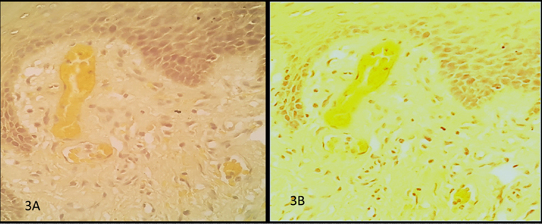
Amongst the two extraction methods, the sections stained by maceration method [Table/Fig-1,2], [Table/Fig-3a] fetched statistically significant results than soxhlet method, [Table/Fig-3b] (22.5%, p=0.04). Therefore, extracts obtained by the former method were considered subsequently in the study.
a) Tissue section stained by maceration method b) Tissue section stained by soxhlet method(40X).
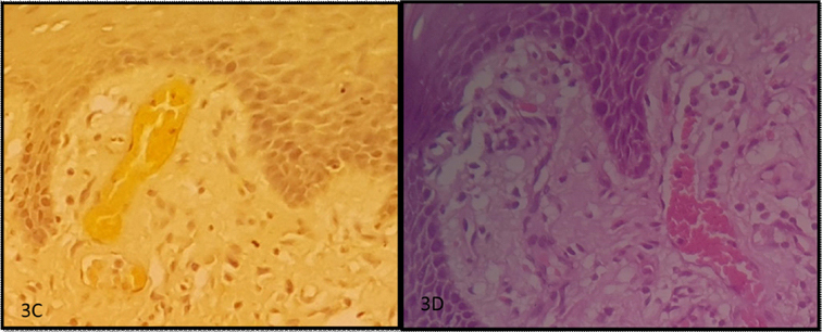
Sections stained with prepared TEM stain [Table/Fig-3c] fetched comparatively better result (2%, p=0.6530) when compared to turmeric stain [Table/Fig-3a].
c) tissue section stained with TEM, d) tissue section stained with H&E in normal tissues(40X).
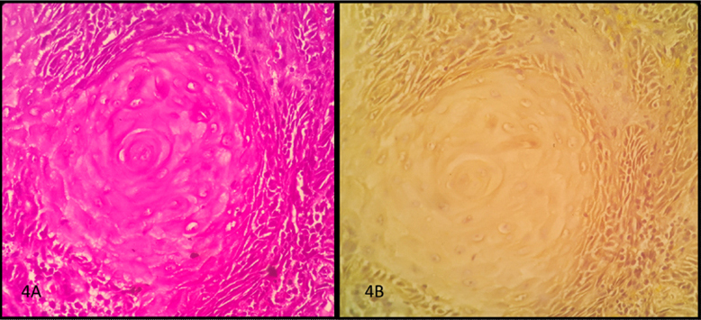
Normal tissue sections stained with TEM [Table/Fig-3c) gave comparable results with H&E stain [Table/Fig-3d] (3%, p=0.3710) similar to pathological tissues [Table/Fig-4a,b] (3.75% p=0.52). In [Table/Fig-5], x-axis components include (A-maceration Vs Soxhlet) A= (H&T) Maceration-68/80=85%, (H&T) Soxhlet-50/80=62.5%, % of difference in staining efficacy between maceration and soxhlet methods=22.5%, p-value = 0.04 value by chi square analysis- statistically significant, (B-Turmeric & TEM) B=(H&T) Maceration-68/80=85%, TEM in normal tissue-70/80=87%-% difference=2%, p=0.6530 value by chi-square analysis- statistically insignificant, (C-TEM & Eosin in normal tissue) C = TEM in normal tissue-70/80=87%, H&E in normal tissue-72/80 =90%, % difference in staining efficacy between (H&T) TEM & H&E=3%, p = 0.3710 value by chi-square analysis-statistically insignificant, (D-TEM & Eosin in pathological tissue) D= (H&T) TEM-66/80=82.5%, H&E-69/80=86.25%, % of difference in staining efficacy between (H&T) TEM and H&E=3.75%, p=0.52 value by chi-square analysis-statistically insignificant; y-axis components include, % of difference in staining efficacy between components A,B,C,D.
Tissue section stained with H&E and TEM in pathological tissues(40X)
Comparative bar graph of all the objectives.
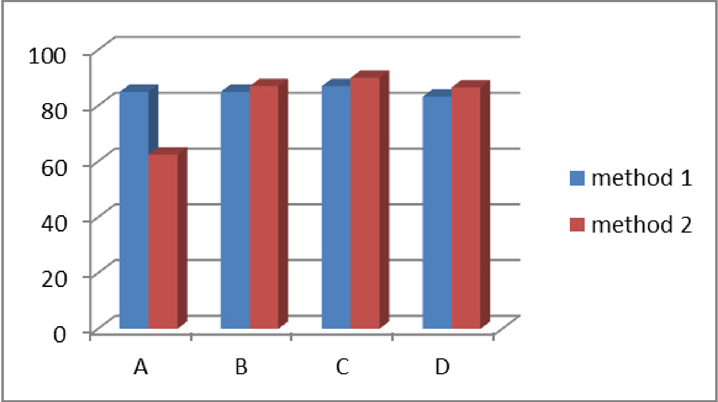
A-maceration versus soxhlet, B-turmeric alone and TEM, C,D-TEM versus Eosin in normal tissue and pathological tissues respectively.
Discussion
The turmeric routinely used for various studies is dried and powdered finely or coarsely [11,12]. Finely ground preparation increases the surface contact between the powdered particles and the solvent; the particles get homogenized which favours an efficient extraction. For these reasons, in the present study, finely ground turmeric was employed.
Principles of staining suggest that there is an ionic bond between the tissue components and the dye, which is associated with electrostatic attraction between the dissimilar ions. In majority of histological staining, an ionic bond can be appreciated between the tissue section and the stain. The pattern of how a tissue binds with the stain depends on the bond between the dye and the tissue components. Factors associated with selective staining are dye concentration, time of action on the solvent, its aqueous or alcoholic nature and its pH. It was observed that the mode of staining and pH of turmeric stain and eosin were similar [13,14,15]. Hence, turmeric extracts were taken to compare with eosin in this present study.
Turmeric contains saponins, tannins, flavonoids-polyphenolic compounds and alkaloids. The tannins and flavonoids are the substances that impart colour. Tannins are the most important ingredients necessary for dyeing. Saponins have the ability to lower the surface tension that benefits the staining efficacy. Flavonoids present in curcumin are primarily the pigments that provide a brightened tinge [1-4].
Traditionally, to obtain extracts from natural sources, maceration and soxhlet methods are used. In the present study, maceration and soxhlet methods were compared to check the better extraction method in relation to their staining efficacy. Tissues stained by maceration fetched better results than the soxhlet method. In maceration technique, greater yield of phytoconstituents like alkaloids, saponins, tannin, flavonoids were obtained, that enhanced the staining efficacy in contrary to the latter technique where the staining constituents could have been destroyed due to heat application [12,13]. Bassey R et al., in 2011, Avwioro OG et al., in 2007 conducted the study with extracts prepared only by soxhlet method, while Kumar S et al., in 2014 did the study with extracts prepared only by maceration method [1,2,3,16,17]. To our knowledge, comparative study between the two methods of extraction was one of the first of its kind.
Earlier studies have shown that acidity, alkalinity and mordant affect some stains. In a study by Elbadawi A in 1976 [18], the need for mordants in certain histochemical reactions has been stressed. Mordant is a substance that binds the stain on tissue sections by forming a coordination complex and the retention property of the stain to the tissue is enhanced [5-9]. It was also reported by Elbadawi A that ferric chloride as mordant was necessary in Verhoeff’s iron haematoxylin stain [18]. In the present study, it was observed that the slides stained with TEM were better appreciable than turmeric alone. Although turmeric stain is a natural one, it tends to achromatize over a period of time. The shelf lives of the slides with mordant were comparable to that of eosin, when reviewed after six months. Bassey R et al., in 2012 Avwioro OG et al., in 2007 and Kumar S et al., in 2014 stated that addition of mordant to Curcuma longa extracts prepared by maceration did not improve its staining qualities [1,2,3]. Avwioro OG et al., in 2007 Kumar S et al., in 2014 and stated that addition of mordant to Curcuma longa extracts prepared by maceration did not improve its staining qualities [2,3]. This finding was in contrast to the present study where it was observed that TEM enhanced the staining efficacy when compared to turmeric stain prepared without mordant.
It is observed that TEM used as a counterstain to hematoxylin gave comparable results to eosin stain in normal tissue and pathological tissues. TEM has shown comparable staining efficacy in normal tissues, which is in favour of studies done by Bassey R et al., in 2011, Aviworo OG et al., in 2007, Kumar S et al., in 2014 [1-3]. However, in the present study, along with normal tissues, pathologic tissues (OSCC) were also stained. The morphological and architectural changes of the cells were well appreciated by TEM staining. Addition of an appropriate natural mordant for better staining and working towards increasing the turmeric stain’s shelf life wasn’t accomplished. To our knowledge, comparative study of turmeric extract (TEM) and eosin on pathological tissues has not been tried earlier. Recently, advanced techniques for extraction like microwave assisted extraction, ultrasound assisted extraction, supercritical fluid extractions have been introduced. These techniques can be incorporated in the upcoming studies in obtaining turmeric stain and can thereby produce promising results where turmeric can be applied as a possible adjunct to eosin.
Conclusion
It was observed that sections stained by maceration method fetched better and statistically significant results than the soxhlet method. The tissue components stained with mordant provided greater clarity and was more appreciable. Normal and pathologic tissues stained with TEM fetched comparable results with eosin.
Curcumin has substantial ability to stain tissue section when applied as a counterstain to hematoxylin.
Abbreviations
H&E-Hematoxylin and Eosin
H&T-Hematoxylin and Turmeric
TEM-Turmeric Extract with Mordant
OSCC-Oral Squamous Cell Carcinoma