Percutaneous Achilles Tenotomy with a Large Gauge Needle in Ponseti Management of CTEV: A Modified Technique
Ritesh Arvind Pandey1, Bobby John2
1 Assistant Professor, Department of Orthopaedics, Christian Medical College and Hospital, Brown Road, Ludhiana, Punjab, India.
2 Professor, Department of Orthopaedics, Christian Medical College and Hospital, Brown Road, Ludhiana, Punjab, India.
NAME, ADDRESS, E-MAIL ID OF THE CORRESPONDING AUTHOR: Dr. Ritesh Arvind Pandey, Assistant Professor, Department of Orthopaedics, Christian Medical College and Hospital, Brown Road, Ludhiana, Punjab-141008, India.
E-mail: riteshpandey8262@yahoo.com
Introduction
Tenotomy of tendo achilles for clubfoot deformity is routinely done percutaneously with a surgical blade. This method though safe and effective, carries risk of complications such as excessive bleeding and injury to nearby neurovascular structures. Alternatively, sectioning of achilles tendon can be done safely by a large gauge needle percutaneously.
Aim
To study the safety and effectiveness of the modified technique.
Materials and Methods
A total of 37 children with 51 congenital clubfoot were included in this observational study. After successful correction of forefoot adduction and heel varus using the Ponseti technique, tendo achilles was sectioned percutaneously with a 16/18 gauge needle. Any complication occurring during the procedure was noted. Completeness of the tenotomy was checked by Thompson’s test and gain in passive dorsiflexion at ankle.
Results
Complete division of tendon was achieved in all 51 feet. No incidence of excessive bleeding, neurovascular injury or formation of pseudoaneurysm was found. However, minor bleeding from the surgical site was noticed in three cases and was managed by applying mild pressure over the involved area.
Conclusion
Percutaneous tenotomy of tendo achilles with a wide gauge needle is simple, safe and effective technique. It causes less morbidity and carries lesser risk of complications when compared to a surgical blade.
Clubfoot, Equinus, Tendo achilles
Introduction
Congenital Talipes Equino Varus (CTEV) is a common congenital anomaly with an incidence of one to three per 1000 live birth [1]. Ponseti’s technique [2] has become the standard and most effective treatment modality for correction of CTEV in newborn [3,4]. It consists of weekly stretching plaster casts followed by percutaneous tenotomy of tendo achilles. Achilles tenotomy is needed in 70-80% of cases after successful correction of forefoot adduction and heel varus [4-6]. Earlier, it was done by open method. This practice was later discontinued because of its invasiveness and high rate of complications. Today, as a part of Ponseti’s method, it is done percutaneously with a surgical blade (no. 11 and 15 being most commonly used). It is a simple and safe technique. However, with the increasing popularity of this procedure, complications are being reported [7,8]. These usually consist of excessive bleeding, formation of pseudoaneurysm and neurovascular injuries. Several modifications of this technique which includes use of modified surgical blade designs [9] and even mini open methods [10,11] have been reported in an attempt to decrease these complications.
Percutaneous tenotomy with a needle was described earlier in literature to correct trigger finger deformity [12-15]. Minkowitz B et al., first described the use of a large gauge hypodermic needle to section the tendo achilles percutaneously as a modification of Ponseti method [9]. Thereafter, similar studies were published by different authors describing this modified technique [16-19]. When compared to other methods, this technique has the possible advantage of being less invasive, simple and causing lesser morbidity. However, being a new method, and very few publications in its support, this surgical technique needs further validation by more similar studies. We aim to report our experience with this method to correct residual equinus in CTEV after successfully correcting the forefoot and midfoot deformities.
Materials and Methods
This observational study was conducted in the Department of Orthopaedics, Christian Medical College and Hospital, Ludhiana, between May 2015 to December 2016. A total of 51 untreated feet with idiopathic CTEV treated by modified method of Ponseti technique were included in the study. Only children below the age of two years were considered for this procedure. Other causes of CTEV like neurological, syndromic and post traumatic were excluded. Children more than two years of age were excluded. Neglected, resistant and relapsed varieties were also kept out of this study. An approval of the Ethics committee was taken and the procedures were in accordance of the standards mentioned in Helsinki Declaration of 1975 and revised in 2000. Pirani score [20] was calculated at presentation for every foot included in the study. Correction was started with weekly stretching and plaster casts. Pirani score was then calculated at every follow up to monitor the deformity correction. This consists of six components and each can score 0, 0.5 or 1. Three components contribute to Midfoot Contracture Score (MCS) i.e., medial crease, curvature of lateral border and position of head of talus. Remaining three components make the Hindfoot Contracture Score (HCS) i.e., posterior crease, empty heel and rigidity of equinus. After successful correction of forefoot adduction and heel varus as per the Pirani recommendation, the children were examined for presence of residual fixed equinus deformity. Those with MCS less than one and HCS more than one were considered for a percutaneous tenotomy of tendo achilles. Parents were informed about the study and surgical technique used in this study. Only those who were ready, were included in the study after signing a written consent.
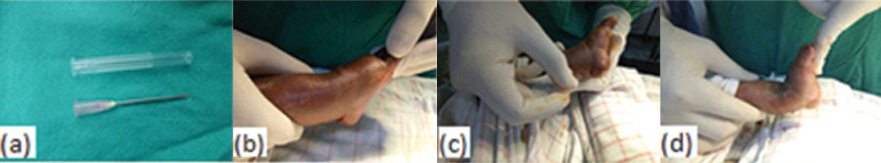
a) 18 gauge needle used for tenotomy: b) preoperative equinus deformity; c) surgical technique; d) deformity correction after tenotomy.
All children were operated with aseptic precautions under short general anaesthesia by the same surgeon. The limb was cleaned with 5% povidone iodine and draped without a tourniquet. Keeping the knee extended, the foot was dorsiflexed to make the tendo achilles tight and was palpated as a tight cord posteriorly. A 16/18 gauge needle chosen to cut the tendon, was introduced near the medial border of the tendon 1 cm above its insertion over the calcaneum. We preferred 18 gauge for children below six months whereas 16 gauge was preferred for those more than six months of age. The tendon was cut by the tip of the needle from medial to lateral direction. While doing so, grating sensation could be felt as fibers of the tendon are severed. The dorsiflexion force over the ankle was continuously maintained. The completion of tenotomy was marked by a snap and visible correction of equinus allowing atleast 10 degrees of dorsiflexion. Thompson’s test was performed in every case to further confirm the completion of section. A complete tenotomy gives a negative Thompson’s test due to absence of transmission of movements from calf to heel. A palpable gap between the two ends of tendon could also be felt. The needle was then removed and passive dorsiflexion rechecked. A knee cast with the knee in 90 degree flexion and foot in maximal abduction and 10 degree dorsiflexion was applied for three weeks. Thereafter, patients were followed up on a monthly basis for minimum of six months and then after every three months.
Results
The study was conducted on 51 feet with clubfoot deformity in 37 children from May 2015 to December 2016, who satisfied the inclusion criteria. There were 29 males and 8 females with a male: female ratio of 3.6:1. Deformity was unilateral in 23 patients whereas in 14 patients, it was bilateral. Those with unilateral deformity, 18 had a right and 5 had a left sided involvement.
The mean age of patients at the start of treatment was 12.4 weeks (3 days to 16 months). Family history of CTEV was present in one (2.7%) child [Table/Fig-2]. The average Pirani score at the beginning of treatment was 4.75 (2 to 6). Tendo achilles tenotomy was performed in all 51 feet for the correction of equinus deformity. The average number of cast required before the tenotomy was 5.26 (4 to 7) and the mean age at tenotomy was 16.42 weeks (5 weeks to 18 months). The average Pirani score after tenotomy was 0.07 (0-2). Mean follow up period after tenotomy was six months. Complete section of the tendon was achieved in all cases and none required an open procedure to complete the tenotomy. At the end of procedure, an increase in dorsiflexion was noted and equinus adequately corrected in all except one patient. This child had bilateral atypical clubfoot and residual equinus even after complete sectioning of the tendon on both sides. The residual deformity was corrected gradually with serial weekly casting. In one foot, control over the needle was lost intraoperatively causing section of the skin posterior and lateral to the tendon. The wound was sutured primarily and further management was uneventful. There was no incidence of excessive bleeding, pseudoaneurysm or neurovascular injury in any patient. However, three patients experienced minor bleeding during the procedure which was controlled successfully by applying pressure over the skin puncture site. Five patients (9.8%) developed plaster related complications which were managed successfully. There was no incidence of local infection in any case [Table/Fig-3].
Demographic profile of the study population.
| Variables | Number | Frequency |
|---|
| Age | 0-6 months | 35 | 94.5% |
| >6 months | 02 | 5.4% |
| Gender | Male | 29 | 78.4% |
| Female | 08 | 21.6% |
| Family history | Positive | 01 | 2.7% |
| Negative | 36 | 97.3% |
| Unilateral | Right | 18 | 48.6% |
| Left | 05 | 13.5% |
| Bilateral | 14 | 37.8% |
| Average Pirani score at presentation | 4.75 |
| Average number of casts before tenotomy. | 5.26 |
| Minimum duration of follow up | 6 Months |
Procedure related complications.
| Complication | N (%) |
|---|
| Plaster related complications | 5 (9.8%) |
| Bleeding (Minor) | 3 (5.8%) |
| Pseudo aneurysms | None |
| Neurovascular injuries | None |
| Infection | None |
| Incomplete tenotomy | 2 (3.9%) |
| Loss of control over needle | 1 (1.9%) |
Discussion
Ponseti method of clubfoot treatment corrects all the deformities effectively except the equinus. This residual equinus deformity does not improve with further manipulation and casting and requires tenotomy of the tendo achilles. Ponseti used an ophthalmic scalpel blade for a percutaneous tenotomy. The long and sharp end of this blade has the potential risk of damaging the structures around the tendon especially, those lateral to it. Obviously, thicker the instrument used to perform tenotomy, more was the risk of damaging nearby structures. Since then, constant efforts have been made to find a safer and less invasive method of doing a tenotomy. Dobbs MB et al., used a shorter ophthalmic blade to minimize this risk [7]. In their study on 219 idiopathic clubfeet, they reported serious bleeding complications following the percutaneous tendo achilles tenotomy in four patients; three due to presumed injury to the peroneal artery and one due to injury to the lesser saphenous vein. Nowadays, a no. 15 surgical blade is most commonly used for the percutaneous tenotomy. However, Burghardt RD et al., reported a case of pseudoaneurysm following the use of even no. 15 blade [8]. Some authors have even suggested an open technique instead of percutaneous method to avoid these complications [7,10].
Many studies have reported vascular anatomical variations in the club foot [21-31]. Burghardt RD et al., described close proximity of the Achilles tendon with the posterior neurovascular bundle [8]. In addition, foot may have a single blood supply through the posterior tibial artery. Insufficiency of anterior tibial artery in clubfoot has been reported in many studies with an incidence up to 85% [7,21,29]. In such circumstances, posterior tibial artery remains the only source of blood supply for the foot and remains at risk during tenotomy. Insufficiency of posterior tibial artery has also been described in clubfoot [25,26,28,30,31]. In these uncommon cases, with the deficiency of the blood supply through both anterior and posterior tibial arteries, the fibular artery becomes dominant and should be carefully protected in clubfoot release surgeries, as well as in Achilles tendon sectioning procedures. The injury to this single vascular supply during tenotomy may severely compromise the vascularity of foot leading to partial or complete necrosis of the foot and risk of amputation. Hence, a technique providing more safe precautions is needed to avoid these complications.
In the present study, 51 clubfeet in 37 children were corrected by Ponseti method using serial stretching casts on weekly basis followed by percutaneous sectioning of tendo achilles. However, tenotomy was done with an 18 gauge needle as recommended by Minkowitz et al., [9]. The technique has already been used for percutaneous release of tendon in trigger finger and has been described to be successful and safe [12-15]. This technique has also been used to release muscle and tendon contracture in adult patients with brain damage [32]. Maranho DAC et al., in their series of 57 feet, reported complication in only two patients in the form of minor bleeding [16]. In a similar study by Rahman et al., complication was seen in nine out of 70 feet [19]. These were in the form of minor bleeding in two, difficult procedure in four and incomplete correction in three feet. Sirsikar et al., used this technique in 49 clubfoot deformities and all were reported as uneventful [18]. Choubey et al., compared the two techniques of doing tenotomy (blade vs needle) and found no significant difference between the two [33]. They did not find any complication associated with percutaneous needle tenotomy technique and reported it as simple, effective and minimally invasive. We didn’t find any incidence of excessive bleeding in any patient with this technique. No incidence of pseudoaneurysm was found in our follow up of 12 months after tenotomy. Post tenotomy infections has been reported in earlier studies [34,35]. In current study, no infection was reported in any of the patients in immediate postoperative period. The result of present and similar previous studies has been compared in [Table/Fig-4].
Results of previous comparative studies.
| Study/year | Number of feet | Needle used for tenotomy | Average Pirani score (pre/post op) | Average follow up | Complications (n) |
|---|
| Minkowitz B et al., [9] | 21 | 16/18 gauge | ---------- | ---------- | None |
| Maranho DAC et al., [16] | 57 | 16 gauge | ---------- | ---------- | Abnormal bleed - 2 |
| Patwardhan S et al., [17] | 600 | 16 gauge | ---------- | ---------- | Not mentioned |
| Sirsikar A and Kiradiya N [18] | 49 | 16/18 gauge | Used Dimeglio score | 7 months | None |
| Rahman MS et al., [19] | 70 | 19 gauge | 4.9/0.75 | 4.5 months | Minor bleed – 2Difficult procedure – 4Incomplete correction – 3 |
| Choubey R and Jain A [33] | 28 | 16 gauge | 5.58/0.31 | 12 months | None |
| Present study | 51 | 18 gauge | 4.75/0.5 | 6 months | Minor bleed – 3Incomplete correction – 2Loss of control - 1 |
Another concern with the percutaneous tenotomy is the risk of incomplete division of the tendon leading to poor correction of equinus deformity and hence early recurrence. Maranho DAC et al., suggested ultrasound guided tenotomy with a wide bore needle to ensure the completeness of this procedure [16]. In the present study, complete section of the tendon was achieved with good dorsiflexion of foot in all patients. However, in the current study, tenotomy was not done under ultrasound guidance and completeness of tenotomy was ensured only on the basis of clinical findings. Furthermore, this finding can also be surgeon dependent with more incidences of incomplete tenotomy by a less experienced surgeon.
Limitation
Our results are from a small patient population (51 feet) with a short follow up period. We recommend further studies in larger population with a longer follow up. In the present study, comparison between the needle and blade techniques was not done and a comparative study between the two techniques is suggested.
Conclusion
We consider the technique to be simple and safe with lower morbidity and equally effective as compared to other methods of tenotomy. The method is less invasive and can be done in a day care surgery under local anaesthesia. With the inherent advantages, we recommend this modified technique of tendo achilles tenotomy for treatment of idiopathic club foot deformity in younger children. With global clubfoot initiative, more and more paramedical staff is being involved in the management of clubfoot. This technique being safe and easier to learn will be useful in these initiatives too.
[1]. Pandey S, Neglected club footFoot 2002 12:123-41. [Google Scholar]
[2]. Ponseti IV, Congenital clubfoot: fundamentals of treatment 1996 New YorkOxford University Press:140 [Google Scholar]
[3]. Goksan SB, Bursali A, Bilgili F, Sivacioglu S, Ayanoglu S, Ponseti technique for the correction of idiopathic clubfeet presenting up to 1 year of age. A preliminary study in children with untreated or complex deformitiesArch Orthop Trauma Surg 2006 126:15-21. [Google Scholar]
[4]. Scher DM, Feldman DS, Van Bosse HJP, Sala DA, Lehman WB, Predicting the need for tenotomy in the Ponseti method for correction of clubfeetJ Pediatr Orthop 2004 24:349-52. [Google Scholar]
[5]. Ponseti IV, Treatment of congenital club footJ Bone Joint Surg Am 1992 74:448-54. [Google Scholar]
[6]. Bor N, Coplan JA, Herzenberg JE, Ponseti treatment for idiopathic clubfoot: minimum 5-year follow upClin Orthop Relat Res 2009 467:1263-70. [Google Scholar]
[7]. Dobbs MB, Gordon JE, Walton T, Schoenecker PL, Bleeding complications following percutaneous tendo achilles tenotomy in the treatment of clubfoot deformityJ Pediatr Orthop 2004 24:353-57. [Google Scholar]
[8]. Burghardt RD, Herzenberg JE, Ranade A, Pseudoaneurysm after Ponseti percutaneous Achilles tenotomy: a case reportJ Pediatr Orthop 2008 28:366-69. [Google Scholar]
[9]. Minkowitz B, Finkelstein BI, Bleicher M, Percutaneous tendo-Achilles lengthening with a large-gauge needle: a modification of the Ponseti technique for correction of idiopathic clubfootJ Foot Ankle Surg 2004 43:263-65. [Google Scholar]
[10]. Dogan A, Kalender AM, Seramet E, Uslu M, Sebik A, Mini-open technique for the achilles tenotomy in correction of idiopathic clubfoot: a report of 25 casesJ Am Podiatr Med Assoc 2008 98:414-17. [Google Scholar]
[11]. Rhett M, William H, Brian S, Garrett L, A mini-open technique for Achilles tenotomy in infants with clubfootJ Child Orthop 2016 10(1):19-23. [Google Scholar]
[12]. Bain GI, Turnbull J, Charles MN, Roth JH, Richards RS, Percutaneous A1 pulley 27. release: a cadaveric studyJ Hand Surg Am 1995 20:781-84. [Google Scholar]
[13]. Saldana MJ, Trigger digits: diagnosis and treatmentJ Am Acad Orthop Surg 2001 9:246-52. [Google Scholar]
[14]. Lorthioir J, Surgical treatment of trigger-finger by a subcutaneous methodJ Bone Joint Surg Am 1958 40:793-95. [Google Scholar]
[15]. Cohen TJ, Tratamento percutâneo do dedo em gatilhoRev Bras Ortop 1996 31:690-92. [Google Scholar]
[16]. Maranho DAC, Nogueira-Barbosa MH, Simão MN, Volpon JB, Use of a large gauge needle for percutaneous sectioning of the Achilles tendon in congenital clubfootActa Ortop Bras 2010 18(5):271-6. [Google Scholar]
[17]. Patwardhan S, Shyam A, Sancheti P, Percutaneous needle tenotomy for tendo-achillis release in clubfoot – technical noteJ Orthop Case Rep 2012 2(1):35-36. [Google Scholar]
[18]. Sirsikar A, Kiradiya N, A prospective study of outcome of percutaneous needle tenotomy for tendo achilles release in congenital talipes equino varusInternational Journal of Medical Science Research and Practice 2014 1(3):84-88. [Google Scholar]
[19]. Rahman MS, Alam MK, Shahiduzzaman M, Rahman A, Percutaneous needle tenotomy for ponseti technique in the management of congenital talipes equinovarus (ctev)J Dhaka Med Coll 2014 23(1):55-59. [Google Scholar]
[20]. Pirani S, Outerbridge H, Moran M, Sawatsky B, A method of evaluating the virgin club foot with substantial interobserver reliability 1995 MiamiPresented at the annual meeting of Pediatric Orthopaedic Society of North America [Google Scholar]
[21]. Edelson JG, Husseini N, The pulseless club footJ Bone Joint Surg Br 1984 66:700-02. [Google Scholar]
[22]. Sodre H, Bruschini S, Mestriner LA, Miranda F, Levinsohn EM, Packard DS, Arterial abnormalities in talipes equinovarus as assessed by angiography and the Doppler techniqueJ Pediatr Orthop 1990 10:101-04. [Google Scholar]
[23]. Ben-Menachem Y, Butler JE, Arteriography of the foot in congenital deformitiesJ Bone Joint Surg Am 1974 56:1625-30. [Google Scholar]
[24]. Greider TD, Siff SJ, Gerson P, Donovan MM, Arteriography in club footJ Bone Joint Surg Am 1982 64:837-40. [Google Scholar]
[25]. Dobbs MB, Gordon JE, Schoenecker PL, Absent posterior tibial artery associated with idiopathic clubfoot. A report of two casesJ Bone Joint Surg Am 2004 86:599-602. [Google Scholar]
[26]. Hootnick DR, Levinsohn EM, Crider RJ, Packard DS, Congenital arterial malformations associated with clubfoot. A report of two casesClin Orthop Relat Res 1982 167:160-63. [Google Scholar]
[27]. Hootnick DR, Packard DS, Levinsohn EM, Crider RJ, Confirmation of arterial deficiencies in a limb with necrosis following clubfoot surgeryJ Pediatr Orthop B 1999 8:187-93. [Google Scholar]
[28]. Kruse L, Gurnett CA, Hootnick D, Dobbs MB, Magnetic resonance angiography in clubfoot and vertical talus: a feasibility studyClin Orthop Relat Res 2009 467:1250-55. [Google Scholar]
[29]. Katz DA, Albanese EL, Levinsohn EM, Hootnick DR, Packard DS, Grant WD, Pulsed color-flow Doppler analysis of arterial deficiency in idiopathic clubfootJ Pediatr Orthop 2003 23:84-87. [Google Scholar]
[30]. Quillin SP, Hicks ME, Absent posterior tibial artery associated with clubfoot deformity: an unusual variantJ Vasc Interv Radiol 1994 5:497-99. [Google Scholar]
[31]. Kitziger K, Wilkins K, Absent posterior tibial artery in an infant with talipes equinovarusJ Pediatr Orthop 1991 11:777-78. [Google Scholar]
[32]. Coroian F, Jourdan C, Froger J, Anquetil C, Choquet O, Coulet B, Percutaneous needle tenotomy for the treatment of muscle and tendon contractures in adults with brain damage: results and complicationsArchives of Physical Medicine and Rehabilitation (APMR) 2017 98(5):915-22. [Google Scholar]
[33]. Choubey R, Jain A, Comparison of percutaneous tenotomy techniques for correction of equinus deformity in Congenital Talipes Equino Varus (CTEV) in children: a randomized clinical trialJournal of Evolution of Medical and Dental Sciences 2015 4(57):9865-70. [Google Scholar]
[34]. Dyer PJ, Davis N, The role of Pirani scoring system in the management of club foot by the Ponseti methodJ Bone Joint Sur [Br] 2006 88(8):1082-84. [Google Scholar]
[35]. Lourenco AF, Morcuende JA, Correction of neglected idiopathic club foot by Ponseti MethodJ Bone Joint Surg [Br] 2007 89(3):378-81. [Google Scholar]