Facial fractures are challenging injuries to treat considering the unpredictability of the pattern of injury, airway issues and associated head injury. Inadequate treatment results in deformity of the face which is difficult to correct secondarily.
ORIF is the gold standard in treatment of displaced facial fractures. Incorporation of primary bone grafts can provide better functional and aesthetic outcome [1] and may reduce operation cost by reducing the number of plates and screws that may be required. Van Meek’ren and Macewen have reported the use of autogenous bone grafts in facial fractures [2,3]. Since then bone grafts have been used in face to fill gaps and correct contour deformity. Primary and secondary bone grafts are used in correcting post-traumatic orbital deformities with variable results [4-6]. Among recent studies from India, Singh et al., has published the spectrum of primary bone grafting in facial fractures and their functional and aesthetic assessment showed low rate of disabilities and high percentage of patient satisfaction [7].
To compare the functional and aesthetic outcome of ORIF in upper and mid-face fractures with and without primary bone grafting.
Materials and Methods
A prospective study after obtaining ethical committee clearance was conducted from January 2012 to December 2013 for a period of 2 years at IPGMER and SSKM hospitals, Kolkata. Thirty patients included in the study were alternatively allotted in two groups. ORIF with bone grafting was done in group 1 patients and group 2 patients underwent only ORIF. Informed consent that included acceptance for participation and use of photographs and radiological investigations for research/publications were obtained from all patients included in the study.
Inclusion criteria: Patients more than 12 years of age admitted in our institute with facial fractures in upper or midface during the study period, who gave consent for the surgery, were included in study. Patients with bilateral upper and midface fractures with undisplaced fractures on one side were also included in the study. Unfractured/undisplaced opposite side was used as a guide to measure postoperative projection deficits in radiological assessment.
Exclusion criteria: Patients with isolated fracture mandible, those with associated CNS injury needing intervention, those with poor general condition making them unfit for early surgery, patients under 12 years, those with bilateral displaced fractures, patients refusing to give consent for surgery and to participate in the study were excluded from the study.
These two groups were again subdivided into those with upper face fractures and those with mid-face fractures to maintain comparability. Bone grafts from calvarium, iliac crest and ribs were used in group I and they were fixed rigidly with screws/plates [Table/Fig-1,2]. Preoperative and postoperative assessments after one and three months were made based on three broad headings:
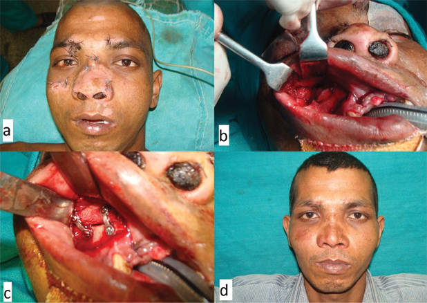
Iliac crest bone grafting for anterior wall of right maxilla. Postoperative picture (a); comminuted anterior wall of right maxilla (b); iliac crest bone graft in place (c); 3 months postoperative picture (d).
Sources of bone graft used and distribution.
| Bone graft source | Number of patients | Percentage of patients |
|---|
| Calvarial | 5 | 33.3% |
| Iliac Crest | 8 | 53.3% |
| Rib | 2 | 13.3% |
| Total | 15 | 100.0% |
Clinical and functional assessment;
Photographic assessment;
Radiological assessment.
Clinical and functional assessment included visual disturbances, frontal sinus/CNS injury, vertical dystopia, enophthalmos and mal-occlusion. Vertical dystopia was measured in millimetres as the patient was sitting erect and in forward gaze. Vertical distance between the mid-pupillary points of both eyes was noted. Enophthalmos was measured with Hertel’s exophthalmometry and compared with the opposite side.
Photographic assessment was made with frontal, bird’s eye view and lateral views with controlled lighting and distance from the lens. Grading of malar asymmetry was done according to classification system proposed by Holmes and Mathews [8] [Table/Fig-3]. Each patient was assigned to one of the following grades:
Photographic assessment of malar asymmetry: grade 1(a), grade 2 (b), grade 3 (c).
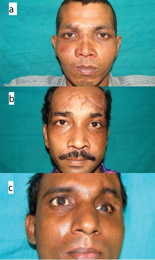
Grade I: excellent cosmetic result, no malar asymmetry;
Grade II: good cosmetic result, malar asymmetry only on careful examination;
Grade III: poor cosmetic result, noticeable malar asymmetry;
Grade IV: gross malar asymmetry.
Radiological assessment was made with postoperative Computerised Tomography (CT) of facial bones. For frontal bone fractures, bone defects were measured in three dimensions. For mid-face fractures, zygomatic complex projection and zygomatic complex height (Furst et al., [9]) was measured as follows:
Zygomatic complex projection: Axial sections of CT of facial bones were used. Horizontal lines representing anterior and posterior zygomatic complex widths were drawn and the distance between the two was measured (A1). Similarly contralateral normal side was measured and the deficit noted (A) [Table/Fig-4a].
Radiological assessment of zygomatic complex projection deficit (a); and zygomatic complex height deficit (b).
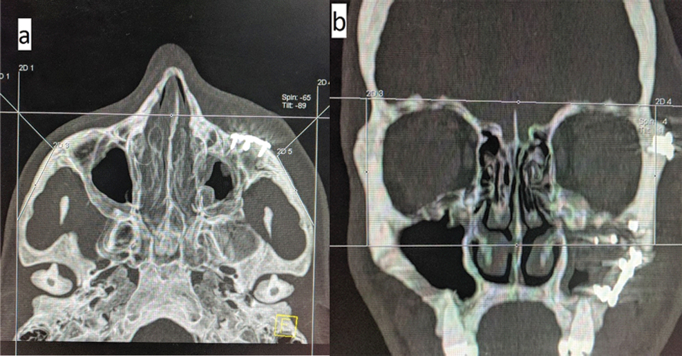
Zygomatic complex height: Coronal sections were used. The distance between the horizontal reference point (orbital roof) and the lateral most point of the curved complex was measured (B1). Similarly normal side was measured and deficit noted (B) [Table/Fig-4b].
Chi-square (χ2) Test was used to test the association of different study variables with the study groups. Z-test (Standard Normal Deviate) was used to test the significant difference between two proportions. T-test was used to compare the means. A p< 0.05 was considered statistically significant.
Results
Most of the patients 18 (60.0%) were in the age group 20-29 years. Five (16.7%), 5 (16.7%) and 2 (6.7%) patients were in age groups 12-20 years, 30-39 years, ≥40 years respectively [Table/Fig-5]. Proportion of males 26 (86.7%) were significantly higher than females 4(13.3%). Road traffic accident (RTA) was the most common aetiology of facial fractures in our study, 25 (83.3%) out of 30 patients. Other causes include assault (13.3%) and fall from height (3.3%) [Table/Fig-6].
Age wise distribution of all patients.
| Age (years) | Frequency | Percentage |
|---|
| 12-20 | 5 | 16.7% |
| 20-29 | 18 | 60.0% |
| 30-39 | 5 | 16.7% |
| ≥40 | 2 | 6.7% |
| Total | 30 | 100.0% |
Distribution of cause of injury.
| Cause of Injury | Number | Percentage |
|---|
| Assault | 4 | 13.3% |
| Fall from height | 1 | 3.3% |
| RTA | 25 | 83.3% |
| Total | 30 | 100 |
Clinical Assessment
Nerve injury/entrapment was found in 50% (15) of our subjects of which infra-orbital nerve was injured in 33.3% (10) and supra-orbital was injured in 16.7%(5). Existing skin lacerations were used in 50%(15) of subjects and in the rest, standard incisions were made. Associated central nervous system injury was present in 13.3%(4) of subjects and no neurosurgical intervention was done in these patients as they were treated conservatively. Visual disturbances and frontal sinus injury were present in 23.3%(7) of patients and malocclusion in 10%(3).
Vertical dystopia: A 23.3%(7) of patients with facial fractures in our study presented with vertical dystopia, the mean value of which was 13 mm (7–18mm) preoperatively. Mean value of vertical dystopia in group 1 after one and three months postoperatively was 1mm and 1.25mm respectively and that of group 2 was 0.67mm and 0.67mm [Table/Fig-7]. Under this parameter group 2 had better result that was statistically significant.
Clinical assessment-vertical dystopia and enophthalmos.
| Vertical Dystopia | 1 months (in mm) | 3 months (in mm) |
|---|
| Group 1 | 1 | 1.25 |
| Group 2 | 0.67 | 0.67 |
| p-value | 0.04 | 0.05 |
| Enophthalmos | 1 months (in mm) | 3 months (in mm) |
| Group 1 | 0.5 | 0.5 |
| Group 2 | 1 | 1 |
| p-value | 0.61 | 0.61 |
Enophthalmos: A 16.7% (5) of patients with facial fractures in our study presented with enophthalmos, the mean value of which was 2.6 mm (2–5mm) preoperatively. Mean value of enophthalmos in group one after one and three months postoperatively was 0.5mm and 0.5mm respectively and that of group 2 was 1mm and 1mm [Table/Fig-7]. Under this parameter group 1 had better result that was not statistically significant.
Photographic Assessment
Frontal asymmetry: Our study has 30 patients of whom, 13 had upper 1/3rd of face fractures and 17 had mid face fractures. Among the 13 of upper face fracture patients, six belong to group 1 and seven belong to group 2. In midface fractures, nine were in group 1 and eight in group 2.
In Group-1 patients, frontal asymmetry after 1 month, 5(83.3%) were in Grade-1and 1(16.7%) was in grade-2 but in Group-2 patients, 3 (42.9%) were in Grade-1and 4 (57.1%) were in Grade-2 (p=0.35).
In Group-1 patients, frontal asymmetry after three months, 4(66.7%) were in grade-1 and 2(33.3%) were in grade-2 but in Group-2 patients, 5(71.4%) were in grade-1 and 2(28.6%) were in grade-2 (p=0.68).
Malar asymmetry: In Group-1(U) patients, malar asymmetry after one month, 8(88.9%) were in grade-1 and 1(11.1%) was in Grade-2 but in Group-2 (U) patients, 7 (87.5%) were in Grade-1and 1 (12.5%) was in Grade-3 (p=0.36).
In Group-1(M) patients, malar asymmetry after three month, 7 (77.8%) were in grade-1 and 2 (22.2%) were in grade-2 but in Group-2 (M) patients, 7 (87.5%) were in Grade-1 and 1 (12.5%) was in Grade-3 (p=0.22) [Table/Fig-8].
Photographic assessment of malar asymmetry.
| | Grade-1 | Grade-2 | Grade-3 | Grade-4 | Total number of patients |
|---|
| At 1 month | Group-1 | 8 | 1 | 0 | 0 | 9 |
| Group-2 | 7 | 0 | 1 | 0 | 8 |
| After 3 months | Group-1 | 7 | 2 | 0 | 0 | 9 |
| Group-2 | 7 | 0 | 1 | 0 | 8 |
Radiological Assessment
Upper face fractures [Table/Fig-9a]
Radiological assessment of upper face fractures.
| Mean Projection Deficit | Group-1 (in mm) | Group-2 (in mm) | p-value |
|---|
| 1 month | 1 | 1 | NS |
| 3 months | 0.67 | 1.28 | <0.05 |
| Mean Height Deficit | Group-1 (in mm) | Group-2 (in mm) | p-value |
| 1 month | 1.33 | 1.71 | <0.05 |
| 3 months | 1.5 | 2.57 | <0.05 |
| Mean Breadth Deficit | Group-1 (in mm) | Group-2 (in mm) | p-value |
| 1 month | 0.33 | 2.14 | <0.01 |
| 3 months | 0.5 | 2.42 | <0.01 |
Mean projection deficit: Mean projection deficit of upper face at the end of one month postoperatively was similar when compared between both the groups (1mm). But at the end of three months, group 2 had more deficit (1.28mm) when compared to group 1 (0.67mm) which was statistically significant (p<0.05).
Mean height deficit: In mean height deficit of upper face at the end of one month postoperatively group 2 had more deficit (1.71mm) when compared to group 1 (1.33mm) which was statistically significant (p<0.50). Similarly after three months, group 2 had more deficit (2.57mm) than group 1 (1.50mm) which was again statistically significant (p<0.05).
Mean breadth deficit: In mean breadth deficit of upper face at the end of 1 month postoperatively group 2 had more deficit (2.14mm) when compared to group 1 (0.33mm) which was statistically significant (p<0.01). Similarly after three months, group 2 had more deficit (2.42mm) than group 1 (0.50mm) which was again statistically significant (p<0.01) [Table/Fig-9a].
Mid face fractures [Table/Fig-9b]:
Radiological assessment of mid face fractures.
| Mean zygomatic complex projection deficit | Group-1 (in mm) | Group-2 (in mm) | p-value |
|---|
| 1 month | 1 | 1.63 | <0.05 |
| 3 months | 0.78 | 1.88 | <0.05 |
| Mean zygomatic complex height deficit | Group-1 (in mm) | Group-2 (in mm) | p-value |
| 1 month | 0.55 | 0.63 | >0.05 |
| 3 months | 0.44 | 0.63 | >0.05 |
Mean zygomatic complex projection deficit in mid face fractures (6.06 mm): In mean zygomatic complex projection deficit of mid face at the end of one month postoperatively group 2 had more deficit (1.63mm) when compared to group 1 (1mm) which was statistically significant (p<0.05). Similarly after three months, group 2 had more deficit (1.88mm) than group 1 (0.78mm) which was again statistically significant (p<0.05).
Mean zygomatic complex height deficit in mid face fractures (5.94 mm): In mean zygomatic complex height deficit of mid face at the end of one month postoperatively group 2 had more deficit (0.63mm) when compared to group 1 (0.55mm) which was not statistically significant (p >0.05). Similarly after three months, group 2 had more deficits (0.63mm) than group 1 (0.44mm) which was again not statistically significant (p >0.05).
Postoperative Complications
Postoperative stay in hospital was more with group 2(7.2 days) when compared to group 1(6.33 days). Postoperative pain was more in the immediate postoperative period with group 2 patients and lesser after one and three months when compared with group 1. But it was not statistically significant.
In Group-1 patients, 2(13.3%) had wound infection. In Group-2 patients, 4(26.7%) had wound infection and this was statistically significant (p=0.05). Surgical site infections in group 1 were treated with removal of screw in one patient and one plate had to be removed in the other. In group 2, of the four cases two patients healed conservatively and plates had to be removed in two patients.
In Group-1 patients, 3(20.0%) had plate related complications. In Group-2 patients, 5(33.3%) had plate related complications and this was statistically significant (p=0.01). Among the three patients in group 1, one had increased bone resorption due to inadequate rigid fixation, screw removal healed the wound in second and plate removal had to be done in the third. In group 2, screws got loosened due to reduced quality of bone fragments in two patients and these patients had to undergo revision surgery with bone grafts (not included in group 1), third patient had plate infection, fourth had nonspecific chronic pain and fifth had definite nerve entrapment.
In Group-1 patients, 5(33.3%) had bone resorption but in Group-2 patients, 6(40.0%) had bone resorption (p=0.70).
Donor site complications: The average score of postoperative pain was 3.73 in a visual analogue scale of (0-9) in the immediate postoperative period, 0.53 after one month and no pain after three months.
Out of 15 cases, 5 patients had scar complications mildest being itching in 2 patients. Hypertrophic scar was seen in 2 patients and one patient complained of unsightly scar.
In Group-1 patients, 3(20.0%) needed secondary procedures but in Group-2 patients, 4(28.6%) needed secondary procedures (p=0.91).
Discussion
Over 4 million people are injured in automobile accidents yearly [10]. The causes of facial injuries include motor vehicle accidents, assaults, altercations, bicycle and motorcycle accidents, home and industrial accidents, domestic violence and athletic injuries. In our study also RTA was the most common cause of facial fractures (83.3%). Other causes were assault and fall from height. Our study states that men are more commonly involved (86.7%) which is comparable with results of Chandra Shekar BR et al., (83%) [11]. This may be attributed to the fact that women drive vehicles less frequently than males in our country.
Fracture involvement of the frontal sinus has been estimated to occur in 2–12% of all cranial fractures and severe fractures occur in 0.7-2% of patients with cranial or cerebral trauma [12]. In our study frontal sinus injury was present in 23.3% of facial fractures. 50% of patients in our study had supra/infra-orbital nerve injury. Whereas the study by Westermark A et al., suggests that preoperative incidence of nerve injury may be up to 80% [13]. The study by Parashar et al., suggests that 68.8% of patients had peri-orbital nerve injuries [14]. The reason for the reduced incidence in our study could be due to the fact that more severe patients with bilateral injuries were not included in the study.
Vertical dystopia and enophthalmos were seen in 23.3% and 16.7% of the patients respectively. Mean deficit in vertical dystopia at the end of three months postoperatively was 0.67 mm in the group without bone graft. And in the group in which bone graft was used it was 1.25 mm. Here group 2 had better results. This value was lesser than the study done by Parashar et al., in which the mean value was 2.06mm [14]. The mean value of enophthalmos after three months was 0.5mm when bone grafts were used and was 1mm when bone grafts were not used. Here use of bone grafts gave better results though not statistically significant. But this value was again lesser than the study by Parashar et al., in which the value was 2.04 [14].
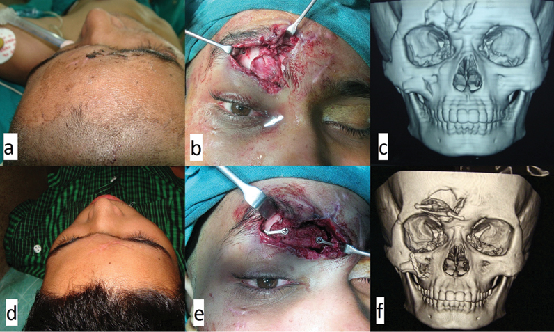
In photographic assessment, among the patients in upper face subgroup, those who had bone grafts had better results in the early postoperative period than those who did not have bone grafts. After three months the difference narrowed though neither was statistically significant. One of the patients had corticocancellous bone graft that got absorbed at a faster rate leading to inferior result [Table/Fig-10]. This was consistent with a study by Dado [15]. Among those who did not receive bone grafts results were better only in patients who had lesser number of fractured segments. In patients who had multiple comminuted fragments, bone resorption rate was higher which led to more frontal asymmetry in this group.
Iliac crest corticocancellous bone graft for frontal sinus defect. Preoperative photograph (a); intraoperative defect (b); preoperative CT scan (c); 3 months postoperative photograph (d); fixation with miniplates (e); postoperative CT scan (f) showing resorption of the cancellous part of the bone graft.
In mid-face, malar asymmetry was comparable between the two groups and had similar results though the group without grafts was marginally better. However it was not statistically different. When analysing individual cases, it revealed similar results like frontal symmetry. More the number of fracture segments in the comminuted fractures, ORIF with bone grafts gave better results.
The most significant comparison was done radiologically. In upper face fractures, mean projection deficit correction though was comparable between the two groups in the early postoperative period, at the end of 3 months, bone graft group showed statistically significant better results. Bone remodeling was better with bone grafts which gave better symmetry when compared to those who had multiple loose bone fragments that had gotten absorbed. Height and breadth deficits were also better corrected in the bone graft group. These suggest that in comminuted fractures in upper face, ORIF with bone grafts provide better results than only ORIF [Table/Fig-11].
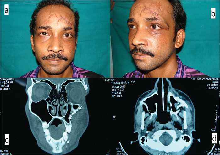
Photographic (a,b) and radiological; (c,d) evidence of left maxillary collapse following ORIF without bone grafts in comminuted fracture left Zygomatic Complex.
In mid-face fractures, mean zygomatic projection deficit was corrected better with bone grafts as better quality bone was used which allowed better purchase for stronger rigid fixation, thus preventing maxillary collapse. Mean zygomatic height deficit was slightly better corrected with bone grafts though not statistically significant.
When postoperative course and complications were compared, patients with bone grafts fared better and had lesser incidence of postoperative complications. Wound infection was present more with the only ORIF group. The reason for this was reduced amount of prosthetic materials employed in the patients in whom bone grafts were used. Rigid fixation and early vascularization of the bone grafts provided better wound stability than the other group. Plate related complications were also significantly higher in the only ORIF group as the incidence of loosening of screws and bone resorption were higher in this group. This also increased the chances of mid-face collapse. Two patients were treated conservatively and plates had to be removed in three other cases. For two more patients, revision surgery had to be done with the help of bone grafts after achieving wound stability.
Limitation
As the study period was restricted, sample size was small. Long-term follow up is needed to assess bone graft remodeling and resorption over extended period of time. Bone scan could have been used as a better indicator to assess structural integrity of the bone grafts used. A double blinded RCT could be done to reduce inter-observer variability.
Conclusion
Though treatment of orbital fractures with only ORIF gave better results and photographic assessment showed no statistical difference between the two groups, radiological assessment revealed that better results may be obtained when bone grafts are employed with ORIF in upper and mid-face fractures. The incidence of postoperative complications were also lesser when bone grafts were used.