Perineural cyst: An Unusual Cause of Radiating Low Backache in Children
Basudev Biswal1, Amit Kumar Satapathy2, Samarendra Mahapatro3, Rashmi Ranjan Das4
1 Senior resident, Department of Pediatrics, AIIMS, Bhubaneswar, Odisha, India.
2 Assistant Professor, Department of Pediatrics, AIIMS, Bhubaneswar, Odisha, India.
3 Associate Professor, Department of Pediatrics, AIIMS, Bhubaneswar, Odisha, India.
4 Assistant Professor, Department of Pediatrics, AIIMS, Bhubaneswar, Odisha, India.
NAME, ADDRESS, E-MAIL ID OF THE CORRESPONDING AUTHOR: Dr, Amit Kumar Satapathy, Sijua, Bhubaneswar-751019, Odisha, India.
E-mail: amitkumar.satapathy@yahoo.co.in
Low back pain, Radicular pain, Tarlov cyst
Dear Editor,
Backache remains a diagnostic challenge across all age groups. In children, low backache has various aetiologies including infections, tumor, rheumatologic conditions and connective tissue disorder. Perineural cyst though usually asymptomatic but rarely can present as radiating back pain in adults. Here, we are reporting a case of perineural cyst, a rare cause of backache in an adolescent boy.
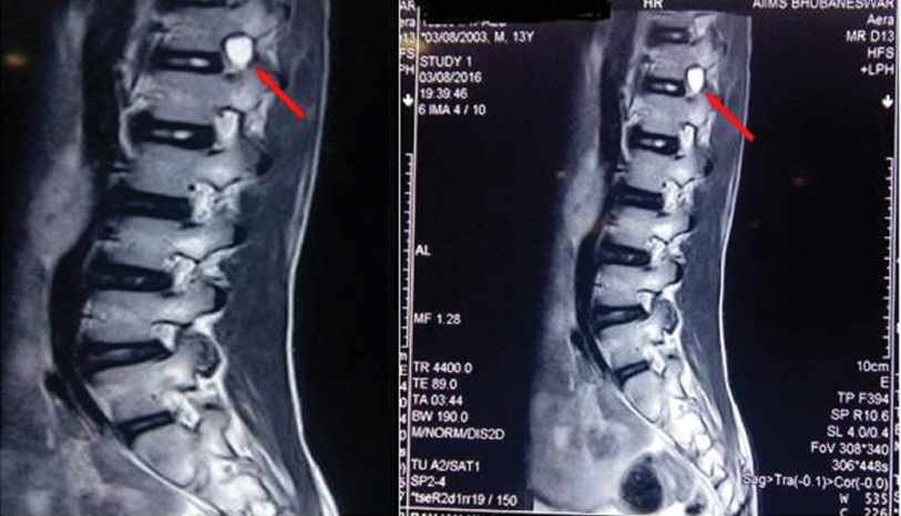
A 13-year-old boy presented with low backache for 2 months along with pain in bilateral hip joint and limping. There was no history of any joint swelling, redness of eye or oral ulcers or history of trauma. General physical examination was essentially normal except for mild pallor and bilateral microcornea which was present since birth. Systemic examination was noncontributory. Initial evaluation revealed hemoglobin of 10.4 gm/dl, total leucocyte count of 9700/cmm with platelet count of 4.1 lac/cmm. Erythrocyte Sedimentation Rate (ESR) was elevated (45 mm in 1st hr) with C-Reactive Protein (CRP) normal. X-Ray spine and hip was normal. USG hip was noncontributory whereas abdomen revealed presence of horseshoe kidney which was a an incidental finding. In view of persistent backache with limping, MRI spine was done which revealed a well defined T2 hyperintense lesion at the neural foramen on right side at D12/L1 level suggestive of perineural cyst [Table/Fig-1a,b]. The child was managed symptomatically with NSAIDs for 2 weeks along with diathermy, after which symptoms improved. Neurosurgery consultation was taken but did not require any urgent intervention. He is currently symptom free on subsequent follow ups.
(a,b). T2 weighted MRI spine showing hyperintensity at the neural foramen on right side at D12/L1 level.
Perineural cyst (also known as Tarlov cyst) usually originates in the perineural space between endoneurium derived from the pia mater and the perineurium formed by the arachnoid mater [1]. They occur along the nerve root near to the dorsal ganglion. The prevalence of Tarlov cyst is as high as 4.6%, mostly detected incidentally on MRI [2]. Usually these cysts are asymptomatic but may cause radicular pain with backache if large enough to cause local compression or stretching of the adjacent nerve roots. Rarely may have involvement of bowel and bladder if located in the sacrum [3]. Though initial aetiology describes as post traumatic by Tarlov in his series but subsequently congenital variants also have been reported without any previous history of trauma [1,4].
There is no definitive management protocol available for perineural cyst. Initial conservative management includes NSAID, oral steroid or epidural steroid injection along with physical therapy for symptomatic cases without any weakness [5,6]. Surgical intervention is warranted if there is failure to medical management or significant neurological involvement in form of motor weakness and bowel bladder involvement [7].
Perineural cyst, though an incidental finding mostly should be considered as a cause of backache with radicular pain in absence of any other aetiologies.
[1]. Tarlov IM, Spinal perineurial and meningeal cystsJ Neurol Neurosurg Psychiatry 1970 33(6):833-43. [Google Scholar]
[2]. Paulsen RD, Call GA, Murtagh FR, Prevalence and percutaneous drainage of cysts of the sacral nerve root sheath (Tarlov cysts)AJNR Am J Neuroradiol 1994 15(2):293-97. [Google Scholar]
[3]. Acosta FL Jr, Quinones-Hinojosa A, Schmidt MH, Weinstein PR, Diagnosis and management of sacral Tarlov cysts: Case report and review of the literatureNeurosurg Focus 2003 15(2):E15 [Google Scholar]
[4]. Schreiber F, Haddad B, Lumbar and sacral cysts causing painJ Neurosurg 1951 8(5):504-09. [Google Scholar]
[5]. Mitra R, Kirpalani D, Wedemeyer M, Conservative management of perineural cystsSpine (Phila Pa1976) 2008 33(16):E565-68. [Google Scholar]
[6]. Zibis AH, Fyllos AH, Arvanitis DL, Symptomatic cervical perineural (Tarlov) cyst: a case reportHippokratia 2015 19(1):76-77. [Google Scholar]
[7]. Landers J, Seex K, Sacral perineural cysts: imaging and treatment optionsBr J Neurosurg 2002 16(2):182-85. [Google Scholar]