Leprosy in Pregnancy: Obstetric Diligence is the Key
Rupali Bhatia1, Sumita Mehta2, Anshul Grover3, Swati Dubey4
1 Senior Resident, Department of Obstetrics and Gynaecology, Babu Jagjivan Ram Memorial Hospital, New Delhi, India.
2 Specialist, Department of Obstetrics and Gynaecology, Babu Jagjivan Ram Memorial Hospital, New Delhi, India.
3 Specialist, Department of Obstetrics and Gynaecology, Babu Jagjivan Ram Memorial Hospital, New Delhi, India.
4 Senior Resident, Department of Obstetrics and Gynaecology, Babu Jagjivan Ram Memorial Hospital, New Delhi, India.
NAME, ADDRESS, E-MAIL ID OF THE CORRESPONDING AUTHOR: Dr. Rupali Bhatia, A-503, Ashoka Apartments, Sector-9, Rohini-110085, New Delhi, India.
E-mail: drrupalibhatia@gmail.com
Leprosy in pregnancy is a rarely encountered event. Out of the 2000 patients detected of leprosy annually, two to three are pregnant women and majority of them are diagnosed in the third trimester. We, hereby, report a case of borderline tuberculoid leprosy with Type I lepromatous reaction in a 26-year-old pregnant woman in early second trimester with a large raised red coloured lesion over the forehead and six other small lesions involving the trunk and limbs with reduced sensory perception over involved skin. Occurrence of leprosy in an obstetric patient belonging to low prevalence area of India is infrequent especially in the post elimination era. However, we do need to have a high index of suspicion in lesions suggestive of the disease.
Antenatal, Hansen’s disease, Multidrug therapy, Skin lesions
Case Report
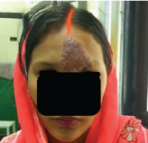
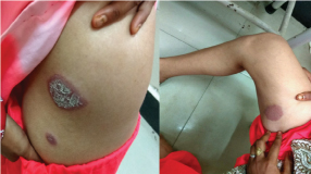
A 26-year-old pregnant woman with previous one live birth and two abortions presented to our antenatal clinic at 16 weeks gestational age, with large red coloured lesions over her face, limbs and trunk, which appeared two weeks back. The lesions first appeared over the face and then progressed to involve the upper limbs, back and lower limbs. These were initially small and skin coloured lesions that increased in size to become large, raised red coloured in two weeks. There was no associated history of localised itching or discharge. No history of generalised symptoms like fever, burning sensation in eyes, nasal symptoms, arthralgia, or myalgia was present. The patient denied any drug intake, preceding traumatic event or similar complaints in past. No history of similar lesions was elicited in the family members. She was illiterate and belonged to low socio-economic group. On examination, the largest lesion was present on the forehead which was 7 cm x 5 cm in size extending from root of the nose to upper part of the forehead in midline. From centre of the forehead, it laterally extended to involve medial two third of her left eyebrow. It was bright red in colour with raised margins and white scales along the periphery [Table/Fig-1]. She had six other similar lesions that were smaller in size over her inner thigh, forearm, feet and lateral trunk [Table/Fig-2]. All the lesions had reduced sensory perception over involved skin, however no motor deficit or thickened nerves were observed. Her general physical and obstetric examination showed no abnormality. To confirm the diagnosis, dermatologist opinion was sought and she was biopsied from two lesions. The slit skin-smear and punch biopsy revealed the diagnosis of borderline tuberculoid leprosy with Type I lepra reaction. She was started on tablets rifampicin (600 mg), clofazimine (300 mg) and dapsone (100 mg) to be taken once a month and clofazimine (50 mg) with dapsone (100 mg) on daily basis for 12 months, as per WHO treatment for multibacillary leprosy. She was also prescribed prednisolone (1 mg/kg/day) for 12 weeks as a management for the lepra reaction. She has entered her third trimester now and the multidrug therapy is being continued. The steroids have been stopped now after tapering the dosage. The lesions have regressed in size and become pale coloured. Her level II obstetric scan shows no congenital anomaly, her antenatal blood investigations are normal and she is on regular antenatal follow up.
Large raised lesion over forehead.
Lesions involving right side of the trunk and inner aspect of thigh.
Discussion
The global prevalence of leprosy is 0.32 per 10000 population and India accounts for 58.8% of the global burden. Around 127,000 confirmed cases of leprosy were registered in India in 2015 but it is rarely encountered in pregnancy. Globally out of the 2000 patients detected of leprosy annually, two to three are pregnant women and majority of them are diagnosed in the third trimester [1].
Leprosy is a chronic infectious disorder caused by bacterium Mycobacterium leprae. It has multi organ manifestations mainly involving the skin, the peripheral nerves, upper respiratory tract mucosa, and the eyes. The bacteria are transmitted through nasal or oral droplets during close contact with untreated cases explaining its higher prevalence in overpopulated and low socioeconomic groups [2]. Timely diagnosis and treatment may cure the disease completely, however if left untreated, it may result in irreversible damage of the involved organs.
WHO distinguishes the disease into paucibacillary and multibacillary leprosy, based on the number of skin lesions and bacterial load. In paucibacillary leprosy, there are few hypopigmented or pale macular lesions with reduced or absent sensory perception, whereas multibacillary leprosy is characterised by more than five skin lesions manifesting as plaques or nodules. Our patient presented with seven lesions as mentioned and hence, categorised as multibacillary leprosy. The disease is also classified as tuberculoid leprosy, borderline tuberculoid leprosy, borderline leprosy, borderline lepromatous leprosy, lepromatous leprosy, histoid leprosy and indeterminate leprosy [3]. The skin biopsy confirmed the presence of borderline tuberculoid leprosy in our patient.
Hypopigmented lesions of leprosy are simulated by other skin diseases such as psoriasis, eczema, dermatitis, tinea and lichenoid eruptions. These conditions are differentiated from leprosy based on distinctive lesions and their histopathology. In our patient, characteristic hypo-anaesthetic lesions without any other clinical symptoms along with the biopsy report paved the diagnosis of leprosy.
Hormonal and metabolic changes in pregnancy such as relative nutrient deficiencies and altered secretion of steroids account for depressed cellular immunity in pregnant women. Appearance of new lesions, reactivation of old disease or even relapse in treated patients can occur during pregnancy. The spectrum of symptoms observed in pregnant females is similar to those seen in general population. Owing to alterations in humoral and cell mediated immunity, leprosy reactions may also be triggered off. Type I or reversal reactions are more frequent in patients with borderline leprosy and presents with cutaneous manifestations as was observed in our patient. Type II reactions (erythema nodosum leprosum) manifest mainly with neuritis and subsequent sensory and motor deficit [3].
Widespread implementation of multidrug therapy and its safe use in pregnancy (with no teratogenicity) have aided in elimination of the disease and normal pregnancy outcomes [4,5]. Gimovsky AC and Macri CJ reported a case of lepromatous leprosy in a 27-year-old woman at 24 weeks gestation after she migrated from Mexico [6]. She had developed subcutaneous nodules on her arms, legs, back, and abdomen four weeks before she conceived. Throughout her pregnancy, she received multidrug treatment and her serial ultrasounds showed consistent foetal growth with no maternal complications. Maurus JN studied clinical course of leprosy in treated pregnant women and reported a low incidence of congenital malformations but an unusually high incidence of twinning and obstetric complications in a small subgroup of patients [7]. The risk for the development of the disease and of disease recurrence in previously treated patients is higher during pregnancy [8]. Contrary to above patients, our case had no previous history of leprosy in self or family and the physical findings did not reveal any positive findings of multiorgan involvement. Also, she belonged to a low prevalence region. However, in lieu of our high index of suspicion, ours being a tropical country, we investigated the case further and the diagnosis was eventually established.
The main determining factor for foetal and maternal outcome is the bacterial load, neuritis being the main complication in mothers [9]. Studies have shown that careful management of reactional states and treatment of women in pregnancy with dapsone therapy prevents nerve damage and in utero adverse effects [6,10]. Similarly, in our case, we found no adverse effects of the disease or its treatment in mother as well as foetus, as evident by level two scan and Doppler.
Conclusion
Occurrence of leprosy in an obstetric patient belonging to low prevalence area of India is infrequent especially in the post elimination era. Therefore, we do need to have a high index of suspicion in lesions suggestive of the disease, especially in absence of any suggestive history as in the above mentioned case. Early detection and treatment with WHO recommended multidrug therapy has no implications on maternal or foetal health.
[1]. World Health Organization. Leprosy. Fact sheet, updated February 2017 [Google Scholar]
[2]. Rodrigues LC, Lockwood DN, Leprosy now: epidemiology, progress, challenges and research gapsLancet Infect Dis 2011 11:464-70. [Google Scholar]
[3]. Sarkar R, Pradhan S, Leprosy and womenInt J Womens Dermatol 2016 2(4):117-21. [Google Scholar]
[4]. Duncan ME, Pearson JMH, Rees RJW, The association of pregnancy and leprosy II: Pregnancy in dapsone-resistant leprosyLeprosy review 1981 52(3):263 [Google Scholar]
[5]. Lockwood DN, Sinha HH, Pregnancy and leprosy: a comprehensive literature reviewInt J Lepr Other Mycobact Dis 1999 67(1):6-12. [Google Scholar]
[6]. Gimovsky AC, Macri CJ, Leprosy in pregnant woman, United StatesEmerg Infect Dis 2013 19(10):1693-94. [Google Scholar]
[7]. Maurus JN, Hansen’s disease in pregnancyObstet Gynecol 1978 52(1):22-25. [Google Scholar]
[8]. Boddinghaus BK, Ludwig RJ, Kaufmann R, Leprosy in a pregnant womanInfection 2007 35(1):37-39. [Google Scholar]
[9]. Duncan ME, Pearson JM, Neuritis in pregnancy and lactationInt J Lepr Other Mycobact Dis 1982 50(1):31-38. [Google Scholar]
[10]. Lyde CB, Pregnancy in patients with Hansen diseaseArch Dermatol 1997 133(5):623-27. [Google Scholar]