A Boy with a Congenital Sagittal Fissure of the Glans Penis Representing an Abortive Isolated Urethral Duplication
Dimitrios Sfoungaris1, Ioannis Valioulis2, Magdalini Mitroudi3, Ioannis Patoulias4, Christina Panteli5
1 Assistant Professor, Department of Paediatric Surgery, Aristotle University of Thessaloniki, Thessaloniki, Greece.
2 Assistant Professor, Department of Paediatric Surgery, Aristotle University of Thessaloniki, Thessaloniki, Greece.
3 Staff Surgeon, Department of Paediatric Surgery, Aristotle University of Thessaloniki, Thessaloniki, Greece.
4 Staff Surgeon, Department of Paediatric Surgery, Aristotle University of Thessaloniki, Thessaloniki, Greece.
5 Staff Surgeon, Department of Paediatric Surgery, Aristotle University of Thessaloniki, Thessaloniki, Greece.
NAME, ADDRESS, E-MAIL ID OF THE CORRESPONDING AUTHOR: Dr. Dimitrios Sfoungaris, Assistant Professor, Department of Paediatric Surgery, General Hospital ‘G. Gennimatas’, Ethnikis Aminis-41, 546 35, Thessaloniki, Greece.
E-mail: surgicalpediatrics@gmail.com
Birth defect, Epispadias, Penile cleft, Supernumerary urethra
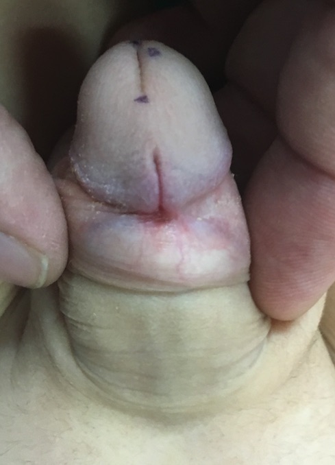
An eight-year-old boy presented with a midline sagittal fissure on the dorsal aspect of the glans penis, which was present since birth and never caused any symptoms. The fissure was 1 cm long extending distally from the corona to almost half way towards the tip of the glans [Table/Fig-1]. Its maximum depth was 2 mm at the coronal side, gradually becoming shallower distally [Table/Fig-2]. The prepuce was complete and intact, with a normal frenulum and was easily retractable. No other abnormality was evident on the penis. No chordee or any other abnormal structure was palpable. The urethral meatus was located at the tip of the glans, inside a normal slit. The urine stream was normal and no urine leak was observed at any site. The child did not have any other health problems and never had any urological complaints. An ultrasonography examination revealed no pathology of the lower or upper urinary system and no abnormal tissue appeared in the penis. The pubic bones showed no dissociation at the symphysis pubis. A diagnosis was reached of an abortive form of duplicate urethra.
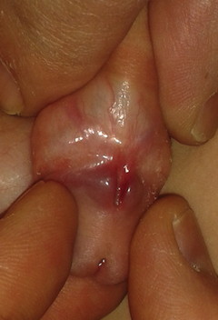
The sides of the fissure are retracted to show the depth of the fissure.
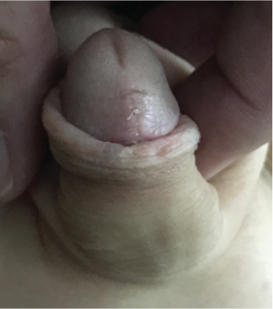
The resection of the fissure was decided for cosmetic reasons. Nevertheless, with the parents’ consent, we decided to deglove the penis dorsally for about 2 cm and explore the dorsal aspect of the corpora cavernosa for any possible anomaly, but no lesion was discovered. At this point, we also performed an artificial erection test during which the penis assumed a normal erect position, excluding the presence of any tethering chordee, a possible remnant of a supernumerary urethra. The glans was reconstituted by suturing the sides around the resected fissure. Histological examination of the resected fissure revealed a stratified epithelial lining with scarce regions of keratinization. The patient was discharged the next day and was examined at three, seven and 23 days post discharge. Healing and cosmesis were satisfactory, and there were no complaints from the parents or the boy [Table/Fig-3].
Compete healing three weeks after surgical correction.
Discussion
Since, the prepuce was intact, and the fissure was positioned exactly along the midline, a pressure disruption deformation from a congenital band was excluded as aetiology. Dorsal midline penile anomalies presenting with a cleft have been described as a part of the bladder-epispadias exstrophy complex [1] or as manifestations of an underlying urethral duplication [2-4]. The presence of a normal functional urethra excludes the diagnosis of an abortive epispadias case.
Urethral duplication presents as a spectrum from simply having a dorsal blind ending sinus, sometimes extending proximally to the symphysis pubis, to a completely duplicated urethra and even a duplication of the bladder, as well [2-4]. No case, as far as we know, has been described with a sagittal fissure of the glans standing alone. Only a photograph of a case resembling ours has been published in a textbook [5]. The presence of a normal functional urethra and the absence of any other signs suggested the diagnosis of an abortive form of urethral duplication, at the most moderate side of the spectrum.
The glanular urethra develops from a nearly solid urethral plate at the tip of the genital tubercle apparrent on the 13th week, 25 mm embryo, when the mesenchyme is still diffuse and not differentiated into glans and corpora cavernosa [6]. At this time, a well-marked urethral groove at the tubercle’s undersurface, that does not reach the tip, represents the origin of the penile urethra. On the 14th week embryo, a glans penis can be identified, with a fine solid indentation at the tip representing the epithelial plate which extends proximally into the glans penis. At the mid glans, the epithelial plate is vacuolated forming a glanular canal. At the level of the proximal glans, the vacuolization reaches the undersurface and thus, a deep glanular urethral groove is created. The distal migration of the glanulo-preputial lamella, which forms the prepuce, causes the fusion of the edges of the glanular groove as well, forming the floor of the glanular urethra under the frenulum.
We believe that the anomaly presented on our image is caused by a duplicated aberrant morphogenesis of the proximal part of the glanular urethra, creating a fissure, much in the same way as the normal ventral proximal glanular urethral groove is created. However, the aberrant groove is not closed to form a tunnel because the fusion of the glanulo-preputial lamella does not happen dorsally.
[1]. Bos EM, Kuijper CF, Chrzan RJ, Dik P, Klijn AJ, De Jong TP, Epispadias in boys with an intact prepuceJ Pediatr Urol 2014 10(1):67-73. [Google Scholar]
[2]. Onofre LS, Gomes AL, Leão JQ, Leão FG, Cruz TM, Carnevale J, Urethral duplication-a wide spectrum of anomaliesJ Pediatr Urol 2013 9(6):1064-71. [Google Scholar]
[3]. Salle JL, Sibai H, Rosenstein D, Brzezinski AE, Corcos J, Urethral duplication in the male: review of 16 casesJ Urol 2000 163(6):1936-40. [Google Scholar]
[4]. Lima M, Destro F, Maffi M, Persichetti Proietti D, Ruggeri G, Practical and functional classification of the double urethra: A variable, complex and fascinating malformation observed in 20 patientsJ Pediatr Urol 2017 13(1):42e1-42e7. [Google Scholar]
[5]. Zaontz MR. Guide to Pediatric Urology and Surgery in Clinical Practice. London, Springer, 2011. Disorders of Male External Genitalia: Hypospadias, Epispadias, Concealed Penis, and Urethral Disorders. Pp. 61-82 [Google Scholar]
[6]. Hadidi AT, Roessler J, Coerdt W, Development of the human male urethra: a histochemical study on human embryosJ Pediatr Surg 2014 49(7):1146-52. [Google Scholar]